Hot Topic
Depression in cancer: The many biobehavioral pathways driving tumor
progression
Beatrice Bortolato
a, Thomas N. Hyphantis
b, Sara Valpione
c,d, Giulia Perini
e, Michael Maes
f,g,h,i,j,
Gerwyn Morris
k, Marta Kubera
l, Cristiano A. Köhler
m, Brisa S. Fernandes
n,o, Brendon Stubbs
p,q,r,
Nicholas Pavlidis
s, André F. Carvalho
m,⇑aDepartment of Mental Health ULSS 10 ‘‘Veneto Orientale”, Venice, Italy
bDepartment of Psychiatry, Division of Medicine, School of Health Sciences, University of Ioannina, Greece cDepartment of Surgery, Oncology and Gastroenterology, University of Padova, Padova, Italy
dMedical Oncology, The Christie NHS Trust, Manchester, United Kingdom eDepartment of Neurosciences, University of Padova, Padova, Italy
fIMPACT Strategic Research Centre, Deakin University, School of Medicine and Barwon Health, Geelong, VIC, Australia gDepartment of Psychiatry, Faculty of Medicine, Chulalongkorn University, Bangkok, Thailand
hDepartment of Psychiatry, Faculty of Medicine, State University of Londrina, Londrina, Brazil iDepartment of Psychiatry, Medical University Plovdiv, Plovdiv, Bulgaria
jRevitalis, Waalre, The Netherlands
kTir Na Nog, Bryn Road Seaside 87, Llanelli SA152LW, Wales, UK
lDepartment of Experimental Neuroendocrinology, Institute of Pharmacology, Polish Academy of Science, Krakow, Poland mDepartment of Clinical Medicine and Translational Psychiatry Research Group, Faculty of Medicine, Fortaleza, CE, Brazil nDeakin University, IMPACT Strategic Research Centre, School of Medicine, and Barwon Health, Geelong, Australia
oLaboratory of Calcium Binding Proteins in the Central Nervous System, Department of Biochemistry, Federal University of Rio Grande do Sul, Porto Alegre, Brazil pPhysiotherapy Department, South London and Maudsley NHS Foundation Trust, Denmark Hill, London SE5 8AZ, United Kingdom
qHealth Service and Population Research Department, Institute of Psychiatry, Psychology and Neuroscience, King’s College London, De Crespigny Park, London Box SE5 8AF, United Kingdom
rFaculty of Health, Social Care and Education, Anglia Ruskin University, Bishop Hall Lane, Chelmsford CM1 1SQ, United Kingdom sDepartment of Medical Oncology, Faculty of Medicine, School of Health Sciences, University of Ioannina, Ioannina 45110, Greece
a r t i c l e
i n f o
Article history:
Received 12 April 2016
Received in revised form 15 October 2016 Accepted 5 November 2016
Keywords:
Major Depressive Disorder Cancer
Inflammation HPA axis Stress Psychiatry
a b s t r a c t
Major Depressive Disorder (MDD) is common among cancer patients, with prevalence rates up to four-times higher than the general population. Depression confers worse outcomes, including non-adherence to treatment and increased mortality in the oncology setting. Advances in the understanding of neurobiological underpinnings of depression have revealed shared biobehavioral mechanisms may contribute to cancer progression. Moreover, psychosocial stressors in cancer promote: (1) inflammation and oxidative/nitrosative stress; (2) a decreased immunosurveillance; and (3) a dysfunctional activation of the autonomic nervous system and of the hypothalamic–pituitaryadrenal axis. Consequently, the prompt recognition of depression among patients with cancer who may benefit of treatment strategies targeting depressive symptoms, cognitive dysfunction, fatigue and sleep disturbances, is a public health priority. Moreover, behavioral strategies aiming at reducing psychological distress and depressive symp-toms, including addressing unhealthy diet and life-style choices, as well as physical inactivity and sleep dysfunction, may represent important strategies not only to treat depression, but also to improve wider cancer-related outcomes. Herein, we provide a comprehensive review of the intertwined biobehavioral pathways linking depression to cancer progression. In addition, the clinical implications of these findings are critically reviewed.
Ó2016 Elsevier Ltd. All rights reserved.
Introduction
Major Depressive Disorder (MDD) is more common among indi-viduals affected by cancer as compared to the general population. While the average rate of MDD is 3.3% in the general population,
http://dx.doi.org/10.1016/j.ctrv.2016.11.004
0305-7372/Ó2016 Elsevier Ltd. All rights reserved.
⇑Corresponding author at: Department of Clinical Medicine, Faculty of Medicine, Federal University of Ceará, Rua Prof. Costa Mendes, 1608, 4o andar, 60430-040 Fortaleza, CE, Brazil. Fax: +55 85 33668054.
E-mail address:andrefc7@terra.com.br(A.F. Carvalho).
Contents lists available atScienceDirect
Cancer Treatment Reviews
the prevalence of MDD among individuals with cancer is approxi-mately 12.5%, which is up to four-times the rate reported in population-based samples[1]. Evidence indicates that co-morbid depression may be associated with a worse prognosis and increased mortality rate in cancer populations[2]. In fact, depres-sion is an independent predictor of more frequent and longer hos-pitalizations, diminished quality of life and decreased compliance to treatments[3]. Meta-analytic evidence indicates that depression triples the risk for non-adherence to medications in women with breast cancer[4]. Moreover, demotivation may promote maladap-tive depressive coping styles with a detrimental effect on survival [5]as well as a possible increase in suicidality[6]. Notwithstanding depression is under-recognized in populations with cancer, limited evidence provides support for the routine screening of distress in cancer populations[7]. Importantly, the treatment of depression has been associated with increased survival in women with meta-static cancer[8]. Furthermore, interventions aiming at improving depression management, education and behavioral activation (i.e. emphasizing pleasant event scheduling and overcoming avoidance behaviors), along with the use of antidepressant treatments when appropriate, have been recognized as effective in reducing depres-sive burden in individuals with cancer[9].
Notwithstanding the burgeoning and stressful potential associ-ated with a diagnosis of cancer, the large prevalence of depression among individuals with cancer is likely to not be entirely explained by the effects of psychological distress. In recent years, advances in the neurobiology of depression and in the physiopathology of can-cer have led to the identification of some shared bio-behavioral mechanisms[10–14]. This field has witnessed an ever increasing accumulation of emerging findings, which deserve a critical review.
Therefore, this comprehensive narrative review aims to: (1) explore epidemiological links between depression and cancer pro-gression; (2) summarize biological mechanisms relevant for the development and recurrence of depressive episodes as well as for cancer progression, with potential mutual reciprocal interactions; and (3) describe behavioral interventions that may target mecha-nistic pathways relevant to both depression and cancer progression.
Search strategy
A comprehensive search for peer-reviewed articles published in English was performed in the PubMed/Medline database including the following search terms: ‘‘Major Depressive Disorder”, ‘‘cancer”, ‘‘tumor”, ‘‘inflammation”, ‘‘oxidative and nitrosative stress”, ‘‘HPA axis”, ‘‘diet”, ‘‘microbiota”, ‘‘physical exercise”, ‘‘sleep dysfunction” up until February 15th, 2016. Bibliographies from selected articles were reviewed to identify additional original reports aligned with our objectives.
Epidemiological links between depression and cancer
Most studies report that the prevalence of depression in onco-logical populations range from 15% to 30%, with variations attribu-table to different screening tools and diagnostic criteria across studies[15]. These heterogeneous results are in part explainable by the fact that only a limited number of studies have assessed the prevalence of depression in cancer samples by means of a val-idated structured diagnostic interview according to criteria estab-lished in the Diagnostic and Statistical Manual of Mental Disorders (DSM) or the International Classification of Diseases (ICD). On the contrary, it is noteworthy that self-rated reports of depression or the detection of depressive symptoms by clinicians do not substan-tiate a reliable diagnosis of MDD. In keeping with this view, it is not
surprising that the prevalence of individuals meeting a diagnosis of MDD according to a structured clinical interview is relatively lower compared to the prevalence of depressive symptoms among indi-viduals with cancer [16]. Meta-analytic evidence of 24 studies involving 4007 subjects from palliative-care settings documented a prevalence of MDD as defined according to the DSM-IV of 16.5% (95% CI 13.1–20.3), while the prevalence of all types of depression was 20.7% (12.9–29.8) and that of minor depression was 9.6% (3.6–18.1) [17]. A recent meta-analysis examining the role of depression as a putative risk of cancer has been recently conducted[18]. This meta-analysis of nine observational studies demonstrated that a DSM or ICD diagnosis of depression was not conclusively associated with cancer risk. In addition, there was a lack of well-designed prospective cohort studies examining this association[18].
Depressive symptoms typically develop during the first year after cancer diagnosis and usually decline as a result of adaptive adjustments to psychological stress related to the diagnosis[19]. For instance, in an epidemiological survey exploring the prevalence of depressive symptoms among subjects of five biennial waves of the Health and Retirement Study, involving 8,387 individuals aged 51–61, subjects diagnosed with cancer had the highest hazard ratio of depressive symptoms within 2 years following initial diagnosis (HR 3.55; 95%, CI, 2.79–4.52), when compared to individuals diag-nosed with diabetes, hypertension, heart disease, arthritis, chronic lung disease or stroke [20]. The chances of developing a new depressive episode following a diagnosis of cancer may be elevated in certain vulnerable groups (e.g., those patients with a previous history of depressive episodes), as suggested by an observational study exploring the relationship between acute stress disorder and subsequent depressive symptoms after a diagnosis of head/-neck/lung cancer[21]. In this study up to 29% of individuals met the criteria for MDD within the first month after the diagnosis, of whom nearly one-third had a positive history for previous depres-sive episodes[21].
The role of stress and inflammation
Tumorigenesis and cancer progression are characterized by the acquisition of six biological attributes, representing the ‘‘hallmarks of cancer” namely (1) resisting cell death; (2) sustaining prolifera-tive signaling; (3) evading growth suppressors; (4) inducing angio-genesis (5) resisting cell death; and (6) activating invasion and metastasis[31]. Continued oxidative stress can propitiate chronic inflammation, which may influence cancer progression. Hence, oxidative stress can activate transcription factors and lead to the expression of over 500 different genes, including those related to growth factors, pro-inflammatory cytokines, chemokines, cell-cycle regulatory molecules, and anti-inflammatory molecules, sug-gesting that oxidative stress, chronic inflammation, and cancer are closely intertwined[32].
Host factors, including cell-mediated immunity, are involved in surveillance and destruction of tumor cells, especially in early disease stages. Stress-induced processes can directly modulate these tumor hallmarks and a large body of preclinical and clini-cal evidence supports the role of inflammation as an essential factor for tumor growth and progression [1]. Pro-inflammatory cytokines are produced by tumor and stromal cells, as well as by tumor-associated macrophages and infiltrating T-cells, and play an important role in tumor angiogenesis, metastatic spread, and possibly resistance to therapy [22,33]. In addition, stress-related effects on angiogenesis may be regulated by neural con-trol mechanisms. In particular, stressors promote angiogenesis via enhanced adrenergic transmission, which stimulate the pro-duction of cytokines (e.g., IL 6, IL-8, TNF-
a
) and vascular endothelial growth factor (VEGF) by the activation of cognate beta-adrenergic receptors on tumor and stromal cells [34]. Fur-thermore, stress-induced activation of the sympathetic nervous system promotes metastatic invasion via an increase in the expression of enzymes endowed with the ability of destroying the cellular matrix surrounding the tumor, e.g. matrix metallo-proteinases (MMP) 2 and MMP-9, which are produced by both tumor cells and macrophages in the tumor microenvironment and may be regulated by stress-triggered factors [35]. On the contrary, the activation of the parasympathetic nervous system appears to down-regulate the immune response and dampen the production of pro-inflammatory cytokines, through cholinergic-mediated decrease in the expression of nuclear factor kappa B (NF-j
B), an effect referred to as the ‘‘cholinergic anti-inflammatory pathway” [36].The production of cytokines in the periphery leads to an amplification of the pro-inflammatory response at the central nervous system (CNS) via an increase in the permeability of the blood–brain barrier (BBB) on circumventricular organs and in the choroid plexus, or via afferent vagal fibers innervating the nucleus of the solitary tract [37]. In addition, growing evidence indicates that perivascular monocytes may infiltrate the brain, promoting the spread of inflammatory stimuli to microglia and astrocytes, which in turn become the main source of immune-inflammatory mediators within the brain [38]. A large body of preclinical and clinical data indicates that an increase in pro-inflammatory cytokines are associated with the so-called ‘‘sick-ness syndrome”, whose manifestations include anhedonia, irri-tability, psychomotor retardation, fatigue, anorexia, insomnia, and increased pain sensitivity[39]. Those symptoms overlap to some extent with the clinical manifestations of depression and are partly responsive to antidepressant treatments. Thus, the immune cross-talk between the central nervous system (CSM) and the periphery is thought to be largely responsible for the development of somatic and affective symptoms of depression under pro-inflammatory conditions, while also possibly contribut-ing to carcinogenesis[40].
Inflammatory aberrations associated with depression
Literature supporting the role of inflammation in depression is extensive. Increased expression of innate immune mediators, including cytokines and acute phase reactants, chemokines and soluble receptor molecules are among the most replicated findings not only in peripheral blood but in the cerebrospinal fluid (CSF) or in post-mortem brain samples of suicide victims[41]. For instance, some meta-analyses have documented higher peripheral levels of pro-inflammatory cytokines namely IL-6, IL-8, IL-1b, TNF-
a
, sol-uble IL-2 receptor (sIL-2R) and C-reactive protein (CRP) in individ-uals with MDD when compared to healthy controls [42,43]. Conversely, in longitudinal population studies, higher CRP levels have been demonstrated to predispose individuals in developing subsequent depressive symptoms, thus supporting that the notion that the activation of the immune system may contribute to the patho-etiology of depression [44]. Further support to this idea derives from evidence that the administration of interferon alpha (IFN)-a
to patients with cancer is associated with the development of depressive symptoms, that only partially respond to conven-tional antidepressant treatments[45]. Polymorphisms in inflam-matory cytokine genes, including those encoding for IL-1b, TNF-a
and CRP, have also been related to antidepressant treatment response[46].In addition to evidence instantiating the role of innate immu-nity in the pathophysiology of depression, other lines of research focused on alterations in adaptive cellular immunity[47]. In fact, increased levels of pro-inflammatory cytokines and reduced signaling of anti-inflammatory mediators, such as IL-10 and trans-forming growth factor beta (TGF-b), may direct the differentiation of T naïve cells towards the T helper 1 (Th1)/Th17 phenotype instead of T regulatory (T reg) cells, further promoting the produc-tion of pro-inflammatory mediators (e.g., IFN, IL-2) in a vicious cir-cle[48,49]. Pro-inflammatory cytokines stimulate the activation of the enzyme indoleamine 2–3 dioxygenase (IDO), which converts tryptophan into kynurenine (KYN), leading to serotonin depriva-tion[50]. In microglial cells, KYN may be further converted into quinolinic acid (QUIN), a powerful N-methyl-D-aspartate (NMDA) agonist and stimulator of glutamate release. QUIN and other tryptophan catabolites induce the further production of pro-inflammatory cytokines and potentiate oxidative and nitrosative stress provoking lipid peroxidation, mitochondrial dysfunction and DNA damage[51]. In this context, it is noteworthy to highlight that recent research also pointed to a role of IDO in the control of inflammation and in peripheral immune-tolerance in cancer patients[52]. In fact, IDO activation can be triggered by innate responses during tumorigenesis, and also by T cell activation, thus representing a potential target for immunotherapy.
Finally, cascades associated with the activation of the afore-mentioned pathways appear to lead to excitotoxic neural damage, a reduced expression of brain derived neurotrophic factor (BDNF) and consequently to a disruption of mechanisms subserving neural plasticity [53,54]. In addition, oxidative stress may reduce the activity of 5,6,7,8-tetrahydrobiopterin (BH4), which is a co-factor for several aromatic amino acid monooxygenases and is rate-limiting for the biosynthesis of the neurotransmitter serotonin and the catecholamines dopamine, epinephrine and nore-pinephrine[55]. Thus, the reduced biosynthesis of catecholamines may also be related to disturbed adrenergic neurotransmitter path-ways under chronic low-grade inflammatory states.
in particular for a subset of individuals with MDD exhibiting low-grade chronic activation of pro-inflammatory pathways. For instance, treatment with the TNF antagonist infliximab was effec-tive in reducing depressive symptoms in individuals with treatment-resistant MDD showing baseline CRP levels >3 mg/dl [57]. In this study, treatment response was associated with tran-scriptional signatures related to glucose and lipid metabolism [58]. Maladaptive habits, including unhealthy diet and smoking, as well as environmental factors, including childhood abuse, have been consistently associated with pro-inflammatory activation [59]. For instance, individuals with a history of childhood abuse exhibit higher plasma CRP, IL-6 and TNF-
a
levels during adulthood in comparison to healthy controls[60]and are exposed to greater risk of chronic diseases in adulthood including cancer, depression, cardiovascular disease and metabolic syndrome [61]. Conse-quently, the characterization of a subset of MDD subjects more marked disruption of pro-inflammatory pathways has emerged as a relevant priority in this field of research.Immune activation and alterations in stress response in depression associated with cancer
The presence of comorbid depression has been demonstrated to have a negative prognostic impact across different cancer popula-tions. For instance, depression has been associated with shorter survival in patients with renal carcinoma[62]. Of note, a subgroup analysis revealed that increased gene expression related to inflam-matory pathways and oxidative stress as well as greater activity of the nuclear transcriptional factor NF-kB in circulating leucocytes, were positively associated with depressive symptoms’ severity [62]. In keeping with this view, elevated concentrations of cytoki-nes (e.g., IL-6 and TNF-
a
) have been associated with later cancer stages, poor differentiation and tumor size/volume across a variety of malignancies. Therefore, the detrimental prognostic impact of depression in comorbidity with cancer may be at least in part mediated by the activation inflammatory cascades[22]. For exam-ple, increased plasma levels of IL-6 were observed in association with depressive symptoms in individuals with advanced ovarian cancer as compared to subjects with tumors of low-grade malig-nant potential[27]. Similarly, IL-6 levels have been associated with depressive symptoms in individuals with cancer[10]. In addition, increased plasma levels of the IL-1 receptor antagonist and soluble TNF receptor 2 (sTNFR2) correlated with fatigue in breast cancer survivors up to 5 years after the diagnosis[63]. Lastly, the blockade of cytokines’ signaling has been associated with reductions in depressive symptoms and better quality of life in patients with advanced cancer[64].Unsurprisingly, abnormalities in the stress responses appear to be closely linked to the immune activation. For instance, pro-inflammatory activation is highly associated with hypothalamic– pituitary–adrenal (HPA) axis dysfunction, notably higher evening cortisol levels [27]. A dysregulated pattern of cortisol secretion has been consistently associated with depression and fatigue in cancer patients[65]. Elevated nocturnal cortisol levels and a flat-tened diurnal cortisol slope have been observed in individuals with breast, ovarian, cervical cancer and lymphoma, and may predict poor survival[66]. These abnormalities in cortisol secretion appear to stimulate the growth and survival of cancer cells, leading to can-cer treatment resistance and inhibiting apoptosis of tumor cells [67]. Moreover, glucocorticoids negatively influence DNA repair mechanisms and modulate the transcription of several genes[68]. The treatment of cancer may in part mitigate pro-inflammatory aberrations and the dysfunctional activation of the HPA axis. For instance, surgical treatment followed by chemotherapy in high-risk ovarian cancer patients was found to be effective in reducing IL-6 levels and nocturnal cortisol and in improving neurovegetative
symptoms of depression, fatigue and quality of life[69]. However, other lines of evidence indicate that cancer treatments may induce per se inflammatory pathways relevant to the development of depressive symptoms. For instance, chemotherapy has been asso-ciated with an increase in the activity of NF-kB as well as an increase in levels of IL-6 and sTNFR2 in parallel to increases in depressive symptoms in women with breast cancer[70].
Further support to the involvement of inflammation in the development of cancer-associated depression derives from the evi-dence that subjects with cancer receiving IFN treatment have a greater likelihood to exhibit depressive symptoms. Up to 50% of individuals with melanoma treated with IFN develop a variety of neurobehavioral symptoms including psychomotor retardation, anorexia, insomnia, and fatigue[71]. The presence of depressive symptoms prior to IFN treatment has been recognized as one of the most robust predictors of depression, and it may be an exclu-sion criterion for IFN therapy because of the substantial risk of depression[72].
Use of antidepressants and cancer
The use of antidepressant treatment is associated with reduc-tions in depressive symptoms across cancer samples, as docu-mented by several randomized controlled trials (RCTs) utilizing mianserin[73], serotonin reuptake inhibitors (SSRIs; e.g. fluoxetine and paroxetine)[71]as well as tricyclic antidepressants (TCAs; e.g. amitriptyline and desipramine)[74]. In addition, some antidepres-sants (e.g. mirtazapine) may also alleviate chemotherapy-induced nausea and cancer-related cachexia-anorexia[75,76]. Prophylactic treatment with paroxetine was effective in reducing depressive symptoms after IFN treatment[71]. Therefore, the assessment of depressive symptoms among individuals with cancer prior to treatment initiation may aid in the identification of vulnerable subjects for whom prophylactic antidepressant treatment may be indicated. However, since only up to 30% of individuals with MDD achieve symptomatic remission following a trial with a first-line antidepressant treatment[77], the identification of novel alternative or complementary treatment strategies is a priority for improving outcomes of depressed individuals suffering from cancer.
On the other hand, several clinical and experimental studies have suggested a link between antidepressant use and cancer [78]. Epidemiological studies on the effects of antidepressants on cancer prognosis suggest that the long-term use of TCAs and SSRIs may increase the risk of breast [79,80]and ovarian cancer [81], whereas the use of TCAs may increase risk of prostate cancer and non-Hodgkin’s lymphoma [82]. However, several further studies did not find a significant association between antidepressant treat-ment and risk towards breast and ovarian cancers[83], and non-Hodgkin’s lymphoma [84]. Moreover, regular use of SSRIs may reduce risk of colorectal[85]and lung[86]cancers.
mechanism of metastatic promotion by pretreatment with antide-pressant drugs before the inoculation of tumor cells deserve fur-ther examination, and may have relevant clinical implications. For example, cancer patients taking antidepressants before surgical manipulation may show an increased risk of cancer cell dissemina-tion during surgery[90], although further studies are required to fully appreciate the benefits and harms of antidepressant use in individuals with malignancies.
Effects of oxidative and nitrosative stress (O&NS)
Chronic inflammation characterized by elevated levels of pro-inflammatory cytokines leads to activation of inducible nitric oxide synthase (iNOS) and increased activity of several transcription fac-tors, such as NF-
j
B (nuclear factorj
B), AP-1 (activator protein 1) and STAT3 (signal transducer and activator of transcription 3) lead-ing in turn to upregulated activity of NADPH oxidase (NOX) and mitochondrial oxidoreductases [91]. The activation of these enzymes and transcription factors lead to the chronic activation of a wide range of inflammatory pathways subsequent to the up-regulation of COX-2 (cyclo-oxygenase-2), and the development of a state of chronic O&NS characterized by excessive production of reactive oxygen species (ROS), such as the superoxide anion, and reactive nitrogen species (RNS), such as nitric oxide (NO), which are known to play a major role in the pathogenesis and pathophys-iology of major depression and inflammatory cancers[92].There is now evidence that depression is accompanied by an increase in O&NS as indicated by an increase in lipid peroxidation, oxidative and nitrosative damage to DNA, nitrosative damage to proteins, hypernitrosylation, autoimmune responses to oxidatively and nitrosatively modified epitopes (neoepitopes, including malondialdehyde (MDA), oxidized LDL and NO-adducts [93,94]. These activated O&NS pathways are partly related to lowered levels of key antioxidants including lowered levels of coenzyme Q10, zinc, vitamin E, glutathione, HDL-cholesterol, albumin, etc. [93]. In addition, increased ROS, RNS, lipid peroxidation, and low-ered levels of antioxidants such as zinc and coenzyme Q10 may aggravate existing inflammatory responses, and may cause autoimmune responses, which may further contribute to immune-inflammatory aberrations [93]. Another dimension of pathology involves the consequences of hypernitrosylation, that is increased nitrosylation of cysteine groups in selected proteins by nitric oxide (NO)[93]. There is some evidence that abnormal levels of this post-transcriptional modification may contribute to neurodegeneration, abnormal hippocampal long-term potentia-tion, impaired synaptogenesis, and lowered neuronal survival [93,94].
An abundant amount of evidence has illustrated that activated O&NS pathways play a role in the pathophysiology and maybe the onset of specific cancers. Firstly, elevated levels of ROS and RNS, in an environment of compromised cellular antioxidant defences, may damage DNA, lipids and proteins and may lead to DNA mutations[92]. Such macromolecular damage leads to com-promised protein functions, mitochondrial impairment, compro-mised cell membrane integrity, genetic instability and the formation of damage associated molecular patterns whose engage-ment with pattern recognition receptors can create a cascade of self-amplifying immune-inflammatory and O&NS pathology as detected in different cancers[95]. ROS signaling plays a major role in the regulation of metabolism, cell fate, tumor cell proliferation, angiogenesis, and metastasis. A second dimension of pathology involves the consequences of abnormal nitrosylation of cysteine groups in selected proteins by NO[96]. S-nitrosylation is now rec-ognized as a major pathway enabling the regulation of redox based protein signal transduction by NO in a cellular environment[97,98] and its dysregulation can produce a plethora of pathogenic
consequences in several disease areas, including cancer [99]. A state of hypernitrosylation caused by excessive levels of NO and dysregulated S-nitrosylation are implicated in the genesis and progression of cancer as well as treatment resistance[100]. For example, tumor growth is regulated by S-nitrosylation of Ras and the epidermal growth factor receptor [101]. Cellular migration and cancer invasiveness may be enhanced by nitrosylation on the integrin alpha chain and nitrosylation of c-Src[102] in prostate and breast cancers. Dysregulated S-nitrosylation also seems to explain, at least in part, the Janus faced role of NO in cancer whereby the molecule exerts pro-tumorigenic or anti-tumorigenic effects depending on cellular context and concentra-tion. The weight of evidence indicates that moderately elevated levels of protein nitrosylation stimulate the development and progression of cancers while excessive levels provoke the activa-tion of enzyme signaling cascades such as the Ras/Erk/MAP kinase pathway leading to cellular apoptosis[103].
Stress as a modulator of immune response in depression and cancer
Psychosocial stressors including low perceived social support, lack of familiar ties as well as history of childhood trauma and adverse life experiences appear to increase the susceptibility to chronic diseases, including MDD and cancer [104]. Moreover, chronic psychosocial stress increases cancer mortality across a diverse array of cancer types (e.g., breast, lung, head and neck, hep-atobiliary, lymphoid, and hematopoietic cancers)[105,106]. A pos-sible behavioral explanation implies the fact that psychological stress exerts detrimental effects on sleep architecture integrity and increases fatigue. In addition, increased threat perception may promote unhealthy lifestyle choices including dietary habits, smoking and alcohol abuse, and lack of physical activity. Further-more, stressors may be responsible for cognitive complaints, espe-cially in the domain of memory, which could have a role in diminishing treatment compliance[107].
However, at a mechanistic level, mounting evidence indicates that chronic stress confer increased vulnerability to chronic dis-eases amplifying inflammatory signals via an increased pro-duction of cytokines and other immune mediators, as well as via a decreased sensitivity to hormonal inhibitory control by the HPA axis, as well altered autonomic responses[108,109]. More-over, a pro-inflammatory milieu promotes oxidative and nitrosa-tive stress that may promote mitochondrial dysfunction and DNA damage[110]. This view is supported by evidence indicating that stressful experiences increase immune responses even in healthy individuals. For instance, the exposure to the Trier Social Stress Test (TSST), a test including tasks of mental arithmetic and public speaking, has been associated with activation of NF-
j
B in periph-eral blood mononuclear cells [111]. However, individuals with MDD with early life stress exhibit an exaggerated pro-inflammatory activation in comparison to healthy individuals to psychosocial stressors[112].transmission is associated with increased invasive potential through increased expression in MMPs[34]. Moreover, stress motes tumor cells resistance to programmed cell death in the pro-inflammatory tumor microenvironment [117]. Lastly, beta-adrenergic effects induce the loss of phagocytic abilities by tumor-associated macrophages and promote the propagation of the inflammatory response[35].
High levels of psychological distress and low social support pre-dict not only impaired activity of the immune innate system, but worse immune cellular response as well. In individuals with stage II and III breast cancer before a surgery intervention, high stress levels as assessed with a self-reported questionnaire (i.e., Impact of Event Scale) predicted subsequent reduced lysis potential by natural killer (NK) cells, as well as diminished response of NK cells to IFN gamma and decreased proliferative response of peripheral blood lymphocytes to a monoclonal antibody stimulating the T-cell receptor[118]. A subsequent small follow-up study involving a subset of patients demonstrated that dysfunctional alterations in NK-cells activity in association with high levels of psychological stress may be long-lasting, although the relevance of stress-induced detrimental effects on cancer survival and recurrence is still unclear[119].
At a molecular level, epigenetic mechanisms have been posited to underlie many of the associations between environmental stress exposure and dysfunctional endocrine and immune responses through its long-lasting impact on gene expression. The most stud-ied epigenetic modifications include changes in the methylation of cytosine-guanine dinucleotides (CpGs), but include as well histone modifications, silencing of the extra copy of the X-chromosome in females, genomic imprinting and alterations in non-coding RNA [120]. Life adversities may influence molecular pathways relevant to the HPA-axis signaling and immune activation, leading to long-lasting immunological and endocrine dysfunction, which confers increased vulnerability to the subsequent development of depres-sion and inflammatory diseases[121].
For instance, early life adversities have been associated with increased cytosine methylation in the exon 1F of the neuron-specific glucocorticoid receptor (NR3C1) promoter, which leads to reduced hippocampal NR3C1 gene expression[122]. Lower glu-cocorticoid receptor (GR) activity is related to the hyper-activation of the HPA axis. In keeping with this, another study showed that male victims of suicide with history of childhood adversities exhibit increased methylation in the promoter of the NR3C1 gene as compared to male victims without history of adversity, thus supporting the idea that stress-induced epigenetic control of GR expression is relevant to the pathophysiology of MDD[123].
Moreover, childhood trauma-dependent DNA demethylation in the FK-506 binding protein 5 (FKBP5) gene, which codes for a heath shock protein (HSP)-90 co-chaperone, has been linked to increased risk of developing psychiatric disorders in adulthood [124]. In fact, when FKBP5 binds HSP-90, the complex decreases GR affinity to cortisol and prevents GR translocation to the cell nucleus and the consequent promotion of the transcription of tar-get genes. Thus, as a result of stress-induced increased demethyla-tion, the subsequent up-regulation of FKBP5 may contribute to reduced GR sensitivity [125]. Another candidate that has been repeatedly investigated for early stress-induced epigenetic changes in depression is theBDNFgene. Increased methylation of the promoter in the BDNF gene has been associated with reduced gene transcription and associated to increased suicidality among individuals with MDD[126]. However, preliminary evidence indi-cates that changes in the methylation of the promoter ofBDNF gene may occur only during the transition from adolescence to adulthood, suggesting the complexity of gene versus environment interactions in shaping developmental trajectories[127].
Stress, gut dysbiosis, and the ‘‘leaky gut”
During the first weeks of life, the gut is progressively colonized by a variety of symbionts, which are important for digestion and a relevant source of nutrients. The most common bacterial phyla present in the gut include theFirmicutes, Bacteroidetes, Actinobacte-ria, and Proteobacteria [128]. The composition of microbiota is highly sensitive to the impact of chronic stress[129]. A growing body of evidence indicates that the development of the CNS and of the HPA axis is highly influenced by alterations in microbiota composition. For example, early stress in the perinatal period, including maternal separation, exerts a huge impact on the micro-biota translating into long-lasting immunological aberrations [130]. Immune challenges in mice during pregnancy, induced with the administration of lipopolysaccharide or with direct injection of IL-6, are associated with increased gut permeability and increased IL-6 expression in the colonic epithelium of the off springs[131]. Preclinical studies demonstrated that the absence of microbiota in germ-free animals is associated with behavioral abnormalities, altered gene expression in several brain areas, as well as with an increased turnover of key neurotransmitters, which may persist into adulthood[132]. Under chronic stressful conditions and also MDD, commensal bacteria and microbial-associated molecular pat-terns in the gut can leak into the peripheral circulation and activate the inflammasome, a cytosolic protein complex that lead to the amplification of pro-inflammatory responses via increased produc-tion of cytokines and other immune mediators [133,134]. Thus, major depression is accompanied by increased bacterial transloca-tion of gram negative bacteria, such asHafnia alvei,Pseudomonas aeruginosa, Morganella morganii, Pseudomonas putida, Citrobacter koseri, andKlebsiella pneumoniaas indicated by increased IgM and IgA responses to the LPS of these commensal gut bacteria [135,136]. Moreover, this increased bacterial translocation in depression is associated with increased inflammatory responses, but especially with activated O&NS pathways, including lipid per-oxidation and hypernitrosylation, and autoimmune responses to oxidative and nitrosative specific neoepitopes [137]. In addition, in another disease strongly comorbid with depression, i.e. chronic fatigue syndrome, highly significant correlations were found between increased bacterial translocation through increased gut permeability (leaky gut) and increased levels of pro-inflammatory cytokines and autoimmune responses directed against serotonin [136,137].
Therefore, increased translocation of Gram-negative bacteria following leaky gut following leaky gut play an important role in the amplification of pro-inflammatory and O&NS signals and Toll-like receptor (TLR) activation and thus the maintenance of chronic activation of immune-inflammatory pathways[95].
Biotin is a vitamin not constitutively produced by humans and sup-plied by intestinal microbiota. Biotinylation of histone proteins is an important epigenetic process resulting in gene repression and relevant to DNA repair processes [142]. Butyrate and acetate, which are fermentation products from dietary fibers metabolized by gut microbiota exert anti-inflammatory activity and regulate the infiltration of macrophages [143]. Thus, alterations in the microbiome induce pro-inflammatory responses that may con-tribute to tumor initiation and development[144]. Moreover, gut microbiota is a major donor of acetyl groups in histone acetylation reactions, which is an epigenetic regulatory mechanism influenc-ing gene expression at the chromatin level[145]. Since oncogenes’ signaling during carcinogenesis may affect acetylation processes and thus chromatin remodeling, gut microbiota manipulations with dietary strategies in cancer studies may increase the arma-mentarium of available treatment options. Interestingly, the mod-ulation of microbiota composition via dietary intervention has recently emerged as a promising strategy for the treatment of major mental disorders, including depression[146,147].
The impact of behavioral strategies on depression associated with cancer
Effects of psychological treatments on depression in patients with cancer
Recent results support the hypothesis that a variety of psycho-logical interventions may be effective in promoting resilience to stress and reinforcing social support, including standard cognitive behavioral therapy and mindfulness-based therapy, but also multi-modal approaches including psychoeducational interventions, anticipatory guidance and measures of psychosocial support [148]. As recently reviewed elsewhere extensively[1], the majority of RCTs were performed on subjects suffering from breast cancer, while literature exploring the impact of psychological treatments in other types of cancer is relatively scarce, albeit rapidly growing. However, despite the scarceness of confirmatory studies, there is a large evidence supporting a model in which behavioral interven-tions may promote biological effects, with a positive impact on the innate and cellular immune response and in the modulation of the HPA axis activity. For instance, psychological treatment was effective in increasing the host T cell response as well as enhancing the cytotoxicity of NK cells [149,150]. Significant increases in the percentage of large granular lymphocytes and NK cytotoxic activity and a small decrease in the percentage of TH CD4+ cells were observed following a 6-week psychiatric group intervention for patients with malignant melanoma, and correlated with improvements in the affective symptoms’ severity [151]. Reductions in cortisol levels or normalization of diurnal cortisol patterns have also been reported [150,152]. The changes in immunological and endocrine responses associated with psycho-logical interventions do not seem only to exert a beneficial impact on coping strategies, stress relieving, affective symptoms and on quality of life measures[153–155], but there is also some evidence of a long-lasting effect on survival. For instance, in a study with a 11-year follow-up of breast cancer patients, psychological treat-ment was effective in reducing the risk of recurrence (hazard ratio [HR] = 0.55; 95% CI 0.28–0.93) and death from breast cancer (HR = 0.44; 95% CI 0.32–0.957) [156]. In addition, among those patients exhibiting a recurrence, a decreased risk of death was reported in the intervention arm as compared to controls (HR = 0.41; 95% CI 0.20–0.83)[157]. Therefore, enhancing positive psychological resources through dedicated programs can be con-sidered an adjunctive useful strategy in developing intervention protocols for decreasing depressive symptoms, and may improve
survival through the amelioration of shared pathways involved in the pathophysiology of both depression and cancer.
The influence of diet on tumorigenesis and depression
The potential of some dietary strategies in influencing biological pathways relevant to carcinogenesis is well supported by epidemi-ological data instantiating that diet enriched with fruits and veg-etables may confer protection from the development of cancer (e.g., colorectal cancer) [158]. In contrast, a diet enriched with refined grains has been associated with higher risk of colorectal cancer[159], as well as with a lowered cancer survival[160]. Con-versely, the supplementation with omega-3-fatty acids with 2 g/day of fish oil for the first 9 week of chemotherapy was effec-tive in contributing to delaying tumor progression and increasing survival in subjects with colorectal cancer[161]. At a mechanistic level, omega-3-fatty acids appear to trigger apoptosis of colon can-cer cells through a mitochondrial pathway [162]. Furthermore, some of these beneficial effects of omega-3 fatty acids may be mediated by alterations in the gut microbiota[163].
Cruciferous vegetables such as cabbage, broccoli, kale, and cau-liflower are rich sources of fiber, lutein, flavonoids, phytosterols, folic acid, sulfur-containing glucosinolates and vitamin C, each of which has been associated with reduced risk of various kinds of cancer[164]. In contrast, high dietary consumption of fat and pro-cessed red meat is associated with increased risk of cancer, possi-bly via increased activity of N-nitroso compounds and heterocyclic aromatic amines[165]. In fact, gut microbiota appears to be pivotal mediators in the production of nitrosative reactive species from red meat consumption. The production of DNA-damaging nitrosa-tive and oxidanitrosa-tive reacnitrosa-tive species by gut commensals has been posited to further increase the risk of cancer[166]. Diet composi-tion appears to influence as well metabolic pathways under hor-monal control, for example in estrogen-driven breast, ovarian and endometrial cancers. The conversion of potentially genotoxic estrogens to their inactive metabolites is mediated by hepatic con-jugation via catechol-O-methyltransferase (COMT). The consump-tion of a diet enriched with vegetables (e.g., tomatoes), natural source of COMT, may thus exert a protective effect reducing the exposure to estrogens[167].
= 1.09–1.46) and worsen prognostic outcome with decreased sur-vival (HR = 1.23, 95% C.I. = 1.02–1.44)[173]. Schoenfeld and Ioanni-dis [174] selected 50 ingredients from random recipes in a cookbook, and reviewed observational studies and meta-analyses performed for each ingredient. The authors found that although 40 (80%) ingredients had articles assessing their association with cancer risk, the epidemiological credibility of those associations was limited, and effect size estimates of relative risks (RR) from meta-analyses were on average null.
The involvement of cancer patients in programs aiming at improving dietary education and promoting healthy lifestyle choices as a complementary treatment strategy may offer alterna-tive opportunities to target both tumor progression and depressive symptoms.
Physical activity
Individuals with cancer are often affected by fatigue, which can also predate the clinical diagnosis and persist for several years. Fatigue significantly limits quality of life, reduces psychosocial functioning and is an independent predictor of decreased survival in individuals with terminal disease[175]. Recent advances in the neurobiology of fatigue revealed that it is more likely to be related to biological pathways promoting neuroinflammation than with primary alterations involving the muscles[176]. For instance, ani-mal rat models of cancer-induced fatigue suggested that behav-ioral alterations occur in parallel with the increased expression of IL-1bmRNA in the cortex and the hippocampus[176].
Exercise plays a vital role in improving physical fitness in cancer survivors[177]. Moreover, physical activity should be considered as a key factor of lifestyle interventions also to reverse negative treatment-related side effects[178]. Since physical exercise has been recognized as a powerful modulator of neuroplasticity and immune response with immunosurveillance-enhancing properties [179], recent investigations started to increasingly explore the potential of additional exercise programs to usual treatment in improving psychosocial functioning as well as fatigue, cognitive functioning and depressive symptoms in individuals with cancer [180,181]. For instance, a small RCT on individuals with colorectal cancer undergoing chemotherapy documented that an adjunctive 18-week supervised exercise program improved fatigue, as assessed by the Multidimensional Fatigue Inventory (MFI) and the Fatigue Quality List (FQL), as well as physical functioning as compared to usual treatment[182]. Similarly, a 12-week exercise program, utilizing a combination of aerobic and resistance exer-cise, demonstrated improved measures of fatigue in patients with hematological cancers[183]. Another RCT on patients with pros-tate cancer undergoing a 12-week exercise program, comprising two supervised gym sessions and one home-based session per week, documented a beneficial impact on depressive symptoms as assessed by the 20-item Center for Epidemiological Studies Depression Inventory (Cohen’sd= 0.35; 95% CI, 0.71 to 0.02) [184]. In keeping with this view, in another RCT on patients with lung cancer, an adjunctive 12-week exercise intervention, consist-ing in moderate-intensity walkconsist-ing for 40 min per day, 3 days per week, was feasible and effective in reducing depressive symptoms and anxiety as assessed by the Hospital Anxiety and Depression Scale (HADS)[185]. Another RCT, examining the differential impact between a high-intensity and a low-moderate regimen of physical exercise in subjects with cancer, demonstrated that both regimens are effective in ameliorating measures of fatigue and physical func-tioning as compared to wait-list controls[186]. Moreover, physical exercise programs improved measures of work functioning. In keeping with this view, a program of low-to-moderate intensity exercise was effective in improving fatigue, quality of life and depression in women with ovarian cancer[187].
Classical yoga programs combining warming, postures and breathing exercises with relaxation are currently studied for their potentially beneficial impact on depressive symptoms, pain per-ception and fatigue in women with breast cancer[188]. Similarly, an additional yoga program to usual treatment (e.g., surgery fol-lowed by adjuvant radiotherapy or chemotherapy) provided signif-icant improvements in depressive symptoms’ severity on the Beck’s Depression Inventory as compared to supportive therapy in a RCT involving patients with stage II and III breast cancer [189]. However, other RCTs did not confirm these data[190]. For instance, a 8-week yoga exercise program with twice-per-week sessions was effective in reducing fatigue in patients with non-metastatic breast cancer undergoing adjuvant chemotherapy but did not reduce depressive symptoms[191].
Preliminary data instantiate also the potential of exercise pro-grams in reversing at least in part some pro-inflammatory alter-ations associated with specific treatments in patients with cancer. For instance, in a RCT examining the effect of physical exer-cise on fatigue and on the levels of inflammatory markers in patients with breast cancer following radiation therapy, a program of progressive resistance exercise was effective in counteracting the increase in IL-6 levels and IL 6/IL-1RA ratio associated with radiation therapy[192]. The impact of physical exercise on fatigue was partially attributable to the reduction in the levels of inflam-matory markers. Moreover, physical exercise interventions exert regulatory effects on the HPA axis activity, with important implica-tions for stress resilience. For instance, a 6-month a combined pro-gram including exercise and hypocaloric diet in women with breast cancer was effective in reducing depressive symptoms on the BDI-II, which was associated to a normalization of HPA axis regulation, as indicated by an increase in morning salivary cortisol at the 6-month follow-up in the intervention group as compared to controls[193].
There is robust evidence from the non-cancer literature that physical activity is effective for reducing depression, including those with MDD[194]. A recent systematic review indicates that these beneficial effects may be partly mediated by a decrease in oxidative stress and (neuro) inflammation, although the evidence remains limited[195].
Taken together, these data indicate that physical exercise is a useful complementary intervention with potential beneficial effects on psychological stress, depression and fatigue among peo-ple with cancer. Further trials with larger sampeo-ples and longer follow-up periods are warranted to evaluate the effects of exercise interventions for people suffering from different malignancies. Fur-thermore, trials with overall survival as the primary outcome are needed to determine whether the suggested benefits will translate into a survival advantage.
Sleep dysfunction as a target in cancer-related depression
including chemotherapy and possibly glucocorticoids and antiemetics significantly contributed to sleep problems, playing a major role in worsening sleep quality. In keeping with this view, in women with breast cancer receiving adjuvant treatment with tamoxifen, the duration of the chemotherapy treatment was signif-icantly associated with the progressive increment of the number of awakenings and sleep disturbances[199].
Notwithstanding these data suggest that the incidence of sleep dysfunction contributes to the severity of depression and fatigue in cancer populations, the direction of causality is highly debated to date, since other data demonstrated that sleep dysfunction is a strong predictor of relapse in MDD and a less powerful predictor of incident MDD in community-based populations[200,201].
Sleep architecture exerts deep influences on the modulation of the immune system’s activity. Sleep dysfunction and sleep depri-vation result in an actidepri-vation of pro-inflammatory response with increased blood levels of IL-6[202]. Additionally, sleep loss is asso-ciated with the upregulation of leukocyte expression of pro-inflammatory cytokine genes and increased NF-kB activity[203]. Additionally, sleep disturbances appear to be associated also with a disrupted cortisol rhythm. For instance, in women with breast cancer, sleep disturbances and feeling less rested in the morning predicted lower awakening cortisol and a slower cortisol decline, suggesting an association between a dysregulation of the HPA axis and sleep dysfunction[204].
Since sleep dysfunction is acknowledged as a remarkable crite-rion for the diagnosis of depression, a derivative consideration from the abovementioned data is that interventions specifically targeting sleep disturbance in cancer might have a remarkable role in reducing subsequent depressive morbidity. Multimodal approaches using sleep hygiene and education, stimulus control, sleep restriction and relaxation and cognitive-behavioral therapy emerged as the gold standard for non-pharmacologic treatment of sleep dysfunction [205]. However, further research is needed to examine the bi-directional relationships between sleep dysfunc-tion and depression occurrence across different cancer popula-tions. Furthermore, more studies are warranted to explore the role of sleep dysfunction in triggering pro-inflammatory
alterations and influencing resilience to stress, with possible detri-mental consequences for cancer-related outcomes.
Concluding remarks
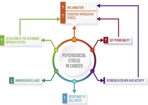
Individual suffering from cancer are at high risk of experiencing major depressive episodes throughout the trajectory of the disease, although this risk appears to be specially prominent within the first year of diagnosis. Recent advances in the understanding of the neurobiological underpinnings of depression indicate that high levels of psychosocial stress and low social support may signifi-cantly contribute to the development of depressive symptoms in cancer populations, not only by increasing psychological distress but also by influencing biological mechanisms relevant for the pathophysiology of depression. A lower resilience to stress may translate into chronic inflammation and altered hormonal and autonomic responses, which are acknowledged factors related to the development and recurrences of depressive episodes. Likewise, stress and depression through shared mechanisms may exert a detrimental effect on cancer-related outcomes.Fig. 1 provides a wide-angle lens of the multiple intertwined biobehavioral path-ways linking stress, depression, and cancer progression. Since antidepressants might insufficiently target depression in cancer populations, the development of alternative or complementary treatment strategies is highly warranted. Behavioral interventions may be feasible adjunctive strategies for targeting depressive symptoms, cognitive dysfunction, fatigue and sleep disturbances among individuals with cancer[206].
Notwithstanding the literature in the field is typically limited by a small number of studies and heterogeneous inclusion criteria, short follow-up periods and heterogeneous assessment tools, which complicates the generalizability of results and complicates comparisons between studies, preliminary data from RCTs have offered encouraging results. In addition, available literature sug-gests that the positive impact of behavioral strategies may be par-tially mediated by their anti-inflammatory potential in association with immunosurveillance-enhancing properties as well as with a modulatory impact on the activity of the HPA axis.
Prospective research may provide clearer causal inferences about the mutual interactions between cancer and depression, and inform about novel possible therapeutic options. In addition, a more profound knowledge base in this emerging field could also offer novel perspectives for the design of preventative strategies for tackling depression in cancer survivors.
Conflicts of interest
The authors declare no conflicts of interest.
Acknowledgments
AFC is supported by a research fellowship award from the Con-selho Nacional de Desenvolvimento Científico e Tecnológico (CNPq; Brazil). CAK is the recipient of a postdoctoral fellowship award from the Coordenação de Aperfeiçoamento de Pessoal de Nível Superior (CAPES; Brazil).
References
[1]Lutgendorf SK, Andersen BL. Biobehavioral approaches to cancer progression and survival: mechanisms and interventions. Am Psychol 2015;70:186–97. [2]Satin JR, Linden W, Phillips MJ. Depression as a predictor of disease
progression and mortality in cancer patients: a meta-analysis. Cancer 2009;115:5349–61.
[3]Pelletier G, Verhoef MJ, Khatri N, Hagen N. Quality of life in brain tumor patients: the relative contributions of depression, fatigue, emotional distress, and existential issues. J Neurooncol 2002;57:41–9.
[4]Fann JR, Thomas-Rich AM, Katon WJ, Cowley D, Pepping M, McGregor BA, et al. Major depression after breast cancer: a review of epidemiology and treatment. Gen Hosp Psychiatry 2008;30:112–26.
[5]Faller H, Bulzebruck H, Drings P, Lang H. Coping, distress, and survival among patients with lung cancer. Arch Gen Psychiatry 1999;56:756–62.
[6]Tang PL, Wang HH, Chou FH. A systematic review and meta-analysis of demoralization and depression in patients with cancer. Psychosomatics 2015;56:634–43.
[7]Meijer A, Roseman M, Delisle VC, Milette K, Levis B, Syamchandra A, et al. Effects of screening for psychological distress on patient outcomes in cancer: a systematic review. J Psychosom Res 2013;75:1–17.
[8]Giese-Davis J, Collie K, Rancourt KM, Neri E, Kraemer HC, Spiegel D. Decrease in depression symptoms is associated with longer survival in patients with metastatic breast cancer: a secondary analysis. J Clin Oncol 2011;29:413–20. [9]Fann JR, Fan MY, Unutzer J. Improving primary care for older adults with
cancer and depression. J Gen Intern Med 2009;24(Suppl. 2):S417–24. [10]Jehn CF, Becker B, Flath B, Nogai H, Vuong L, Schmid P, et al. Neurocognitive
function, brain-derived neurotrophic factor (BDNF) and IL-6 levels in cancer patients with depression. J Neuroimmunol 2015;287:88–92.
[11]Lebena A, Vegas O, Gomez-Lazaro E, Arregi A, Garmendia L, Beitia G, et al.
Melanoma tumors alter proinflammatory cytokine production and
monoamine brain function, and induce depressive-like behavior in male mice. Behav Brain Res 2014;272:83–92.
[12]Hughes S, Jaremka LM, Alfano CM, Glaser R, Povoski SP, Lipari AM, et al. Social support predicts inflammation, pain, and depressive symptoms: longitudinal relationships among breast cancer survivors. Psychoneuroendocrinology 2014;42:38–44.
[13]Kurz K, Schroecksnadel S, Weiss G, Fuchs D. Association between increased tryptophan degradation and depression in cancer patients. Curr Opin Clin Nutr Metab Care 2011;14:49–56.
[14]Wei YC, Zhou FL, He DL, Bai JR, Ding H, Wang XY, et al. Oxidative stress in
depressive patients with gastric adenocarcinoma. Int J
Neuropsychopharmacol 2009;12:1089–96.
[15]Cardoso G, Graca J, Klut C, Trancas B, Papoila A. Depression and anxiety symptoms following cancer diagnosis: a cross-sectional study. Psychol Health Med 2015;1–9.
[16]Coyne JC, Schwenk TL, Fechner-Bates S. Nondetection of depression by primary care physicians reconsidered. Gen Hosp Psychiatry 1995;17:3–12. [17]Mitchell AJ, Chan M, Bhatti H, Halton M, Grassi L, Johansen C, et al. Prevalence
of depression, anxiety, and adjustment disorder in oncological,
haematological, and palliative-care settings: a meta-analysis of 94 interview-based studies. Lancet Oncol 2011;12:160–74.
[18]Ahn HK, Bae JH, Ahn HY, Hwang IC. Risk of cancer among patients with depressive disorder: a meta-analysis and implications. Psychooncology 2016. [19]Schag CA, Ganz PA, Polinsky ML, Fred C, Hirji K, Petersen L. Characteristics of women at risk for psychosocial distress in the year after breast cancer. J Clin Oncol 1993;11:783–93.
[20]Polsky D, Doshi JA, Marcus S, Oslin D, Rothbard A, Thomas N, et al. Long-term risk for depressive symptoms after a medical diagnosis. Arch Intern Med 2005;165:1260–6.
[21]Kangas M, Henry JL, Bryant RA. The course of psychological disorders in the 1st year after cancer diagnosis. J Consult Clin Psychol 2005;73:763–8. [22]Schrepf A, Lutgendorf SK, Pyter LM. Pre-treatment effects of peripheral
tumors on brain and behavior: neuroinflammatory mechanisms in humans and rodents. Brain Behav Immun 2015;49:1–17.
[23]Cosci F, Fava GA, Sonino N. Mood and anxiety disorders as early manifestations of medical illness: a systematic review. Psychother Psychosom 2015;84:22–9.
[24]Green AI, Austin CP. Psychopathology of pancreatic cancer. A psychobiologic probe. Psychosomatics 1993;34:208–21.
[25]Wedding U, Koch A, Rohrig B, Pientka L, Sauer H, Hoffken K, et al. Requestioning depression in patients with cancer: contribution of somatic and affective symptoms to Beck’s Depression Inventory. Ann Oncol 2007;18:1875–81.
[26]Vine MF, Calingaert B, Berchuck A, Schildkraut JM. Characterization of prediagnostic symptoms among primary epithelial ovarian cancer cases and controls. Gynecol Oncol 2003;90:75–82.
[27]Lutgendorf SK, Weinrib AZ, Penedo F, Russell D, DeGeest K, Costanzo ES, et al. Interleukin-6, cortisol, and depressive symptoms in ovarian cancer patients. J Clin Oncol 2008;26:4820–7.
[28]Van Esch L, Roukema JA, Ernst MF, Nieuwenhuijzen GA, De Vries J. Combined anxiety and depressive symptoms before diagnosis of breast cancer. J Affect Disord 2012;136:895–901.
[29]Eskelinen M, Ollonen P. Beck Depression Inventory (BDI) in patients with breast disease and breast cancer: a prospective case-control study. In Vivo 2011;25:111–6.
[30]Liu L, Rissling M, Natarajan L, Fiorentino L, Mills PJ, Dimsdale JE, et al. The longitudinal relationship between fatigue and sleep in breast cancer patients undergoing chemotherapy. Sleep 2012;35:237–45.
[31]Hanahan D, Weinberg RA. Hallmarks of cancer: the next generation. Cell 2011;144:646–74.
[32]Reuter S, Gupta SC, Chaturvedi MM, Aggarwal BB. Oxidative stress, inflammation, and cancer: how are they linked? Free Radic Biol Med 2010;49:1603–16.
[33]Valpione S, Martinoli C, Fava P, Mocellin S, Campana LG, Quaglino P, et al. Personalised medicine: development and external validation of a prognostic model for metastatic melanoma patients treated with ipilimumab. Eur J Cancer 2015;51:2086–94.
[34]Armaiz-Pena GN, Cole SW, Lutgendorf SK, Sood AK. Neuroendocrine influences on cancer progression. Brain Behav Immun 2013;30(Suppl.): S19–25.
[35]Sica A, Mantovani A. Macrophage plasticity and polarization: in vivo veritas. J Clin Invest 2012;122:787–95.
[36]Tracey KJ. Reflex control of immunity. Nat Rev Immunol 2009;9:418–28. [37]Qin L, Wu X, Block ML, Liu Y, Breese GR, Hong JS, et al. Systemic LPS causes
chronic neuroinflammation and progressive neurodegeneration. Glia 2007;55:453–62.
[38]Torres-Platas SG, Cruceanu C, Chen GG, Turecki G, Mechawar N. Evidence for increased microglial priming and macrophage recruitment in the dorsal anterior cingulate white matter of depressed suicides. Brain Behav Immun 2014;42:50–9.
[39]Dantzer R, O’Connor JC, Freund GG, Johnson RW, Kelley KW. From inflammation to sickness and depression: when the immune system subjugates the brain. Nat Rev Neurosci 2008;9:46–56.
[40]Najjar S, Pearlman DM, Alper K, Najjar A, Devinsky O. Neuroinflammation and psychiatric illness. J Neuroinflamm 2013;10:43.
[41]Miller AH, Maletic V, Raison CL. Inflammation and its discontents: the role of cytokines in the pathophysiology of major depression. Biol Psychiatry 2009;65:732–41.
[42]Dowlati Y, Herrmann N, Swardfager W, Liu H, Sham L, Reim EK, et al. A meta-analysis of cytokines in major depression. Biol Psychiatry 2010;67:446–57. [43]Liu Y, Ho RC, Mak A. Interleukin (IL)-6, tumour necrosis factor alpha
(TNF-alpha) and soluble interleukin-2 receptors (sIL-2R) are elevated in patients with major depressive disorder: a meta-analysis and meta-regression. J Affect Disord 2012;139:230–9.
[44]Valkanova V, Ebmeier KP, Allan CL. CRP, IL-6 and depression: a systematic review and meta-analysis of longitudinal studies. J Affect Disord 2013;150:736–44.
[45]Capuron L, Gumnick JF, Musselman DL, Lawson DH, Reemsnyder A, Nemeroff CB, et al. Neurobehavioral effects of interferon-alpha in cancer patients: phenomenology and paroxetine responsiveness of symptom dimensions. Neuropsychopharmacology 2002;26:643–52.
[46]Bufalino C, Hepgul N, Aguglia E, Pariante CM. The role of immune genes in the association between depression and inflammation: a review of recent clinical studies. Brain Behav Immun 2013;31:31–47.
[47]Haapakoski R, Ebmeier KP, Alenius H, Kivimaki M. Innate and adaptive immunity in the development of depression: an update on current knowledge and technological advances. Prog Neuropsychopharmacol Biol Psychiatry 2016;66:63–72.
[48]Leonard B, Maes M. Mechanistic explanations how cell-mediated immune activation, inflammation and oxidative and nitrosative stress pathways and their sequels and concomitants play a role in the pathophysiology of unipolar depression. Neurosci Biobehav Rev 2012;36:764–85.