PROGRAMA DE PÓS-GRADUAÇÃO EM ODONTOLOGIA MESTRADO EM ODONTOLOGIA
CAMILA DE ATAIDE E FERRAZ
EFETIVIDADE DE DIFERENTES MÉTODOS MECÂNICOS DE REMOÇÃO DE DENTINA CARIADA: AVALIAÇÃO IN VITRO ATRAVÉS DE MICRO-CT E
FOTOGRAFIA DIGITAL
EFETIVIDADE DE DIFERENTES MÉTODOS MECÂNICOS DE REMOÇÃO DE DENTINA CARIADA: AVALIAÇÃO IN VITRO ATRAVÉS DE MICRO-CT E
FOTOGRAFIA DIGITAL
Dissertação submetida ao Programa de Pós-Graduação em Odontologia da Faculdade de Farmácia, Odontologia e Enfermagem da UFC, como requisito parcial para a obtenção do Título de Mestre em Odontologia.
Área de concentração: Clínica Odontológica. Orientadora: Profa. Dra. Monica Yamauti.
Coorientador: Prof. Dr. Carlos Augusto de Oliveira Fernandes.
Dados Internacionais de Catalogação na Publicação Universidade Federal do Ceará
Biblioteca de Ciências da Saúde
F432e Ferraz, Camila de Ataide e.
Efetividade de diferentes métodos mecânicos de remoção de dentina cariada: avaliação in vitro através de micro-CT e fotografia digital. / Camila de Ataide e Ferraz. – 2013.
68 f. : il. color., enc. ; 30 cm.
Dissertação (mestrado) – Universidade Federal do Ceará; Centro de Ciências da Saúde; Faculdade
de Farmácia, Odontologia e Enfermagem; Departamento de Odontologia; Programa de Pós-Graduação em Odontologia; Mestrado em Odontologia, Fortaleza, 2013.
Área de Concentração: Clínica Odontológica. Orientação: Profa. Dra. Mônica Yamauti.
1. Cárie Dentária. 2. Preparo da Cavidade Dentária. 3. Fotografia Dentária. I. Título.
EFETIVIDADE DE DIFERENTES MÉTODOS MECÂNICOS DE REMOÇÃO DE DENTINA CARIADA: AVALIAÇÃO IN VITRO ATRAVÉS DE MICRO-CT E
FOTOGRAFIA DIGITAL
Dissertação submetida à Coordenação do Programa de Pós-Graduação em Odontologia da Faculdade de Farmácia, Odontologia e Enfermagem, da Universidade Federal do Ceará, como requisito parcial para a obtenção do Título de Mestre em Odontologia com Área de concentração em Clínica Odontológica.
Aprovado em _____/_____/_____.
BANCA EXAMINADORA
____________________________________________________ Profa. Dra. Monica Yamauti. - (Orientadora)
Universidade Federal do Ceará (UFC)
____________________________________________________ Prof. Dr. Sérgio Lima Santiago
Universidade Federal do Ceará (UFC)
____________________________________________________ Profa. Dra. Paula Borges Jacques
Ao Tiago, meu grande amor e companheiro de todas as horas, por sempre me apoiar e estar presente durante mais esta caminhada.
A Deus, pelo dom da vida, por me guiar pelos caminhos certos e por permitir a realização de mais este sonho.
À Profa. Monica Yamauti, por todas as orientações necessárias à realização deste trabalho, por toda a dedicação, apoio e incentivo. Por todo o carinho e atenção, muito obrigada! “Deus, realmente, coloca as pessoas certas na nossa vida e Ele é quem decide o rumo dos nossos passos”.
Ao Prof. Carlos Augusto de Oliveira Fernandes, por me acolher no Mestrado, por seu carinho e por suas valiosas orientações.
Ao Prof. Juliano Sartori Mendonça, pela realização da estatística e por toda a atenção com que sempre me tratou, desde a época da graduação.
À Universidade Federal do Ceará, na pessoa do seu Magnífico Reitor Prof. Dr. Jesualdo Pereira Farias.
À Faculdade de Farmácia, Odontologia e Enfermagem, em nome de sua diretora, Profa. Maria Goretti Rodrigues de Queiroz.
Ao Prof. Sérgio Lima Santiago, vice-diretor da Faculdade de Farmácia, Odontologia e Enfermagem, pela atenção e carinho.
À coordenadora do Programa de Pós-graduação em Odontologia da Universidade Federal do Ceará, Profa. Lidiany Karla Azevedo Rodrigues, por sua dedicação e incentivo.
Ao corpo docente do Programa de Pós-Graduação em Odontologia da Universidade Federal do Ceará, por todo o aprendizado adquirido nesta etapa.
Aos professores da FOP-UNICAMP, Marinês Nobre dos Santos e Wander José da Silva, por todas as orientações e incentivo e às alunas de doutorado Bruna Raquel Zancopé e Daniele Rodrigues Picco por me acolherem em Piracicaba.
À Patrícia Lima Thé, por sua amizade e colaboração durante a elaboração deste trabalho e à Lidiane Costa, por ter dividido comigo diversos dias de muito trabalho e dedicação na FOP, em Piracicaba.
Aos colegas da turma de mestrado pelos bons momentos vividos durante o curso.
A todos os funcionários da Faculdade de Farmácia, Odontologia e Enfermagem da UFC, pela atenção com que sempre me trataram.
À FUNCAP e à CAPES, pela concessão da bolsa de mestrado no programa de Pós-graduação do qual fiz parte.
A remoção seletiva de dentina cariada é um desafio para a Odontologia contemporânea, já que os métodos tradicionais de escavação podem remover, além do tecido cariado, dentina sadia. Objetivou-se determinar a efetividade de remoção de cárie (ERC) e o potencial minimamente invasivo (PMI) de três métodos mecânicos de escavação. Doze molares humanos com lesões de cárie oclusal em dentina foram distribuídos, de forma randomizada, em três grupos de acordo com o método de remoção: broca carbide, cureta de dentina e broca de polímero. Todos os dentes, antes de serem submetidos à etapa de escavação da cárie, tiveram suas raízes embutidas em resina acrílica. Seccionaram-se os dentes uma única vez no sentido longitudinal, tendo como referência visual o centro da lesão. Cada uma das secções foi submetida a fotografias digitais padronizadas e escaneamento por microtomografia computadorizada de raios-X (micro-CT) (Skyscan 1174, Skyscan, Kontich, Belgium) em dois momentos distintos, antes e após a remoção de cárie. Utilizou-se um programa de imagens de domínio público, ImageJ, para análise das imagens obtidas. Definiram-se as áreas de dentina cariada inicial (CI), da cavidade preparada (CP) e da cárie residual (CR) nas fotografias e nas imagens de micro-CT de acordo com critério visual. Determinou-se a ERC entre os métodos com base na relação CR/CI, enquanto o PMI foi determinado através da relação entre CP/CI. Analisaram-se os dados obtidos através de Análise de Variância a Um Critério e teste-t de Student ou Kruskal-Wallis e teste Student-Newman-Keuls. Tanto para a análise de imagem digital quanto para a análise de micro-CT, a broca carbide apresentou maior efetividade em relação à cureta (p=0,0063; p=0,0263, respectivamente) e em relação à broca de polímero (p=0,0028; p=0,0005, respectivamente), enquanto que cureta de dentina e broca de polímero mostraram resultados semelhantes para os dois tipos de imagens (p>0,05). Em relação ao PMI, para a análise de imagem digital, a broca de polímero apresentou-se como método com potencial invasivo similar à cureta de dentina (p=0.1240), porém com invasividade menor do que a broca carbide (p=0,0028). Para a análise de micro-CT, os valores de PMI de todos os grupos foram significantemente diferentes, e a broca de polímero também demonstrou o método mais conservador, tanto em relação à cureta de dentina (p=0,0030), quanto em relação à broca carbide (p<0.001). Portanto, a broca carbide foi o método mais efetivo para remoção de cárie, porém o menos conservador; e a broca de polímero apresentou baixo potencial invasivo, porém não foi capaz de remover toda a dentina cariada.
Selective removal of carious dentin is a challenge in contemporary Dentistry as traditional methods of excavation can remove, besides caries, healthy dentin. The aim of the study was to determine the caries removal effectiveness (CRE) and minimally invasive potential (MIP) of 3 mechanical caries removal methods. Twelve human molars with occlusal caries lesions in dentin were randomly divided into 3 groups according to the caries removal method: carbide bur, hand instrument and polymer bur. All teeth, before caries excavation, had their roots embedded in acrylic resin. These specimens were sectioned longitudinally into two halves through the center of the carious lesion. Each section was subjected to digital photographs that were standardized and to computed microtomography X-ray (micro-CT) (Skyscan 1174, Skyscan, Kontich, Belgium) scanning at two different times, before and after caries excavation. The public image program ImageJ was used for analysis of all obtained images. Initial carious dentin (IC), prepared cavity (PC) and residual caries (RC) areas from digital and micro-CT images were defined according to visual criteria. CRE among caries removal methods was determined based on the ratio RC/IC, whereas MIP was determined by the ratio PC/IC. Data were analyzed using One-Way ANOVA and Student's t-test or Kruskal-Wallis and Student-Newman-Keuls test. Either for digital image or micro-CT analysis, carbide bur showed more CRE method than hand excavation (p=0,0063; p=0,0263, respectively) and more than polymer bur (p=0,0028; p=0,0005), while hand instrument and polymer bur presented similar results. Regarding MIP, for digital image analysis, polymer bur were different from carbide bur (p=0,0028), but were not different from those of hand instrument (p=0.1240). For micro-CT analysis, the MIP values of all groups were significant different, and polymer bur was more conservative than hand excavation (p=0,0030) and carbide bur (p<0.001). Therefore, carbide bur was the most effective method for removal of caries, but was the least conservative, and polymer bur presented low invasive potential, but was not able to remove all carious dentin.
1. INTRODUÇÃO GERAL... 9
2. PROPOSIÇÃO... 14
Objetivo Geral... 15
Objetivos Específicos... 15
3. CAPÍTULO... 16
CAPÍTULO 1: Effectiveness of different mechanic methods on dentin caries removal: micro-CT and digital image evaluation……… 18 4. CONCδUSÃO GERAδ………... 44
REFERÊNCIAS………... 46
ANEXOS……….. 51
ANEXO A - Apreciação do Comitê de Ética em Pesquisa... 52
1 INTRODUÇÃO GERAL
A odontologia minimamente invasiva advoga que a remoção de tecido cariado seja eficaz, porém deve eliminar ou minimizar a perda de tecido sadio, hígido, seja ele esmalte ou dentina (TYAS et al., 2000). A escavação de cárie deve limitar-se à remoção de apenas tecido necrótico e amolecido (NEVES et al., 2011a). O limite da quantidade de tecido cariado a ser removido, ainda hoje, é, contudo, controverso, pois nenhuma ferramenta de diagnóstico até agora desenvolvida e utilizada é capaz de definir clinicamente qual o limite ideal em que se deve parar a remoção de tecido durante o processo de remoção de cárie, sem que o tecido hígido seja atingido (NEVES et al., 2011a).
Teoricamente, apenas a camada mais superficial do tecido cariado, ou seja, a camada demoninada infectada, que apresenta grande presença de micro-organismos viáveis e que apresenta desnaturação irreversível das fibras colágenas (FUSAYAMA; OKUSE; HOSODA, 1966), é que deve ser removida. Clinicamente, isso é, porém, muito difícil de ser realizado. Por outro lado, a camada mais interna de tecido dentinário cariado, denominada de dentina afetada por cárie, além de ser livre de bactérias, apresenta limitada desnaturação do colágeno e ainda é passível de remineralização. Portanto, deve ser conservada durante os preparos cavitários (BANERJEE; KIDD; WATSON., 2003; BANERJEE, KIDD, WATSON, 2000; FUSAYAMA; OKUSE; HOSODA, 1966).
disso, como a dureza pode variar entre os grupos de dentes e a localização e profundidade da lesão, uma remoção inadequada (sobre ou subextensão) de tecido pode ser, erroneamente, realizada (CELIBERTI; FRANCESCUT; LUSSI, 2006), resultando na remoção desnecessária de dentina hígida ou de tecido dentinário cariado ainda passível de remineralização, como a camada de dentina afetada por cárie (BANERJEE; KIDD; WATSON., 2003; BANERJEE, KIDD, WATSON, 2000; FUSAYAMA; OKUSE; HOSODA, 1966).
Métodos alternativos de remoção de cárie, tais como brocas de polímeros, abrasão a ar e métodos químico-mecânicos têm sido propostos para permitir uma remoção de dentina cariada menos invasiva, destrutiva e mais conservadora com relação à quantidade de tecido a ser removido. Além disso, propõem-se a ser métodos mais confortáveis, que proporcionem mínimas alterações térmicas teciduais, menor vibração e dor ao paciente, e que sejam capazes de remover apenas dentina infectada por cárie (NEVES et al., 2011b; SILVA et al., 2006; BOSTON, 2003; WHITE; EAKLE, 2000; ERICSON et al., 1999).
As brocas de polímeros, inicialmente descritas por BOSTON (2003), são constituídas por um polímero de poliamida/imida, com propriedades físico-mecânicas, como a dureza, ligeiramente inferiores à da dentina hígida. Assim, quando a broca atinge dentina hígida, ou a dentina afetada por cárie, ela perde o corte e começa a produzir vibrações que impossibilitam que mais cortes ou desgastes teciduais sejam realizados. Em oposição às bocas carbide, suas lâminas de corte são retas ao invés de em forma de espiral, já que foram desenvolvidas para remover a dentina através de uma pressão localizada. Ao pressionar o tecido ao longo da superfície dentária, há um momento em que ele se rompe e é removido, deslocado, da cavidade (BOSTON, 2003). De acordo com o fabricante, as brocas de polímero possuem material mais duro do que a dentina infectada e menos duro do que a dentina hígida, esclerótica ou pigmentada, o que permite uma remoção de cárie de forma bastante seletiva. Pelo fato de que a dentina hígida não é cortada, os túbulos dentinários não são expostos e, por isso, menos ou nenhuma dor chega a ser relatada pelos pacientes durante o seu uso (CELIBERTI; FRANCESCUT; LUSSI, 2006). No entanto, a efetividade (quantidade de tecido removido) dessas novas brocas na seletividade de remoção de dentina cariada em dentes permanentes ainda não está bem estabelecida.
raio-X que gera imagens em alta resolução. Esse método tem sido, atualmente, bastante utilizado em pesquisas com osso (BURGHARDT et al., 2008; SCHWEIZER et al., 2007; DAVIS; WONG, 1996). Contitui-se também em uma técnica bastante promissora para pesquisas em dentes, em casos quando se necessita mensurar a espessura do esmalte, a morfologia do canal radicular, e avaliar a qualidade dos preparos endodônticos, a engenharia ou densidade mineral dos tecidos dentais duros (LO; ZHI; ITTHAGARUN, 2010; ANDERSON et al., 1996; KINNEY et al., 1995). Mais recentemente, estudos avaliando lesões de cáries em esmalte, detecção de cáries proximais, técnicas de remoção de tecido cariado e tratamentos restauradores têm tornado a micro-CT uma técnica promissora para estudos em Cariologia (BEDINI et al., 2012; COCHRANE et al., 2012; HAMBA et al., 2012; MELEO et al., 2012; SOVIERO et al., 2012; NEVES et al., 2011a; NEVES et al., 2011b; NEVES et al., 2010). Com o uso da micro-CT, é possível comparar a estrutura interna dos dentes antes e após um procedimento experimental, quantas vezes forem necessárias (WILLMOTT; WONG; DAVIS, 2007; CLEMENTINO-LUEDEMANN et al., 2006), antes e após o procedimento de remoção de cárie dentária, por exemplo (NEVES et al., 2011b; NEVES et al., 2011c), ou mudança no conteúdo mineral das lesões de cárie após procedimento de des/remineralização artificial (DAVIS; WONG, 1996).
Da mesma forma que a micro-CT, a fotografia digital é uma técnica não destrutiva de avaliação experimental que permite a mensuração e a avaliação dos tecidos dentais duros, de forma que a amostra pode ser avaliada por várias vezes sem que haja de ser alterada, possibilitando, inclusive, avaliações de tratamentos realizados (SWAIN, XUE, 2009; KIM; PAIK, LEE, 2007; CELIBERTI; FRANCESCUT; LUSSI, 2006; ELLIOTT; DOVER, 1982).
2 PROPOSIÇÃO
2.1 Objetivo Geral
Analisar a efetividade e o potencial de invasão de três métodos mecânicos na remoção de cárie dentinária, através de análise de área em fotografia digital e em micro-CT.
2.2 Objetivos Específicos
1. Testar a hipótese nula de que todos os métodos mecânicos utilizados serão igualmente capazes de remover dentina cariada, quando avaliados por meio de análises em fotografia digital e em micro-CT.
3 CAPÍTULO
Esta dissertação está baseada no Artigo 46 do Regimento Interno do Programa de Pós-Graduação em Odontologia da Universidade Federal do Ceará, que regulamenta o formato alternativo para dissertações de Mestrado e teses de Doutorado e permite a inserção de artigos científicos de autoria ou coautoria do candidato, publicados ou ainda não submetidos para publicação em periódicos científicos, escritos no idioma exigido pelo veículo de divulgação. Por se tratar de pesquisas envolvendo seres humanos, ou parte deles, o projeto de pesquisa foi submetido à apreciação do Comitê de Ética em Pesquisa, tendo sido aprovado (Anexo A). Assim, esta dissertação é composta de um capítulo contendo um artigo científico que será submetido para publicação no periódico Operative Dentistry, conforme descrito abaixo:
Capítulo 1:
“Effectiveness of different mechanical methods on dentin caries removal: micro-CT and digital image evaluation”
Authors: Camila Ferraz, Juliano Sartori Mendonça, Juan de la Cruz Cardona Perez,
CAPÍTULO 1
Title: Effectiveness of different mechanical methods on dentin caries removal:
micro-CT and digital image evaluation.
Authors: Camila Ferraz1, Juliano Sartori Mendonça2, Juan de la Cruz Cardona Perez3, Alexandre Rodrigues Freire4, Patrícia Lima Thé1, Carlos Augusto de Oliveira Fernandes5, Monica Yamauti6*
1
DDS, Graduate Student, Graduate School of Dentistry, Faculty of Pharmacy, Dentistry and Nursing, Federal University of Ceara, Rua Capitão Francisco Pedro, S/N - Rodolfo Teofilo, 60430-170, Fortaleza-CE-Brazil.
2
DDS, MS, PhD, Adjunct Professor, Faculty of Pharmacy, Dentistry and Nursing, Federal University of Ceara, Rua Capitão Francisco Pedro, S/N - Rodolfo Teofilo, 60430-170, Fortaleza-CE-Brazil.
3
PhD, Assistant Professor, Optics Department, Faculty of Sciences, University of Granada, Campus Fuente Nueva s/n, 18071, Granada-Spain.
4
DDS, Graduate Student, Graduate School of Dentistry, Piracicaba Dental School - UNICAMP, Av. Limeira, 901 Caixa Postal 52, 13414-903, Piracicaba -SP-Brazil.
5
DDS, MS, PhD, Professor, Graduate School of Dentistry, Faculty of Pharmacy, Dentistry and Nursing, Federal University of Ceara, Rua Capitão Francisco Pedro, S/N - Rodolfo Teofilo, 60430-170, Fortaleza-CE-Brazil.
6
DDS, MS, PhD, Professor, Graduate School of Dentistry, Faculty of Pharmacy, Dentistry and Nursing, Federal University of Ceara, Rua Capitão Francisco Pedro, S/N - Rodolfo Teofilo, 60430-170, Fortaleza-CE-Brazil.
*Corresponding author: Monica Yamauti
Visiting Professor
Graduate School of Dentistry, Department of Restorative Dentistry, Faculty of Pharmacy, Dentistry and Nursing, University Federal of Ceara
Rua Capitão Francisco Pedro, S/N - Rodolfo Teófilo - Fortaleza-CE-Brazil, 60430-170 Phone/Fax: +558533668232
Title: Effectiveness of different mechanical methods on dentin caries removal: micro-CT
and digital image evaluation
Running title: Caries removal with different mechanical methods.
Clinical significance:
SUMMARY
Purpose: To determine caries removal effectiveness (CRE) and minimally invasive potential
(MIP) of caries excavation methods using digital imaging and microtomography analyses. Methods: Twelve human molars with occlusal caries lesions in dentin were randomly divided
into 3 groups (carbide bur, hand instrument and polymer bur). They were sectioned mesio-distally, and standardized digital and computed microtomography X-ray (micro-CT) images were taken from each section before and after caries excavation. On each image, initial carious dentin (IC), prepared cavity (PC) and residual caries (RC) were defined according to visual criteria using ImageJ software. CRE was determined based on the ratio RC/IC, whereas MIP was determined by the ratio PC/IC. Data were analyzed using One-Way ANOVA and Student's t-test or Kruskal-Wallis and Student-Newman-Keuls test. The level of significance was set at 0.05.
Results: Either for digital image or micro-CT analysis, carbide bur showed higher CRE
values than hand excavation (p=0.0063; p=0.0263, respectively) and polymer bur (p=0.0028; p=0.0005), while hand instrument and polymer bur presented similar results (p>0.05). Regarding MIP, for digital image analysis, polymer bur were different from carbide bur (p=0.0030), but were not different from those of hand instrument (p=0.1240). For micro-CT analysis, the MIP values of all groups were significant different, and polymer bur was the most conservative method than the other methods (p<0.05). Conclusions: Carbide bur was the most effective method for caries removal, but was not totally conservative. Polymer bur and hand excavation presented low invasive potential, but were not able to remove all carious dentin.
INTRODUCTION
In light of minimally invasive dentistry, caries excavation has become more conservative.1 Theoretically, only the superficial layer, i.e. infect dentin, that is strongly infected with viable microorganisms, and as presents irreversible denaturation and disorganization of the collagen fibers1 should be removed. Clinically, this is a hard task to achieve. Traditionally, carious dentin is removed mechanically with hand excavation and/or slow-speed round diamond, tungsten carbide or carbon-steel burs.2,3 However, these techniques are indiscriminant and non-selective in removing carious tissues. Often during excavation, clinicians tend to include all soft, discolored and stained tissue to ensure complete elimination of the infected layer.4,5 In such cases, tactile and optical judgment is used to evaluate the consistency and color of the dental tissue. These criteria was shown to be adequate to ensure removal of the most of the infected dentin,5 but they are still clinical and subjective parameters, dependent on the operators´ experience.1 Thus, their use frequently results in unnecessary removal of sound dentin or tissue with reduced mineral content, like affected-caries dentin,4,3 that is still remineralizable organic matrix of the lesion and, therefore, must be preserved.4,1
Alternative caries removal methods such as polymer burs, air abrasion and chemomechanical have been proposed to allow a less invasive/less destructive dentin caries excavation and conserving tooth substance, a more gentle, comfortable and conservative caries excavation to provide a minimal thermal change, less vibration and pain, and removal of infected dentin only.6-10
cavity.10 As opposed to conventional carbide burs, their cutting edges are not spiralled but straight. According to the manufacturer, this material is harder than infected dentin but softer than normal, sound dentin and also even softer than sclerotic, discoloured dentin, thus allowing a very selective caries removal. As dentin tubules are not opened, less or no pain is also suggested.11 However, the efficacy of these new burs on selectively and efficiently removing natural carious dentin in permanent teeth is not well established.
X-ray computed microtomography (micro-CT) is a microscopic version of computed tomography that uses an X-ray focused beam providing higher resolution to investigate samples in vitro at a resolution of a few micrometres. It constitutes a non-destructive imaging method where individual projections (radiographs) could be recreated in any plane, and data could be represented as 2D or rendered 3D images and structures.12 Images could be assessed qualitatively and quantitatively. The method has been frequently used in experiments exploring bone.13,14 More recently, studies regarding evaluation of enamel lesion caries, detection of proximal carious lesion, caries-excavation techniques and restorative treatments have been developed with this technique, which seems to be promising in Cariology.2,6,12,15-17 Despite its many advantages, micro-CT is not yet a reliable method for caries detection, and for this reason, more studies are necessary to evaluate and define its feasibility as method of diagnostic.
MATERIAL AND METHODS
Sample Preparation
Twelve extracted human molars with dentin caries on their occlusal surfaces were collected from the Human Research ethics Committee of the Institution. Immediately after extraction, the teeth were properly cleaned and stored in 0.1% (w/v) thymol solution for a period no longer than 3 months. The roots of all teeth were embedded in acrylic resin in a mould (dimensions: 2 x 2 x 2 cm), and were sectioned longitudinally in two halves through the center of the carious lesion using a low-speed diamond saw, thickness γ00 μm (IsoεetTM Low Speed Saw, Buehler, Lake Bluff, IL, USA) under running water. Visual inspection was performed to determine the limits of the caries lesion. All teeth presented occlusal caries reaching dentin either confined to outer half of dentine or reaching the inner half of dentin. The teeth were numbered and classified according to lesion size as small (caries lesion extending over up occlusal dentin surfaces) or large (caries lesion extending over more than half of the occlusal surface). Teeth were randomly allocated to three experimental groups of four samples each. Small and large lesions were equally and randomly distributed between each experimental group.
Each section of teeth was digitally photographed (Sony αγ00, Sony Corp, Japan), and
- Group 1 (n=4): Conventional round carbide bur. Brand new burs #4, #6 and #8 (Dentsply Maillefer, Ballaigues, Switzerland) were used in a slow-speed handpiece without water cooling. Carious dentin was excavated with circular movements starting from the periphery to the center of the lesion.11 Caries removal ended when hard dentin was detected using a non-flexible probe (SS White Duflex, Rio de Janeiro, Brazil). Dentin was considered hard when, under a firm pressure, the probe was not able to penetrate into tissue.4 For each tooth, a new carbide bur was used.
- Group 2 (n=4): Hand excavator. Carious dentin was removed using brand-new excavators #14 and #19 (SS White Duflex, Rio de Janeiro, Brazil). During excavation, dentine hardness was checked and carious dentin removal completed when hard tissue was detected with the probe (SS White Duflex, Rio de Janeiro, Brazil) as described for Group 1. For each tooth, a new hand instrument was used.
- Group 3 (n=4): Polymer bur. SmartBurs® II #4, #6 and #8 (SS White, Lakewood, NJ, USA) were used with a slow-speed handpiece without water cooling. Carious dentin was removed with circular movements starting from the center of the lesion to the periphery as recommended by the manufacturer. Excavation ended when the instrument became macroscopically abraded and blunted, and was no longer able to remove tissue.9 A probe was also used to check the hardness of left tissue. For each tooth a new burs were used.11
During the time between procedures, the samples were stored in physiological solution with a relative humidity of H=100%. After finishing the caries excavation, tooth halves were separated. Digital and micro-CT were taken and assigned to the “post-rank”.
Digital images
irradiance of the CIE D65 standard illuminant. For each sample, both before and after the caries removal procedure, 20 consecutive digital images were taken using a Digital Single δens Reflex (DSδR) commercial camera (αγ00, Sony Corp., Japan) operating in manual
mode with fixed parameters. The camera was calibrated using a spectroradiometer (PR704, Photo Research Inc, Chatsworth, USA) and a Color Checker (Gretag Macbeth, New Windsor, USA). The geometry of measuring/illumination used for camera calibration was 0˚/diffuse,
and the CIE 19γ1 β˚ Standard observer (CIE Bureau, β004) was used to calculate color. The
image acquisition set-up is schematically shown in Figure 1.
X-ray computed microtomography (micro-CT)
All halves surfaces were scanned before and after the carious removal, at 50kV, 800 µA, and 14.1 µm pixel size using a 0.5 mm Al filter to eliminatw low-energy X-rays in a Skyscan 1174 high-resolution descktop micro-CT scanner (Skyscan, Kontich, Belgium). Rotation step was set to 0.70º, resulting in 264 two-dimensional projections over a 180º rotation of the specimen. Flat-field reference was taken before start of time section scanner to improve the acquisition settings. The reconstruction program NRecon (NRecon, SkyScan, Kontich, Belgium) reads angular shadow 16-bit projection images saved by the control program and reconstruct virtual slices (cross sections). Before the reconstruction, beam-hardening correction was performed at 20%, with the same scanning parameters aiming input of optimal contrast limits, based on prior scanning and reconstruction of the tooth.
After reconstruction, a Dataviewer software (SkyScan, Kontich, Belgium) was used to select the most similar 2D image to the respective digital image sample, and it was saved as a single image to further analyses and comparisons.
All the measurement steps, including the image processing, image analysis and area measuring were performed using the software ImageJ (NIH, Bethesda, USA). For each specimen, two areas of interest (Initial Carious Dentin - IC - from “pre-rank” and Prepared Cavity - PC from “post-rank”) were measured (Figure β). The Residual Carious Dentin - RC, and Removed Sound Dentin - RS - areas were also determined (Figure 2). Digital image scaling and calibration was used to calculate the area values. The final result for each sample was the average over 20 images.
Performance assessment of caries removal procedures
After obtaining all area measurements, performance assessment of the three different caries removal procedures was carried out by evaluating the caries-removal effectiveness (CRE) and the minimal-invasiveness potential (MIP) indexes, supported in Neves et al. (2011b).6
1. Caries-removal effectiveness (CRE): the effectiveness of caries removal of the three excavation methods was evaluated by means of the ratio RC/IC. The values of this parameter vary from 0 to 1, and high values represent less effective caries excavation.
2 Minimal-invasiveness potential (MIP): the invasiveness of sound dentin of the three excavation methods was evaluated by means of the ratio PC/IC. The higher the MIP value, the more invasive is the method.
Statistical Analysis
RESULTS
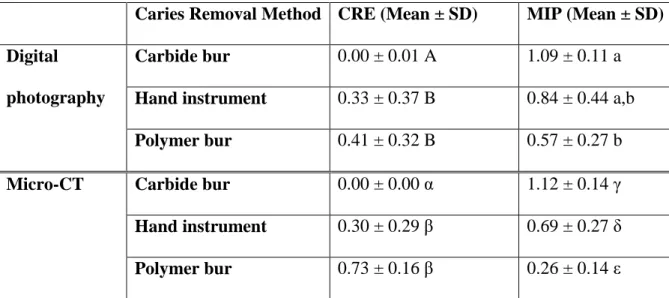
Results of Caries Removal Effectiveness (CRE) and Minimally Invasive Potential (MIP) are shown in Table 1 and Figure 3.
According to digital imaging analysis, for CRE there was statistical differences between groups (p=0.0034). Carbide bur was statistically more effective in removing caries (0.00 ± 0.01) than was hand instrument (p=0.0063) and polymer bur (p=0.0028). Hand instrument and polymer bur showed similar effectiveness (p=0.8957). For MIP, statistical differences were found between groups (p=0.0122). Carbide bur and hand instruments did not show significant difference regarding their minimally invasive potential to remove sound dentin (p=0.2092). MIP values of polymer bur (0.57 ± 0.27) were statistically different from those of carbide bur (p=0.0030) but they were not statistically different from hand instrument (p=0.1240).
DISCUSSION
Minimal invasive dentistry advocates that elimination of the heavily infected dentin and preservation of the residual affected dentin are defined as prerequisites for effectively arresting the carious process without harming the long-term survival of the pulp and the restoration.18 However, this is a difficult task to achieve as caries removal endpoint is still controversial and subjective.19 Current concept for caries excavation has raised discussion about moving toward to more objective and more conservative approaches to selectively remove carious dentin than traditional methods.3,6
polymer burs may not be acute enough to detect the subtleties between remineralizable dentin with minimal collagen degradation.21
In addition, polymer bur also presented lower MIP than carbide bur, and this value was lower than 0 (Table 1 and Fig. 3). When this ration is lower than 1, it means that prepared cavity was smaller than initial carious lesion, and residual carious dentin could have been left. This could be another evidence that polymer bur do not excavate into sound dentin tissue, but was not able to totally remove caries. In fact, these results are in agreement with other studies,10,11 which showed that polymer burs excavation led to one of the largest coincidence between caries removed and caries lesion limits and the smallest area of overpreparation; they also exhibited the biggest underprepared area of all methods evaluated. Residual carious dentin might be left at the cavity walls due to the presence of a compact and thick smear layer, produced during the use of polymer bur. Unlikely, when carbide burs are used the smear layer is different and not so compact.21 This is the so-called compacting effect on carious dentin.3 Such effect could hinder and compromise the action of polymer burs despite its self-limiting properties. We speculate that this similar effect could also happen during teeth storage, which is different from a clinical situation. Vital teeth present dentin fluid that exudes from pulpal blood vessels to dentin surfaces prepared to keep it moist. We speculate that laboratorial conditions could lead to more compact and dense carious dentin layer as a compacting smear layer.
in the same tooth, its assessment usually varies between operators and erroneous removal of dentine (over or underpreparation) may occur.11 Hand instrument is considered a suitable method, combining good excavation time and effectiveness caries removal,11 but its action still depends on clinical judgment and operator´s experience to be really effective.3
On the other hand, carbide bur presented the lowest CRE (0.00 for both digital and micro-CT images) (Table 1 and Fig. 3). These low values mean that carbide burs almost did not leave residual carious dentin, being effective when removing caries. Prior studies also reported that steel and carbide bur and hand excavator showed similar demineralized dentin removal effectiveness.11
The development of nondestructive methods to validate caries diagnosis and removal in vitro is a requirement in dental research, and micro-CT has been cited as an appropriate and promising instrument.23 Micro-CT is increasingly becoming popular in Dentistry, as it enables collecting detailed full-quantitative and qualitative data of the substrate before and after a specific treatment.15,17,24-27 It is possible to observe dimensional differences, gap formation, or other alterations induced on the same sample after application of treatments.17 Its application to study caries-excavation techniques has recently been demonstrated, by comparing the internal tooth structure before and after caries removal.6,16,25,28 However, the use of micro-CT as an accurate tool for caries detection is controversial. On the one hand, accuracy, highest values of sensitivity and highest correlation of micro-CT with histology at both diagnostic thresholds could be observed.15 Notwithstanding, Mitropoulos et al. (2010)29 reported that micro-CT diagnostic was strong correlated with some visual criteria to but was not a reliable alternative to histological examination for caries research. The lack of correlation between these two diagnostic methods was attributed to inherent deficiency of microtomography technology, such as radiography, to detect early signs of demineralization.29 Our results showed that micro-CT is able to detect caries before and after its excavation, though high precision in caries lesion limits could not be observed.
account, analysis of carious dentin could be considered the evaluation of the features of a caries lesion once it has been detected. Such features include optical, physical, chemical or biochemical parameters, such as color, size or surface integrity. Therefore, to assess caries removal methods, we used digital image analysis tool to diagnose carious lesions. Our results demonstrate that digital photography seems to be a viable method to analyze mineralized and carious dental tissues, as well as color alterations before and after caries excavation.
Although this study did not aim to compare caries detection methods, it is possible to note that there was a slight tendency for similarity between digital image and micro-CT analysis.
CONCLUSIONS
REFERENCES
1. Fusayama T, Okuse K, & Hosoda H (1966) Relationship between hardness, discoloration, and microbial invasion in carious dentin Journal of Dental Research 45(4) 1033-1046.
2. Neves AA, Coutinho E, Munck JD, Lambrechts P, & Meerbeek BV (2011a) Does diagnodent provide a reliable caries-removal endpoint? Journal of Dentistry 39(5) 351-360. 3. Banerjee A, Kidd EAM, & Watson T (2000) In vitro evaluation of five alternative methods of carious dentine excavation Caries Research 34(2) 144-150.
4. Banerjee A, Kidd EAM, & Watson T (2003) In vitro validation of carious dentin removed using different excavation criteria American Journal of Dentistry 16(4) 228-230. 5. Kidd EAM, Joyston-Bechal S, & Beighton D (1993) Microbiological validation of assessments of caries activity during cavity preparation Caries Research 27(5) 402-408. 6. Neves AA, Coutinho E, Munck JD, & Meerbeek BV (2011b) Caries-removal effectiveness and minimal-invasiveness potential of caries-excavation techniques: A micro-CT investigation Journal of Dentistry 39(2) 154-162.
7. Neuhaus KW, Ciucchi P, Donnet M, Lussi A (2010) Removal of enamel caries with an air abrasion powder Operative Dentistry 35(5) 538-546.
8. Motisuki C, Lima LM, Bronzi ES, Spolidorio DM, Santos-Pinto L (2006) The effectiveness of alumina powder on carious dentin removal Operative Dentistry 31(3) 371-376.
9. Silva NRFA, Carvalho RM, Pegoraro LF, & Tay FR, Thompson VP (2006) Evaluation of a Self-limiting Concept in Dentinal Caries Removal Journal of Dental Research 85(3) 282-286.
10. Boston DW (2003) New device for selective dentin caries removal Quintessence International 34(9) 678-685.
11. Celiberti P, Francescut P, & Lussi A (2006) Performance of four dentine excavation methods in deciduous Teeth Caries Research 40(2) 117-123.
12. Hamba H, Nikaido T, Sadr A, Nakashima S, & Tagami J (2012) Enamel Lesion Parameter Correlations between Polychromatic Micro-CT and TMR Journal of Dental Research 91(6) 586-591.
13. Burghardt AJ, Kazakia GJ, Laib A, & Majumdar S (2008) Quantitative assessment of bone tissue mineralization with polychromatic micro-computed tomography Calcified Tissue International 83(2) 129-138.
15. Soviero VM, Leal SC, Silva RC, & Azevedo RB (2012) Validity of microCT for in vitro detection of proximal carious lesion in primary molars Journal of Dentistry 40(1) 35-40. 16. Neves AA, Coutinho E, Cardoso MV, Jaecques SV, & Meerbeek BV (2010) Micro-CT based quantitative evaluation of caries excavation Dental Materials 26(6) 579-588.
17. Meleo D, Manzon L, Pecci R, Zuppante F, & Bedini R (2012) A proposal of microtomography evaluation for restoration interface gaps Annali Dell'istituto Superiore Di Sanità 48(1) 83-88.
18. Kidd EA (β004) How ‘clean’ must a cavity be before restoration? Caries Research 38(3) 305-313.
19. Neves AA, Coutinho E, Cardoso MV, Lambrechts P, & Meerbeek BV (2011c) Current concept and techniques for caries excavation and adhesion to residual dentin Journal of Adhesive Dentistry 13(1) 7-22.
20. Toledano M, Cabello I, Yamauti M, & Osorio R (2012) Differential resin-dentin bonds created after caries removal with polymer burs Microscopy and Microanalysis 18(3) 497-508.
21. Meller C, Welk A, Zeligowski T, & Splieth C (2007) Comparison of dentin caries excavation with polymer and conventional tungsten carbide burs Quintessence International 38(7) 565-9.
22. Yip HK, Stevenson AG, & Beeley JA (1994) The specificity of caries detector dyes in cavity preparation British Dental Journal 176(11) 417-421.
23. Huysmans MC, & Longbottom C (2004) The challenges of validating diagnostic methods and selecting appropriate gold standards Journal of Dental Research 83(Spec No C) C48–52.
24. Lee HS, Berg JH, Garcia-Godoy F, & Jang KT (2008) Long-term evaluation of the remineralization of interproximal carieslike lesions adjacent to glass-ionomer restorations: a micro-CT study American Journal of Dentistry 21(2) 129-132.
25. Clementino-Luedemann TN, & Kunzelmann KH (2006) Mineral concentration of natural human teeth by a commercial micro-CT Dental Materials Journal 25(1) 113-119. 26. Olejniczak AJ, & Grine FE (2006) Assessment of the accuracy of dental enamel thickness measurements using microfocal X-ray computed tomography Anatomical Record Part A: Discoveries in Molecular, Cellular, and Evolutionary Biology 288(3) 263-275.
28. Willmott NS, Wong FSL, & Davis GR (2007) An x-ray microtomography study on the mineral concentration of carious dentine removed during cavity preparation in deciduous molars Caries Research 41(2) 129-134.
29. Mitropoulos P, Rahiotis C, Stamatakis H, & Kakaboura A (2010) Diagnostic performance of the visual caries classification system ICDAS II versus radiography and micro-computed tomography for proximal caries detection: an in vitro study Journal of Dentistry 38(11) 859-867.
Captions of figures
Figure 1: Experimental set-up used to obtain digital images.
Figure 2: Schematic representation of digital image and micro-CT analysis.
Table 1 – Mean ± SD of Caries Removal Effectiveness (CRE) and Minimal Invasive Potential (MIP) according to different carious removal methods using digital photography and Micro-CT.
Caries Removal Method CRE (Mean ± SD) MIP (Mean ± SD)
Digital
photography
Acknowledgements
4 CONCLUSÃO GERAL
REFERÊNCIAS
1. ANDERSON, P.; ELLIOTT, J. C.; BOSE, U.; JONES, S. J. A comparison of the mineral content of enamel and dentine in human premolars and enamel pearls measured by x-ray microtomography. Arch Oral Biol., v. 41, n. 3, p. 281-290.Mar. 1996.
2. BANERJEE, A.; KIDD, E. A. M.; WATSON, T. In vitro evaluation of five alternative methods of carious dentine excavation. Caries Res., v. 34, n. 2, p. 144-150.Mar./Apr. 2000.
3. BANERJEE, A.; KIDD, E. A. M.; WATSON, T. In vitro validation of carious dentin removed using different excavation criteria. Am J Dent., v. 16, n. 4, p. 228-230,Aug. 2003.
4. BEDINI, R.; PECCI, R.; NOTARANGELO, G.; ZUPPANTE, F.; PERSICO, S.; DICARLO, F. Microtomograghy evaluation of dental tissue wear surface induced by in vitro simulated chewing cycles on human and composite teeth. Ann Ist Super Sanita., v. 48, n. 1, p. 65-70. 2012.
5. BOSTON, D. W. New device for selective dentin caries removal. Quintessence Int., v. 34, n. 9, p. 678-685.Oct. 2003.
6. BURGHARDT, A. J.; KAZAKIA, G. J.; LAIB, A.; MAJUMDAR, S. Quantitative assessment of bone tissue mineralization with polychromatic micro-computed tomography. Calcif Tissue Int., v. 83, n. 2, p. 129-138.Aug. 2008.
7. CELIBERTI, P.; FRANCESCUT, P.; LUSSI, A. Performance of four dentine excavation methods in deciduous Teeth. Caries Res., v. 40, n. 2, p. 117-123. 2006.
8. CLEMENTINO-LUEDEMANN, T. N. R.; ILIE, A. D. N.; HICKEL, R.;
KUNZELMANN, K. H. Micro-computed tomographic evaluation of a new enzyme solution for caries removal in deciduous teeth. Dent Mater J., v. 25, n. 4, p. 675-683.Dec. 2006.
10.DAVIS, G. R.; WONG, F. S. X-ray microtomography of bones and teeth. Physiol Meas., v. 17, n. 3, p. 121-146.Aug. 1996.
11.ELLIOTT, J. C.; DOVER, S. D. X-ray microtomography. J Microsc., v. 126, n. 2, p. 211-213.May. 1982.
12.ERICSON, D.; ZIMMERMAN, M.; RABER, H.; GOTRICK, B.; BORNSTEIN, R.; THORELL, J. Clinical evaluation of efficacy and safety of a new method for
chemomechanical removal of caries. A multi-centre study. Caries Res., v. 33, n. 3, p. 171-177.May./June. 1999.
13.FUSAYAMA T.; OKUSE K.; HOSODA H. Relationship between hardness, discoloration, and microbial invasion in carious dentin. J Dent Res., v. 45, n. 4, p. 1033-1046, July./Aug. 1966.
14.HAMBA, H.; NIKAIDO, T.; SADR, A.; NAKASHIMA, S.; TAGAMI, J. Enamel Lesion Parameter Correlations between Polychromatic Micro-CT and TMR. J Dent Res., v. 91, n. 6, p. 586-591.June. 2012.
15.KIDD, E. A. M.; JOYSTON-BECHAL, S.; BEIGHTON, D. Microbiological
validation of assessments of caries activity during cavity preparation. Caries Res., v. 27, n. 5, p. 402-408. 1993.
16.KIM, I.; PAIK, K. S.; LEE, S. P. Quantitative evaluation of the accuracy of micro-computed tomography in tooth measurement. Clin Anat., v. 20, n. 1, p.27-34.Jan. 2007.
17.KINNEY, J. H.; BALOOCH, M.; HAUPT, D. L. JR.; MARSHALL, S. J.;
MARSHALL, G. W. JR. Mineral distribution and dimensional changes in human dentin during demineralization. J Dent Res., v. 74, n. 5, p. 1179-1184.May. 1995.
18.LO, E. C.; ZHI, Q. H.; ITTHAGARUN, A. Comparing two quantitative methods for studying remineralization of artificial caries. J Dent., v. 38, n. 4, p. 352-359.Apr. 2010.
20.NEVES, A. A.; COUTINHO, E.; CARDOSO M. V.; LAMBRECHTS, P.; MEERBEEK, B. V. Current concept and techniques for caries excavation and adhesion to residual dentin. J Adhes Dent., v. 13, n. 1, p. 7-22.Feb. 2011a.
21.NEVES, A. A.; COUTINHO, E.; CARDOSO, M. V.; JAECQUES, S. V.;
MEERBEEK, B. V. A micro-CT based quantitative evaluation of caries excavation. Dent Mater., v. 26, n. 6, p. 579-588.June. 2010.
22.NEVES, A. A.; COUTINHO, E.; MUNCK, J. D.; LAMBRECHTS, P.; MEERBEEK, B. V. Does diagnodent provide a reliable caries-removal endpoint? J Dent., v. 39, n. 5, p. 351-360.May. 2011c.
23.NEVES, A. A.; COUTINHO, E.; MUNCK, J. D; MEERBEEK, B. V. Caries-removal effectiveness and minimal-invasiveness potential of caries-excavation techniques: a micro-CT investigation. J Dent., v. 39, n. 2, p. 154-162.Feb. 2011b.
24.SCHWEIZER, S.; HATTENDORF, B.; SCHNEIDER, P.; AESCHLIMANN, B.; GAUCKLER, L.; MULLER, R.; GÜNTHER, D. Preparation and characterization of calibration standards for bone density determination by micro-computed tomography. Analyst., v. 132, n. 10, p. 1040-1045.Oct. 2007.
25.SILVA, N. R. F. A.; CARVALHO, R. M.; PEGORARO, L. F.; TAY, F. R.; THOMPSON, V. P. Evaluation of a Self-limiting Concept in Dentinal Caries Removal. J Dent Res., v. 85, n. 3, p. 282-286.Mar. 2006.
26.SOVIERO, V. M.; LEAL, S. C.; SILVA, R. C.; AZEVEDO, R. B. Validity of micro-CT for in vitro detection of proximal carious lesion in primary molars. J Dent., v. 40, n. 1, p. 35-40.Jan. 2012.
27.SWAIN, M. V.; XUE, J. State of the art of micro-ct applications in dental research. Int J Oral Sci., v. 1, n. 4, p. 177-188.Dec. 2009.
28.TYAS, M. J.; ANUSAVICE, K. J.; FRENCKEN, J. E.; MOUNT, G. J. Minimal intervention dentistry: A review - fdi commission project. Int Den J., v. 50, p.1-12. 2000.
ANEXO B - MANUAL DE NORMALIZAÇÃO PARA DEFESA DE
DISSERTAÇÃO DE MESTRADO E TESE DE DOUTORADO NO FORMATO
ALTERNATIVO DO PROGRAMA DE PÓS-GRADUAÇÃO EM
ODONTOLOGIA UNIVERSIDADE FEDERAL DO CEARÁ
ESTRUTURA DO TRABALHO
As teses e dissertações apresentadas ao Programa de Pós-Graduação em Odontologia da Universidade Federal do Ceará poderão ser produzidas em formato alternativo ou tradicional de acordo com o artigo 46 do Regimento Interno do Programa de Pós-Graduação em Odontologia da Universidade Federal do Ceará. O formato alternativo estabelece, a critério do orientador e com a aprovação da Coordenação do Programa, que os capítulos e os apêndices poderão conter cópias de artigos de autoria ou coautoria do candidato, publicados ou ainda não submetidos para publicação em periódicos científicos, escritos no idioma exigido pelo veículo de divulgação.
§1º - O orientador e o candidato deverão verificar junto às editoras a possibilidade de inclusão dos artigos na dissertação ou tese, em atendimento à legislação que rege o direito autoral, obtendo, se necessária, a competente autorização, devendo assinar declaração de que não estão infringindo o direito autoral transferido à editora.
Os formatos padrão e alternativo das dissertações de mestrado e teses e doutorado da UFC deverão obrigatoriamente conter:
a) capa – cobertura externa de material flexível ou rígido que oferece melhor proteção ao trabalho. Usa-se a cor preta para dissertações e teses com os caracteres dourados. Nela devem constar, na seguinte ordem:
- nome da instituição, seguido do centro ou faculdade, departamento e curso, todos centralizados a partir da primeira linha do texto, em letras maiúsculas;
- nome do autor, centralizado e colocado após o cabeçalho inicial, em letras maiúsculas;
- título em letras maiúsculas e centralizado, colocado após o nome do autor; - subtítulo (se houver) em letras maiúsculas, separado por dois pontos do título;
- local (cidade) da instituição onde vai ser apresentado o trabalho, em letras maiúsculas, na margem inferior e centralizado na penúltima linha;
- ano de entrega, seguindo o local, na margem inferior e centralizado na última linha. b) lombada (opcional) - de acordo com a NBR 12225/1992, é a parte da publicação que reúne as margens internas ou dobras das folhas, sejam elas costuradas, grampeadas, coladas ou mantidas juntas de outra maneira:
- último sobrenome do autor e título do trabalho escrito longitudinalmente e legível do alto para o pé da lombada. Dessa forma, possibilita a leitura quando a publicação estiver no sentido horizontal, com a face voltada para cima;
- ano de publicação colocado logo após o título;
- quando necessário, identifica-se com outros elementos alfanuméricos, por exemplo: v. 2.
c) folha de rosto (obrigatório) – contém elementos essenciais que identificam o trabalho:
O anverso da folha de rosto deve conter, na seguinte ordem:
- nome do autor, responsável intelectual do trabalho, centralizado na primeira linha do texto, em letras maiúsculas;
- título principal do trabalho em letras maiúsculas e centralizado, colocado após o nome do autor;
- subtítulo (se houver) em letras maiúsculas, separado por dois pontos do título;
- número de volumes (se houver mais de um, deve constar em cada folha de rosto) centralizado e colocado logo após o título ou o subtítulo acompanhado da respectiva especificação;
- nota explicativa contendo a natureza e objetivo do trabalho, nome da instituição e área de concentração, transcrita em espaço simples e em letras normais, alinhada a partir do centro da folha em tipo menor que o usado para o texto;
- nome do orientador e do coorientador (se houver) iniciando e finalizando nas mesmas margens da nota explicativa, distante desta por uma linha em branco;
- local (cidade) da instituição onde será apresentado o trabalho, em letras maiúsculas e centralizado na penúltima linha;
- ficha catalográfica no tamanho 7,5 cm x 12,5 cm, elaborada de acordo com o Código de Catalogação Anglo-Americano vigente e localizada na parte inferior da folha. A ficha deve ser feita pelo (a) bibliotecário (a) da biblioteca que serve ao curso em questão.
d) errata (de acordo com a necessidade) – constituída pela referência do trabalho e pelo texto da errata. Pode ser apresentada em papel avulso ou encartado acrescido ao trabalho depois da impressão do mesmo. Deve ser inserida após a folha de rosto.
Página 6 de 21
e) folha de aprovação (obrigatório para teses e dissertações) – colocada em folha distinta logo após a folha de rosto, contém:
- autor, centralizado na primeira linha do texto, em letras maiúsculas;
- título por extenso e subtítulo (se houver), centralizados e em letras maiúsculas, colocados logo após o autor;
- o subtítulo deve ser separado do título por dois pontos;
- nota explicativa contendo a natureza e objetivo do trabalho, nome e área de concentração, transcrita em espaço simples e em letras normais, alinhada a partir do centro da folha em tipo menor que o usado para o texto;
- data de aprovação, colocada logo após a nota;
- nome, titulação e assinatura dos componentes da banca examinadora e instituição a que pertencem, ocupando a metade inferior da folha. Os trabalhos defendidos em formato alternativo têm como exigência mínima:
em revista científica com classificação Qualis A Nacional ou superior;
revista científica com classificação Qualis C Internacional ou superior. Obs.: A lista Qualis válida é a mais recente disponível no site da CAPES http://qualis.capes.gov.br/webqualis/ConsultaListaCompletaPeriodicos.
Em formato alternativo: Capa
Folha de rosto (primeira folha interna) Ficha catalográfica (verso da folha de rosto) Folha de aprovação
Agradecimentos (Opcional) Epígrafe (Opcional)
Resumo Abstract
Lista de Abreviaturas e Siglas (Opcional) Sumário
1. Introdução Geral 2. Proposição 3. Capítulos
4. Conclusão Geral Referências Bibliografia (Opcional) Glossário (Opcional) Apêndice (Opcional) Anexo (Opcional) RESUMO
O Resumo é a síntese dos pontos relevantes do documento, em linguagem clara, concisa e direta. Ele transmite informações e fornece elementos para decidir sobre a consulta do texto completo. Seis itens são essenciais para a elaboração de um resumo: a) situar o trabalho; b) expor os objetivos; c) descrever a metodologia utilizada; d) expor a própria experiência; e) apresentar os resultados obtidos; f) conclusão. Usar, de preferência, a terceira pessoa do singular e empregar o verbo na voz ativa. Localizado em folha separada, limita-se a um parágrafo. Deve conter no mínimo 250 e no máximo 500 palavras. Logo abaixo do resumo, indicam-se as palavras-chave. Deve obrigatoriamente estar relacionado a todos os capítulos do trabalho, sejam eles experimentais ou não.
ABSTRACT
É a tradução fiel do resumo para a língua inglesa, dessa forma segue a mesma orientação do resumo. Logo abaixo do abstract, devem ser indicadas as key-words.
É a indicação do conteúdo do documento, refletindo as principais divisões e seções na mesma ordem e grafia em que se apresentam no texto. O sumário deve oferecer ao leitor uma visão global do estudo realizado, e deve:
• Ser localizado após todos os elementos pré-textuais, e não devem constar no sumário.
• Ser transcrito em folha distinta, com o título centrado.
• O título do capítulo ou seção deve aparecer no sumário com o mesmo tipo de letra utilizado no texto.
• Cada parte é seguida pelo número da página em que se inicia.
• Usa-se o termo "sumário" (e. não a palavra índice ou lista) para designar esta parte. 1 INTRODUÇÃO GERAL
Parte inicial do texto, a introdução apresenta a formulação clara e simples do tema investigado; deve constar a delimitação do assunto tratado, sua justificativa, objetivos da pesquisa, rápida referência a trabalhos anteriormente realizados e outros elementos necessários para situar o tema do trabalho. A introdução, como primeira seção do texto, receberá o indicativo 1 (um), não sendo aconselhada a inclusão de figuras e/ou tabelas.
2. PROPOSIÇÃO
Trata-se da descrição dos objetivos da investigação – o propósito da pesquisa científica. Constitui a segunda parte do texto, recebendo o indicativo 2. Nesta parte será(ão) apresentado(s) o(s) objetivo(s) da pesquisa que será(ão) concernente(s) ao(s) capítulo(s) apresentado(s) subsequentemente.
3. CAPÍTULOS
Deve(m) ser inserida(s) a(s) cópia(s) de artigo(s) de autoria ou coautoria do candidato, já publicado(s) em periódicos científicos ou ainda não publicado(s). Cada capítulo deve conter sua indicação, seguido do número (em arábico) correspondente. Ex.: Capítulo 1, Capítulo 2, e assim sucessivamente, e deverá informar o nome do periódico onde o artigo foi submetido para publicação. O idioma e as normas de referências e de escrita devem ser as da revista à qual o artigo foi submetido para publicação.
4. CONCLUSÃO GERAL
a inclusão de dados novos neste capítulo. Não deve ser uma repetição dos resultados, deve constar o que foi resolvido, comprovado, justificado, atingido, dificuldades encontradas, mudanças que se fizeram necessárias, novas indagações que surgiram durante o transcorrer do trabalho, que contribuições esse trabalho trouxe e sugestões de novas pesquisas. Devem ser referentes a todos os capítulos apresentados.
PÓS-TEXTUAIS
São elementos complementares que mantêm relação com o texto, mas que, para torná-los menos densos e não prejudicá-torná-los, costumam vir apresentados após a parte textual.
REFERÊNCIAS GERAIS
Consistem numa listagem de todo o material bibliográfico utilizado para a produção da parte geral do trabalho, permitindo a identificação de publicações, no todo ou em parte. Inclui apenas referências das citações utilizadas no texto e não indicadas em nota de rodapé. Esta lista permite ao leitor comprovar fatos ou ampliar conhecimentos, mediante consulta às fontes referenciadas. As comunicações pessoais não fazem parte da lista de referências, sendo colocadas apenas em nota de rodapé. É válido ratificar que não devem ser inseridas as referências já relacionadas nos trabalhos apresentados nos capítulos, apenas deve conter as referências usadas na introdução geral e na discussão geral. As referências nos trabalhos apresentados à FFOE/UFC deverão ser baseadas nas normas apresentadas no Guia para Normatização de Trabalhos Acadêmicos da Biblioteca Universitária.
APRESENTAÇÃO GRÁFICA Formato
a) papel branco, formato A4 (210 mm x 297 mm);
b) digitação em fonte tamanho 12 para o texto (Times New Roman ou Arial);
c) digitação em fonte tamanho 10 (Times New Roman ou Arial) para citações longas, notas de rodapé, paginação, legendas de ilustrações e tabelas;
d) a digitação é feita no anverso da folha com exceção para a folha de rosto;
e) opcionalmente pode-se digitar no anverso e no verso da folha dependendo do tipo de papel utilizado;
f) a digitação é feita na cor preta;
g) o projeto gráfico é de responsabilidade do autor do trabalho. Margem
c) parágrafo inicial de 2 cm a partir da margem esquerda;
d) a citação longa é destacada com recuo de 4 cm da margem esquerda. Espacejamento
a) todo o texto deve ser digitado com 1,5 cm de entrelinhas;
b) as citações longas, as notas, os resumos, as referências, as legendas das ilustrações e tabelas, a ficha catalográfica, a natureza do trabalho, o objetivo, o nome da instituição e a área de concentração devem ser digitados em espaço simples;
c) as referências ao final do trabalho devem ser separadas entre si por espaço duplo; d) os títulos das seções e subseções devem ser separados do texto que os precede ou os sucede por um espaço duplo ou dois espaços simples;
e) as notas de rodapé devem ser digitadas dentro das margens, separadas do texto por um espaço simples de entrelinhas e por filete de 3 cm, a partir da margem esquerda;
f) na folha de rosto e na folha de aprovação, a natureza do trabalho, o objetivo, o nome da instituição e a área de concentração devem ser alinhados do centro da folha para a margem direita.
Indicativos de seção
a) indicativo numérico de uma seção antecede seu título, alinhado à esquerda, separado por um espaço de caractere;
b) os títulos sem indicativo numérico, como errata, agradecimentos, resumo, listas de ilustrações, listas de abreviaturas e siglas, lista de símbolos, sumário, glossário, apêndices, anexos e índices devem ser centralizados conforme a NBR 6024/1989.
Paginação
a) todas as folhas do trabalho são contadas a partir da folha de rosto, sequencialmente; b) a numeração é colocada a partir da primeira folha da parte textual;
c) a numeração é em algarismos arábicos, no canto superior direito da folha a 2
cm da borda superior, ficando o último algarismo a 2 cm da borda direita da folha, em tamanho menor que o do texto;
d) em caso de digitação no anverso e no verso da folha, a numeração das páginas deve ser em algarismos arábicos no canto superior esquerdo (para páginas pares) e no canto superior direito (para páginas ímpares);
f) a numeração de apêndices e anexos, quando utilizados, deve ser contínua à do texto principal.
Numeração progressiva, de acordo com a NBR 6024/1989 a) evidencia e sistematiza o conteúdo do trabalho em seções;
b) as seções são partes em que se divide o texto de um documento e contêm as matérias consideradas afins na exposição ordenada do assunto;
c) as seções primárias são as principais divisões do texto de um documento e devem iniciar-se em folha distinta;
d) as seções primárias podem ser divididas em seções secundárias; as secundárias, em terciárias; as terciárias, em quaternárias, e assim por diante;
e) os títulos das seções são destacados gradativamente, usando-se racionalmente os recursos de negrito, itálico ou grifo, caixa alta ou maiúsculas etc., conforme a NBR 6024, no sumário e, de forma idêntica, no texto;
f) quando uma seção tem título, ele é colocado na mesma linha do respectivo indicativo, e a matéria da seção pode começar na linha seguinte da própria seção ou em uma seção subsequente;
ANEXO C - NORMAS DO PERIÓDICO OPERATIVE DENTISTRY
INSTRUCTIONS TO AUTHORS
Dear Authors,
We are so grateful to each of you for continuing to provide Operative Dentistry with such outstanding manuscripts to consider. We have seen a steady increase each year in the number of manuscripts that are sent to us for publication consideration. For example, in 2012 we received 505 manuscripts—a 238% increase over 2002 and a 742% increase over 1992, 80 were printed for a 15% acceptance rate. Because of the costs of maintaining a manuscript submission system that can deal with this kind of traffic, our costs are increasing as fast as our submission rate. Unfortunately, it has come time that we need to pass a small portion of these costs on to you, our submitting authors. Operative Dentistry is charged by our submission vendor 25.00USD per manuscript that goes through our system, whether it is accepted for publication or not. Beginning with the first submission of 2013, that cost will now be charged to our authors to submit a manuscript into our system; it is a one time cost per manuscript, meaning that even if you are asked to submit several revisions of your paper, only the original submission will be charged. This 25.00USD fee will be required for a manuscript to be considered in any way. Please understand that this fee is charged to us by our vendor, and will be non-refundable. Paying the submission fee will have no bearing on whether or not your manuscript will be accepted either for review, or for publication. We thank you for understanding the necessity of this step. Should you have any questions about this new policy, please contact our offices at editor@jopdent.org.
Sincerely,
Operative Dentistry Office Staff
All submitted manuscripts will be subject to the possibility of e-publication only. We now have the option of assigning 3-5 articles to each issue that will be published exclusively at our on-line journal www.jopdentonline.org. These e-pub articles will be paginated with an "e" prefix and will carry a fully citable DOI number. If you are not interested in the possibility of having your paper published only electronically, please do not submit your manuscript to us. Your authorization to allow us to e-publish will help us to publish manuscripts even faster than we have in the past. Our goal is to have a manuscript through the review process (submission to acceptance) in 2 months and from acceptance to publication within 2 months. Please feel free to send any questions about this policy to editor@jopdent.org.
Operative Dentistry requires electronic submission of all manuscripts. All submissions must be sent to Operative Dentistry using the Allen Track upload site. Your manuscript will only be considered officially submitted after it has been approved through our initial quality control check, and any problems have been fixed. You will have 6 days from when you start the process to submit and approve the manuscript. After the 6 day limit, if you have not finished the submission, your submission will be removed from the server. You are still able to submit the manuscript, but you must start from the beginning. Be prepared to submit the following manuscript files in your upload:
A Laboratory or Clinical Research Manuscript file must include: o title
o running (short) title
o clinical relevance statement o concise summary (abstract)
o introduction, methods & materials, results, discussion and conclusion o references (see Below)
o The manuscript MUST NOT include any: identifying information such as:
Authors
Acknowledgements
Correspondence information