All rights reserved
Pediatric Diabetes
Review Article
In vitro (re)programming of human bone
marrow stromal cells toward insulin-producing
phenotypes
Limbert C, Seufert J. In vitro (re)programming of human bone marrow stromal cells toward insulin-producing phenotypes.
Pediatric Diabetes 2009: 10: 413–419.
Catarina Limbertaand
Jochen Seufertb
aDivision of Pediatric Endocrinology and Diabetology, Children’s University Hospital Dona Estef ˆania, Lisbon, Portugal; and bDivision of Endocrinology and Diabetology, Department of Internal Medicine II, University Hospital Freiburg, Freiburg, Germany
Key words:β-cell replacement – BM stromal cells – cell therapy – T1D Corresponding author:
Prof. Jochen Seufert, MD Schwerpunkt Endokrinologie/ Diabetologie
Abteilung Innere Medizin II Universit ¨atklinikum Freiburg Hugstetterstrasse 55 79106 Freiburg Germany. Tel: (+49) 0761-270-3634; fax: (+49) 0761-270-3413; e-mail: jochen.seufert@ uniklinik.freiburg.de
Submitted 6 September 2008. Accepted for publication 16 January 2009
Diabetes mellitus is a chronic disease with great social and economical impact. In all its forms, it affects nearly 200 million people worldwide (1). Type 1 diabetes (T1D) represents 10% of all cases. Because of the increase in obesity in developed countries, the prevalence of type 2 diabetes (T2D) has been rising very rapidly, affecting children and adolescents.
T1D and advanced T2D are caused by a progressive loss of functional pancreatic β-cell mass within the pancreatic islets. The extraordinary results of the Edmonton protocol have shown that human pancreatic islet transplantation can normalize glycemic control of insulin-dependent diabetic patients (2). Nevertheless, this therapeutic option does not represent
a significant clinical benefit for all diabetic patients; the demand of islets for transplantation is very high and human donor pancreas for isolation of islet grafts is limited. Furthermore, transplants do last no longer than 2 yr and are accompanied by significant side effects as a consequence of lifelong aggressive immunosuppression (3). Therefore, intensive search for new sources for β-cell replacement has been undertaken. Regeneration of existing mature β-cells and replacement of insulin-producing cells are current research lines that could possibly solve the problem of islet shortage. Differentiation of embryonic stem (ES) cells has been thoroughly investigated; however, results are not yet satisfactory (4–6). Adult stem/progenitor
cells of pancreatic and extrapancreatic origin have been demonstrated to differentiate in vitro into insulin-producing phenotypes (7).
Bone marrow stromal cells (BM-MSC) display adult stem cell character (8). Despite their mesodermal origin, the capacity of BM-MSC to restore β-cell function has been demonstrated by several groups (9, 10). Notwithstanding, the fate and function of in vitro cultivated BM-MSC and in vivo transplanted BM-MSC appear to diverge.
This article focuses on recent experiments on BM-MSC as a suitable source forβ-cell replacement and its putative therapeutic potential in diabetes mellitus.
Potential sources for cell-based therapy in diabetes mellitus
ES cells
ES cells are non-committed cells with infinite self-renewal capacity, which, under specific conditions, are able to differentiate into cells of any tissue. It has been shown that ES cells may provide an unlimited supply of pancreaticβ-cells (11). In the past 2 yr, important steps have been taken toward differentiation and isolation of endodermal pancreatic precursor cells from rodent and also human ES cells with subsequent generation of insulin-producing phenotypes (4–6). Regrettably, differentiation efficacy is low and, so far, no glucose-responsive insulin secretion was observed in these phenotypes.
In spite of the enormous potential of ES cells, major ethical concerns regarding the harvesting of human ES cells, together with the associated tumorigenic risk of developing teratocarcinoma (12), have been delaying the breakthrough of ES cells for the development of new forms of regenerative medicine. Nevertheless, in the diabetes field, ES cell investigation has been crucial to uncover developmental steps of human pancreatic endocrine cells in vitro because almost all data available are based on animal models.
Adult stem cells within and outside the pancreas
Identification, isolation, and in vitro proliferation of stem/progenitor cells within the endocrine pancreas might represent an ideal solution forβ-cell replacement in T1D. Previous experiments in vitro and/or in vivo described the existence of precursor cells with poten-tial to differentiate into insulin-producing phenotypes in the pancreatic islets (13, 14), ducts (15), and acini (16), as well as within the adult and fetal pancreas. Both mechanisms of bona fide proliferation and neogenesis have been implicated in the generation of newβ-cells in response to specific stimuli. Yet, the existence of a ‘true’ pancreatic endocrine stem cell has been strongly ques-tioned. Genetic lineage tracing studies have demon-strated that physiologicalβ-cell regenerative processes
occur through replication of existing matureβ-cells and not via differentiation of pancreatic progenitor cells (17). This conflicts with recent studies that corrobo-rate the existence of pancreatic precursor cells (18, 19). Using a unique pancreatic injury model, Xu et al. has convincingly demonstrated the de novo generation of β-cells from pancreatic progenitor β-cells through reactiva-tion of neurogenin 3, a marker of pancreatic endocrine precursor cells in the developing pancreas (20).
After intensive investigation in the last few years, it is now well accepted that populations of adult cells residing in a specific tissue are not necessarily com-mitted to a specific cell lineage. Despite discussion on mechanisms and pathways responsible for differenti-ation (neogenesis, differentidifferenti-ation, transdifferentidifferenti-ation, or cell fusion) (21), in vitro and in vivo experiments have shown that adult stem cells from different origins could be induced to replace or regenerate injured tissues by overwhelming germ layer boundaries (7).
Extrapancreatic stem/progenitor cells from liver (22, 23), central nervous system (24), and bone marrow (BM) (9, 10) have been described to express and produce insulin in vitro, when subjected to specific biological conditions or genetic manipulation strategies. While some of these studies claimed the potential of differentiated cells to ameliorate hyperglycemia in diabetic animal models, the in vitro insulin secretion is limited and glucose-sensing response is reduced compared with nativeβ-cells, which means that complete differentiation process into mature β-cell-like phenotypes could not be achieved ex vivo and the mechanisms of in vivo amelioration are unexplained yet. Umbilical cord-derived mesenchymal stem cells (UC-MSC) have been suggested as an attractive source for the cell-based therapy of diabetes mellitus. In previous experiments, isolated UC-MSC have been found to reduce insulitis and ameliorate glycemic control when transplanted into T1D mouse models (25), but no study has so far reported differentiation of UC-MSC into insulin-producing phenotypes.
Studies on peripheral blood cells with the capac-ity to transdifferentiate into insulin cell types are few and could never be reproduced (26). BM-derived hematopoietic stem cells (HSC), which have been a valuable source for the cellular treatment of several hematological diseases, showed controversial results in diabetic mice models (27, 28). The observation that HSC differentiate in vivo, into insulin-positive cells, was never replicated (28). Moreover, it has been sug-gested that the most probable effect of infused HSC is to induce regeneration of the injured islets in diabetic pancreas (27).
In humans, only one clinical trial has described a successful therapeutic outcome after autologous non-myeloablative HSC transplantation in newly diagnosed T1D. Indeed, improved peak stimulated C-peptide
levels, decreased glutamic acid decarboxylase (GAD) antibodies, and prolonged insulin independence were observed in a significant number of patients (29). Because of the administration of immunosuppressive agents and antithymocyte globulin to these patients, it is impossible, in a context of autoimmune disease, to evaluate the beneficial effects of HSC transplantation vs. immunomodulation. Moreover, it is questionable if such an intervention with well-known acute and long-term complications is ethically acceptable in a treatable disorder like T1D.
Mesenchymal stem cells
Mesenchymal cells are part of the ‘stroma’, which, in turn, corresponds to the supporting framework of each tissue or organ (Fig. 1). Stromal cells ensure major support functions, including a three-dimensional structure (fibroblasts, collagen, and vascular cells), nutritional source (blood supply), and tissue repair (endothelial cells, immune cells, mesenchymal cells, growth factors, and cytokines). These properties have raised the interest of investigators on stromal cells for regenerative medicine.
Mesenchymal stromal cells are multipotent cells
The multilineage differentiation capacity of mesenchy-mal stromesenchy-mal cells (MSC) has been initially described in the BM (8). BM includes at least two distinct popula-tions of cells, both with high plasticity: hematopoietic CD34+stem cells (HSC) –the precursors of all mature blood cells (30) –and mesenchymal CD34− stem
cells (BM-MSC). BM-MSC are a specific population within marrow stromal cells that have been shown to differentiate into mesodermal lineages, including fat, bone, cartilage, muscle and also toward epithelial, neuroectodermal, and endodermal phenotypes. More recently, cells with MSC properties have been identi-fied in adipose tissue and umbilical cord, placenta, and synovium (31).
Characteristics of BM-MSC
Because of their migration and homing capacity, mes-enchymal cells play a fundamental role in embryonic morphogenesis, tissue repair, and regeneration (32). In BM-MSC, expression of several chemokine receptors, such as Cxrc4 receptor, for stem cell factor 1 strongly suggest this property (33). Furthermore, extensive in vitro studies have demonstrated that BM-MSC can exert profound immunosuppressive effects via mod-ulation of cellular immune pathways (34–36). Addi-tionally, the immunophenotype of BM-MSC (MHCI+, MHCII−, CD80−, CD86−, and CD40−) is considered non-immunogenic. This feature may allow for allo-genic transplantation of the cells with no need for immunosuppression. On the other hand, the stromal supporting nature of MSC offers an additional advan-tage for maintenance of the grafts or regeneration of injured tissues.
Isolation of BM-MSC
According to the International Society for Cellular Therapy, MSC can be distinguished by in vitro plastic adherence, self-renewal, specific surface markers, and
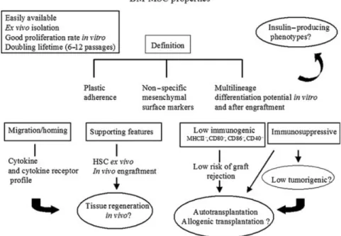
Fig. 1. The bone marrow-derived mesenchymal stem cell (BM-MSC) properties: most important features of BM-MSC, which renders these cells very attractive for cell-based therapies and in particular forβ-cell replacement in type 1 diabetes..
differentiating capacity into multiple mesenchymal lineages following engraftment (37). Yet, so far, there is no consensus marker panel that allows for prospective identification of MSC from the in vitro marrow stromal cell population. Recently, it has been reported that neuro ganglioside GD2 surface marker is expressed in BM-MSC and not in other marrow stromal cells (38). Moreover, SSEA-4, a glycolipid antigen marker of human ES cells (39) as well as the nucleolar protein nucleostemin are suggested as adult MSC markers (40).
Are adult BM-MSC a promising source for β-cell replacement?
In vitro and in vivo reprogramming of adult cells. Repro-gramming adult somatic cells implicates induction or repression of key regulatory genes, which are able to switch cells from one lineage into another or to dedif-ferentiate a mature phenotype back to a more primitive state to obtain pluripotency.
In recent pioneering experiments, mouse and human fibroblasts have been successfully reprogrammed into ES-like stem cell lines. These induced pluripotent stem cells (iPS) were generated by in vitro retroviral trans-duction of four transcription factors (Oct3/4, Sox2, c-Myc, and Klf4) that completely modified the epige-netic signature and phenotype of these cells (41, 42). Using the same techniques, murine liver and stomach cells have also been reprogrammed into iPS cells (43).
Moreover, reprogramming of adult exocrine pancre-atic cells into functionalβ-cells was recently achieved by re-expressing a combination of key regulatory genes (Ngn3, Pdx1, and MaFA) in living adult animals. A great advantage of this in vivo strategy was to maintain the viral transduced exocrine cells in their native envi-ronment, which may favor maturation and survival of newly reprogrammedβ-cells (44).
Because of their multipotent properties, MSC may have an almost unlimited differentiation potential once they have been reprogrammed. This might be achieved by specific culture conditions, genetic induction of key transcriptions factors, or more recently through epigenetic modifications.
Reprogramming BM-MSC into insulin-producing
phenotypes
In recent years, an increasing body of evidence suggests that BM-MSC have the potential to differentiate in vitro into insulin-producing cells in mouse (9, 10) and human systems (45, 46) and to revert hyperglycemia at least in diabetic mouse models (9, 10, 45). Initial results have been achieved by ex vivo expansion of plastic adherent BM-MSC and prolonged endocrine cultivation conditions (9, 10). More recently, the endocrineβ-cell-like phenotype was obtained by forced expression of a pancreatic endocrine key regulator,
Pdx-1, in primary BM-MSC, with higher differentiation efficiency (45). Furthermore, epigenetic modification could induce differentiation of mouse BM-MSC into insulin-producing cells, even in a glucose-sensing manner (47). Nevertheless, efficacy of BM-MSC differentiation is low and highly variable. The most important limitation at the current state of knowledge is the insulin secretion capacity of these cells, which is far from native β-cells. While several studies have demonstrated the potential of endocrine-differentiated BM-MSC to produce and secrete insulin in a glucose-sensing manner, insulin amount is still significantly lower compared with matureβ-cells (9, 10, 45, 47).
This might be related to donor-specific variabil-ity, heterogeneity of MSC preparations, and non-optimized differentiation protocols. Also, there might be unknown BM-MSC populations with suitable endocrine commitment to be found. In an attempt to overcome such issues, we used a well-characterized cell line, the telomerase-immortalised human adult bone marrow-derived mesenchymal stem cell line (hMSC-TERT), as a human BM-MSC model to investigate the molecular mechanisms of endocrine differentiation in MSC (Limbert C, P¨ath P, Ebert R, Rothham-mer V, Kassem M, Jakob F, Seufert J, unpublished data). These cells were able to induce insulin in response to specific culture conditions or via ectopic expression of two major pancreatic endocrine genes, Ngn3 and/or Pdx1. Specifically, our data indicate that Ngn3 transcription factor is involved in Pdx1 and insulin expression in hMSC-TERT, thus mimicking the pancreatic endocrine transcription factor cascade in the developing pancreas. Still, insulin secretion was reduced and no response to glucose stimulation was observed, which indicates reduced endocrine differ-entiation capacity. Although hMSC-TERT display ‘endodermal prepatterning’ features, molecular tools for the glucose-sensing machinery are missing in this cell line. In vivo experiments could favor the endocrine maturation of modified hMSC-TERT. Moreover, a different combination of key regulatory genes might be more adequate to obtain functionalβ-cell-like pheno-types out of this population.
Anyhow, it is well accepted that independently of the protocol and of the MSC population or donors, reprogramming BM-MSC into functional insulin-producing phenotypes in vitro requires several important biological steps presented in Table 1.
Transplantation of adult BM-MSC into diabetic
mouse models
The direct transplantation of donor BM-MSC into diabetic mice have convincingly demonstrated that the major effect of BM-MSC infusion is to enhance the number of endogenous mouse β-cells with increase of mouse insulin and amelioration of
Table 1. Major biological steps for reprogramming BM-MSC into functional insulin cell-types
1. Gene expression markers for:
(i) Endodermal patterning of MSC: ABCG2, Cxcr4, Thy-1, Foxa2, Sox17, Isl1, Hlxb9
(ii) Endocrine precursor cells: Ngn3, NeuroD, Nkx6.1, Pax4
(iii) Matureβ-cell
Key regulators of insulin expression-Pdx1 and Maf-A Proinsulin (Ins)
Glucose transporter-Glut2
Glucose sensing-machinery- Glucokinase (Gck) 2. Protein synthesis
Insulin and/or C-peptide
3. Insulin secretion in response to glucose challenge in vitro 4. Improvement of hyperglycemia in diabetic animal
models and/or humans after engraftment of insulin-producing phenotypes
5. Excision of the implanted cells leads to hyperglycemia in the diabetic models
Expression of early endodermal markers in isolated adult BM-MSC indicates the presence of suitable BM-MSC cell sub-population. After the endocrine differentiation process, expression of major regulatory transcription factors is mandatory to recognize endocrine precursor phenotypes or matureβ-cells. The function of insulin-producing phenotypes is assessed by the expression of insulin gene, specific glucose transporter and glucose sensing factors, as well as production, storage, and secretion of insulin protein in a glucose-dependent manner. In vivo, these cells should revert hyperglycemia in overt diabetic animal models and excision of the engrafted cells triggers hyperglycemia.
hyperglycemia (48, 49). In contrast to previous findings (28), differentiation of donor BM-MSC into insulin-producing cells seems to be rare. Interestingly, transplantation of in vitro endocrine-differentiated BM-MSC into streptozotocin injured non-obese diabetic mice increases insulin and C-peptide levels as well as improves glycemic control. Nevertheless, as shown by hyperglycemia after excision of the transplant, there is no regeneration of endogenous mouseβ-cells (10, 45). These observations strongly suggest that, in vivo, fate and function of endocrine-differentiated BM-MSC are distinct from that of directly infused BM-MSC. Yet, both can either revert or ameliorate diabetes.
At this point, some reflection should be made to establish which mechanism is more suitable to be adopted in cell-based strategies: cell reprogramming with consecutive cell replacement or in situ induction ofβ-cell regeneration.
Other promising approaches to cure T1D mellitus
In vivo restoration ofβ-cell mass might prove beneficial for the reversal of hyperglycemia, but protection of the expanded β-cells from ongoing autoimmune destruction is equally important to cure T1D mellitus.
The efficacy of immunosuppressive agents such as prednisone, azathioprine, and cyclosporine to suppress T lymphocytes in T1D patients has been thoroughly evaluated (50, 51). Although slower decline in C-peptide levels could be observed in these patients, withdrawal of the drugs led to progression ofβ-cell mass destruction, which would implicate lifelong immuno-suppression with all known chronic complications.
More targeting immunomodulatory approaches have been developed and are now on early phase clinical trials. Treatment with either a peptide of the heat-shock protein 63, Diapep277® (Andromeda Biotech Ltd.’s, Yavne, Israel), an autoantigen with immunomodulatory properties, or anti-CD3 antibody has been effective in reducing insulitis and insulin requirements in newly diagnosed T1D mice and humans (52, 53). Results of a most recent clinical trial revealed that vaccination with two dose of recombinant GAD65 peptide, in early diagnosed type 1 diabetics (< 6 months), induces preservation of residual β-cell mass. This seems to result from stimulation of an antigen-specific T-cell population and modulation of B-cell memory (54). Similar to anti-CD3 treatment, GAD vaccination slowed the decline of stimulated C-peptide level, conferring a metabolic benefit after 18 months of trial (53). In contrast to anti-CD3 therapy, no reduction in insulin dose requirements was observed in treated patients and no side effects were detected.
Yet, none of the described immunomodulatory approaches has been sufficient for inducing insulin independence.
Conclusion and perspectives
While cell-based therapy has still a fairly long way to go in order to deliver a full response to T1D, there is still room for exogenous insulin strategies developments. Using nanoparticle systems, new concepts of glucose-sensing insulin release are being tested on diabetic animal models. SmartInsulin aims at being a once-a-day subcutaneous insulin formulation liberated in a glucose-regulated manner. Then again, the existence of molecular signals other than glucose that could unnecessarily release insulin into the circulation must be certified to avoid dangerous bursts of insulin. Whereas Smart particle delivery systems might represent a relevant alternative in the treatment of T1D, it does not comprise the key to insulin independence.
Because of their intrinsic properties, MSC display great potential to be used as a real alternative source for cellular therapy in many degenerative diseases, including T1D.
Much has been achieved in the past 5 yr in the field of stem cells. However, there is no reliable cell source that could be used for the cell-based therapy of diabetes mellitus, so far. In what concerns reprogramming of MSC into functional insulin-producing phenotypes,
there are still a few resources to be explored. Once adequate mechanisms for cell-based therapy are established and tools are successfully applied, it will be possible to obtain cells capable of regenerating or replacing the missing pancreaticβ-cells.
It is quite clear that the cure of diabetes will require a combination of distinct strategies. Cell-based thera-pies may recover endogenous insulin production; yet, modulation of the autoimmune disease behind T1D is essential to achieve an effective and long-lasting ther-apeutic intervention. Also,β-cell proliferating agents, such as incretin analogs and islet neogenesis-associated protein, might be of benefit when used simultaneously to cell-based therapies, more so on advanced stages of β-cell destruction. Hence, to achieve the goal of permanent insulin independence, work on distinct but complementary research lines has still to go further.
Acknowledgements
C. L. was supported by a Research Fellowship from the European Society for Pediatric Endocrinology sponsored by NovoNordisk, a Visiting Fellowship from the International Society for Paediatric and Adolescent Diabetes and Doctoral fellowship supported by the Foundation for Science and Technology (European Community, Portugal). J. S. is supported by Deutsche Forschungsgemeinschaft and by the Bundesministerium f ¨ur Bildung und Forschung within the consortium and Advance Knowledge for Restoration of Beta Cells and Diabetes Therapy in the German competence network diabetes mellitus.
References
1. WILDS, ROGLICG, GREENA, SICREER, KINGH. Global prevalence of diabetes: estimates for the year 2000 and projections for 2030. Diabetes Care 2004: 27: 1047–1053.
2. SHAPIROAM, LAKEYJR, RYANEA et al. Islet trans-plantation in seven patients with type 1 diabetes mellitus using a glucocorticoid-free immunosuppressive regimen. N Engl J Med 2000: 343: 230–238.
3. RYANEA, PATYBW, SENIORPA et al. Five-year follow-up after clinical islet transplantation. Diabetes 2005: 54: 2060–2069.
4. D’AMOURKA, BANGAG, ELIAZERS et al. Production of pancreatic hormone-expressing endocrine cells from human embryonic stem cells. Nat Biotechnol 2006: 24: 1392–1401 [Epub 2006 October 19].
5. JIANGJ, AUM, LUK et al. Generation of insulin-producing islet-like clusters from human embryonic stem cells. Stem Cells 2007: 25: 1940–1953 [Epub 2007 May 17].
6. SHIMJH, KIMSE, WOODH et al. Directed differentia-tion of human embryonic stem cells towards a pancreatic cell fate. Diabetologia 2007: 50: 1228–1238 [Epub 2007 April 18].
7. LIMBERTC, PATHG, JAKOBF, SEUFERTJ. Beta-cell replacement and regeneration: strategies of cell-based therapy for type 1 diabetes mellitus. Diabetes Res Clin Pract 2008: 79: 389–399 [Epub 2007 September 12].
8. PITTENGERMF, MACKAYAM, BECKSC et al. Multi-lineage potential of adult human mesenchymal stem cells. Science 1999: 284: 143–147.
9. CHOIKS, SHINJS, LEEJJ, KIMYS, KIMSB, KIMCW. In vitro trans-differentiation of rat mesenchymal cells into insulin-producing cells by rat pancreatic extract. Biochem Biophys Res Commun 2005: 330: 1299–1305. 10. TANGDQ, CAOLZ, BURKHARDTBR et al. In vivo
and in vitro characterization of insulin-producing cells obtained from murine bone marrow. Diabetes 2004: 53: 1721–1732.
11. SORIAB, ROCHEE, BERNAG, LEON-QUINTOT, REIGJA, MARTINF. Insulin-secreting cells derived from embryonic stem cells normalize glycemia in streptozotocin-induced diabetic mice. Diabetes 2000: 49: 157–162.
12. FUJIKAWAT, OHSH, PIL, HATCHHM, SHUPET, PETERSENBE. Teratoma formation leads to failure of treatment for type I diabetes using embryonic stem cell-derived insulin-producing cells. Am J Pathol 2005: 166: 1781–1791.
13. GUZY, NASIRI, TEITELMANG. Regeneration of pan-creatic beta cells from intra-islet precursor cells in an experimental model of diabetes. Endocrinology 2001: 142: 4956–4968.
14. GAOR, USTINOVJ, KORSGRENO, OTONKOSKIT. In vitro neogenesis of human islets reflects the plasticity of differentiated human pancreatic cells. Diabetologia 2005: 48: 2296–2304.
15. BONNER-WEIRS, TANEJAM, WEIRGC et al. In vitro cultivation of human islets from expanded ductal tissue. Proc Natl Acad Sci U S A 2000: 97: 7999–8004. 16. MINAMIK, OKUNOM, MIYAWAKIK et al. Lineage
tracing and characterization of insulin-secreting cells generated from adult pancreatic acinar cells. Proc Natl Acad Sci U S A 2005: 102: 15116–15121 [Epub 2005 October 6].
17. DORY, BROWNJ, MARTINEZOI, MELTONDA. Adult pancreatic beta-cells are formed by self-duplication rather than stem-cell differentiation. Nature 2004: 429: 41–46.
18. BAEYENSL, DEBREUCKS, LARDONJ, MFOPOUJK, ROOMANI, BOUWENSL. In vitro generation of insulin-producing beta cells from adult exocrine pancreatic cells. Diabetologia 2005: 48: 49–57 [Epub 2004 December 23]. 19. SEABERGRM, SMUKLERSR, KIEFFERTJ et al. Clonal identification of multipotent precursors from adult mouse pancreas that generate neural and pancreatic lineages. Nat Biotechnol 2004: 22: 1115–1124.
20. XUX, D’HOKERJ, STANGEG et al. Beta cells can be generated from endogenous progenitors in injured adult mouse pancreas. Cell 2008: 132: 197–207.
21. WAGERSAJ, WEISSMANIL. Plasticity of adult stem cells. Cell 2004: 116: 639–648.
22. ZALZMANM, ANKER-KITAIL, EFRATS. Differentia-tion of human liver-derived, insulin-producing cells toward the beta-cell phenotype. Diabetes 2005: 54: 2568–2575.
23. SAPIRT, SHTERNHALLK, MEIVAR-LEVYI et al. Cell-replacement therapy for diabetes: generating functional insulin-producing tissue from adult human liver cells. Proc Natl Acad Sci U S A 2005: 102: 7964–7969.
24. HORIY, GUX, XIEX, KIMSK. Differentiation of insulin-producing cells from human neural progenitor cells. PLoS Med 2005: 2: e103.
25. ENDEN, CHENR, REDDIAS. Effect of human umbilical cord blood cells on glycemia and insulitis in type 1 diabetic mice. Biochem Biophys Res Commun 2004: 325: 665–669.
26. RUHNKEM, UNGEFRORENH, NUSSLERA et al. Differ-entiation of in vitro-modified human peripheral blood monocytes into hepatocyte-like and pancreatic islet-like cells. Gastroenterology 2005: 128: 1774–1786.
27. HESSD, LIL, MARTINM et al. Bone marrow-derived stem cells initiate pancreatic regeneration. Nat Biotech-nol 2003: 21: 763–770 [Epub 2003 June 22].
28. IANUSA, HOLZGG, THEISEND, HUSSAINMA. In vivo derivation of glucose-competent pancreatic endocrine cells from bone marrow without evidence of cell fusion. J Clin Invest 2003: 111: 843–850.
29. VOLTARELLIJC, COURICE, STRACIERIAB et al. Autol-ogous nonmyeloablative hematopoietic stem cell trans-plantation in newly diagnosed type 1 diabetes mellitus. JAMA 2007: 297: 1568–1576.
30. KONDOM, WAGERSAJ, MANZMG et al. Biology of hematopoietic stem cells and progenitors: implications for clinical application. Annu Rev Immunol 2003: 21: 759–806.
31. PHINNEYDG, PROCKOPDJ. Concise review: mesenchy-mal stem/multipotent stromesenchy-mal cells: the state of transdif-ferentiation and modes of tissue repair –current views. Stem Cells 2007: 25: 2896–2902 [Epub 007 September 27].
32. RIDLEYAJ, SCHWARTZMA, BURRIDGEK et al. Cell migration: integrating signals from front to back. Science 2003: 302: 1704–1709.
33. KOBLAST, ZACHAROVOVAK, BERKOVAZ et al. Iso-lation and characterization of human CXCR4-positive pancreatic cells. Folia Biol (Praha) 2007: 53: 13–22. 34. BEYTHS, BOROVSKYZ, MEVORACHD et al. Human
mesenchymal stem cells alter antigen-presenting cell maturation and induce T-cell unresponsiveness. Blood 2005: 105: 2214–2219 [Epub 004 October 28].
35. TIANY, DENGYB, HUANGYJ, WANGY. Bone marrow-derived mesenchymal stem cells decrease acute graft-versus-host disease after allogeneic hematopoietic stem cells transplantation. Immunol Invest 2008: 37: 29–42.
36. GOTHERSTROMC. Immunomodulation by multipotent mesenchymal stromal cells. Transplantation 2007: 84: S35–7.
37. DOMINICIM, LEBLANCK, MUELLERI et al. Minimal criteria for defining multipotent mesenchymal stromal cells. The International Society for Cellular Therapy position statement. Cytotherapy 2006: 8: 315–317. 38. MARTINEZC, HOFMANNTJ, MARINOR, DOMINICIM,
HORWITZEM. Human bone marrow mesenchymal stromal cells express the neural ganglioside GD2: a novel surface marker for the identification of MSCs. Blood 2007: 109: 4245–4248 [Epub 2007 January 30].
39. GANGEJ, BOSNAKOVSKID, FIGUEIREDOCA, VISSERJW, PERLINGEIRORC. SSEA-4 identifies mesenchymal stem cells from bone marrow. Blood 2007: 109: 1743–1751 [Epub 2006 October 24].
40. KAFIENAHW, MISTRYS, WILLIAMSC, HOLLAN
-DERAP. Nucleostemin is a marker of proliferating
stromal stem cells in adult human bone marrow. Stem Cells 2006: 24: 1113–1120 [Epub 2005 November 10]. 41. MEISSNERA, WERNIGM, JAENISCHR. Direct
repro-gramming of genetically unmodified fibroblasts into pluripotent stem cells. Nat Biotechnol 2007: 25: 1177–1181 [Epub 2007 August 27].
42. PARKIH, ZHAOR, WESTJA et al. Reprogramming of human somatic cells to pluripotency with defined factors. Nature 2008: 451: 141–146 [Epub 2007 December 23].
43. AOIT, YAEK, NAKAGAWAM et al. Generation of pluripotent stem cells from adult mouse liver and stomach cells. Science 2008: 14: 14.
44. ZHOUQ, BROWNJ, KANAREKA, RAJAGOPALJ, MELTONDA. In vivo reprogramming of adult pan-creatic exocrine cells to beta-cells. Nature 2008: 455: 627–632.
45. KARNIELIO, IZHAR-PRATOY, BULVIKS, EFRATS. Generation of insulin-producing cells from human bone marrow mesenchymal stem cells by genetic manipulation. Stem Cells 2007: 5: 5.
46. MORISCOTC, DE FRAIPONTF, RICHARDMJ et al. Human bone marrow mesenchymal stem cells can express insulin and key transcription factors of the endocrine pancreas developmental pathway upon genetic and/or microenvironmental manipulation in vitro. Stem Cells 2005: 23: 594–603.
47. TAYARAMMAT, MAB, ROHDEM, MAYERH. Chro-matin-remodeling factors allow differentiation of bone marrow cells into insulin-producing cells. Stem Cells 2006: 24: 2858–2867 [Epub 006 September 21].
48. HASEGAWAY, OGIHARAT, YAMADAT et al. Bone marrow (BM) transplantation promotes beta-cell regen-eration after acute injury through BM cell mobilization. Endocrinology 2007: 148: 2006–2015 [Epub 7 January 25].
49. LEERH, SEOMJ, REGERRL et al. Multipotent stromal cells from human marrow home to and promote repair of pancreatic islets and renal glomeruli in diabetic NOD/scid mice. Proc Natl Acad Sci U S A 2006: 103: 17438–17443 [Epub 2006 November 6].
50. THE CANADIAN-EUROPEAN RANDOMIZED CONTROL
TRIALGROUP. Cyclosporin-induced remission of IDDM
after early intervention. Association of 1 yr of cyclosporin treatment with enhanced insulin secretion. Diabetes 1988: 37: 1574–1582.
51. SILVERSTEINJ, MACLARENN, RILEYW, SPILLARR, RADJENOVICD, JOHNSONS. Immunosuppression with azathioprine and prednisone in recent-onset insulin-dependent diabetes mellitus. N Engl J Med 1988: 319: 599–604.
52. RAZI, ELIASD, AVRONA, TAMIRM, METZGERM, COHENIR. Beta-cell function in new-onset type 1 diabetes and immunomodulation with a heat-shock protein peptide (DiaPep277): a randomised, double-blind, phase II trial. Lancet 2001: 358: 1749–1753.
53. KEYMEULEN B, VANDEMEULEBROUCKE E, ZIEGLER
AG et al. Insulin needs after CD3-antibody therapy in new-onset type 1 diabetes. N Engl J Med 2005: 352: 2598–2608.
54. LUDVIGSSONJ, FARESJOM, HJORTHM et al. GAD treatment and insulin secretion in recent-onset type 1 diabetes. N Engl J Med 2008: 359: 1909–1920.