Neurosurgery
Manuscript Draft
Manuscript Number: WNS-18-888R1
Title: How does minimally invasive TLIF (transforaminal lumbar interbody fusion) influence lumbar radiological parameters?
Article Type: Original Article
Keywords: Lumbar lordosis; Minimally Invasive Surgery; Lumbar radiological outcomes; Single level fusion; Transforaminal Lumbar Interbody Fusion
Corresponding Author: Mrs. Cláudia Maria Pereira, MB
Corresponding Author's Institution: Faculty of Medicine, University of Porto
First Author: Cláudia Maria Pereira, MB
Order of Authors: Cláudia Maria Pereira, MB; Pedro S Silva, MD; Marisa Cunha, MD, MSc; Rui Vaz, MD, PhD; Paulo Pereira, MD, PhD
Abstract: Background: Minimally Invasive Transforaminal Lumbar Interbody Fusion (MIS-TLIF) has become a popular method of interbody fusion.
Clinical outcomes after MIS-TLIF have been reported but few studies have focused on the radiological changes in the segmental parameters of the operated and adjacent segments and in lumbar lordosis, for single level MIS-TLIF.
Methods: From March 2009 to September 2016, 117 patients who underwent a single-level MIS-TLIF surgery for lumbar degenerative disease were
enrolled in this retrospective study.
Anterior disc height (ADH), posterior disc height (PDH) and segmental angle (SA) of the operated and adjacent levels and lumbar lordosis (LL) were evaluated on X-rays obtained preoperatively and postoperatively at 6-12 months follow-up visits. Cage related parameters including fusion and subsidence rates were analyzed on postoperative CT scans. Clinical assessment used validated outcome scores such as Oswestry Disability Index (ODI) questionnaire and Odom's criteria.
Results: ADH and PDH of the operated segment increased significantly after surgery, but no significant changes were seen in SA of that level. Statistically significant decreases were observed in PDH of both adjacent segments and increases in adjacent superior SA. Lumbar lordosis showed a slight but statistically significant improvement after surgery that was mostly correlated with the postoperative increase in adjacent superior SA (r: 0.58; p < 0.001). No significant correlations were found between clinical and radiological results.
Conclusions: Single-level MIS-TLIF increased disc height but not the segmental angle at the operated level. Lumbar lordosis improvement after surgery was mainly associated with the increase of the cranial segmental
Dr Edward C. Benzel
Editor-in-Chief
World Neurosurgery
27 February 2018
Dr Edward C. Benzel,
I am submitting the paper entitled “How does minimally invasive TLIF (transforaminal
lumbar interbody fusion) influence lumbar radiological parameters?” for consideration of
publication in World Neurosurgery.
In this study, we observed that single-level MIS-TLIF increased both anterior and posterior
disc heights of the operated segment, but no significant changes were seen in the segmental
angle at the same level. Small yet statistically significant decreases were observed in
posterior disc height of both adjacent segments and increases in adjacent superior
segmental angle. Lumbar lordosis increased on average 2 degrees after surgery and this was
mostly correlated with the postoperative gain in the cranial segmental angle. No significant
correlations were found between clinical and radiological results.
I, Cláudia Pereira, certify that this manuscript is a unique submission and is not being
considered for publication, in part or in full, with any other source in any medium.
Please address correspondence to:
Cláudia Pereira
Serviço de Neurocirurgia
Centro Hospitalar S. João, EPE
Alameda Professor Hernâni Monteiro
4200-319 Porto, Portugal
[email protected]
Yours sincerely,
Cláudia Pereira
Pereira
Conflict of interest
The authors declare no conflict of interest.
1 May 2018
Re: Resubmission of manuscript “How does minimally invasive TLIF (transforaminal
lumbar interbody fusion) influence lumbar radiological parameters?”, WNS-18-888
Dr Edward C. Benzel
Editor-in-Chief
WORLD NEUROSURGERY
Dear Editor
Thank you for the opportunity to revise this manuscript, “How does minimally invasive
TLIF (transforaminal lumbar interbody fusion) influence lumbar radiological
parameters?”. I very much appreciate the careful review and suggestions and I believe
that the manuscript is significantly improved after making the suggested edits.
Following this letter are the reviewer comments with the responses in italics. Changes
made in the manuscript are marked using track changes. The revision has been
developed in consultation with all coauthors, and each author has given approval to
the final form of this revision.
Thank you for your consideration.
Please address correspondence to:
Cláudia Pereira
Serviço de Neurocirurgia
Centro Hospitalar S. João, EPE
Alameda Professor Hernâni Monteiro
4200-319 Porto, Portugal
I had the pleasure to review your very interesting paper, current theme, very well
done, very clear. Just maybe the article would be better with some radiological image.
We are grateful for your kind comments and your appreciation of our work. We agree
that this paper will benefit from explanatory radiologic images and so we added some
to the article (Figures 1 to 3).
Reviewer #2
�
The authors refer daily use of a non-narcotic drug reported by 20.9% of the patients
and daily use of narcotic/opioid drugs required by 9.9% of patients; this could
influence the clinical outcome and then the results of clinical evaluation. This patients
could be excluded.
We agree with the statement of the reviewer that the use of analgesic medication is a
measure of clinical outcome. In fact, in our series, the correlation between ODI and
“use of medication” (Stanford criteria) was negative (r= -0.380, p<0.001, Spearman),
meaning that patients who take stronger analgesics have higher ODI scores (not
presented data).
However, the primary endpoint of this investigation was a difference in radiological
parameters at the operated segment with surgery, in particular with the use of straight
cages in minimally invasive TLIF. Hence, the use of medication is not a confounding
variable regarding the primary endpoint of this study, so we think that there is no
benefit in excluding these patients from the study.
�
The percentages reported are not precise.
Thank you for pointing this. In fact, there was an error in table 5, which was eliminated
from the current revised version of the paper. We also reviewed all the percentages in
segmental angle.
We decided to add explanatory radiological images to better illustrate the parameters
measured and we believed that it clarifies this point.
�
The authors say that the expected primary endpoint was a difference of PDH and SA
between pre and postoperative times at the operated level, without mentioning the
other radiological parameters measured; this point must be clarified.
We agree with this remark and thank the reviewer for bringing it to us. Thus, we have
added a section, “secondary endpoints” to overcome this issue so that the methods are
reflected in the results and support the conclusions.
�
The sample could be too heterogeneous, including all symptomatic lumbar
degenerative diseases; different etiologies could influence rate of fusion, radiological
changes, but most of all clinical outcome.
We agree with the reviewer that the heterogeneity of the sample could influence the
clinical outcomes. However, the study population included all patients operated in our
institution after application of the inclusion/exclusion criteria and so it was intended to
be representative of the “real world” population who undergoes minimally invasive
TLIF for degenerative pathologies. The research questions of this study were about
radiological differences in the operated segment and adjacent levels after surgery, and
how the cage position can influence the segmental and lumbar sagittal alignment.
Neither of these questions were influenced by the surgical indication, as we report in
the last sentence of the “radiological evaluation” section of the “Results”.
�
The authors report the use of straight cages of 32x10mm in 81% of patients without
mentioning the characteristics of the device used for the remaining 19%. This point
should be clarified.
�
Pain and functional disability were quantitatively measured using Low back and Leg
pain numeric rating scale (NRS-11), Oswestry Disability Index (ODI) questionnaire,
Odom's criteria and Stanford's score, but in the paragraph "Clinical evaluation" and in
Table 5 not all of them are reported. This point must be clarified.
We removed Table 5 and have ensured that the information related to the clinical
evaluation is fully reported in the text.
�
It lacks of explanatory radiological images.
We agree with the reviewer and added some explanatory radiological images for a
better understanding of the radiological measures taken.
�
The format of the references is not uniform. It should be corrected.
We noticed the mistake and corrected the format of the references.
�
The References section needs to be updated and upgraded, adding at least the
following:
1. Clinical and Radiologic Comparison of Minimally Invasive Surgery With Traditional
Open Transforaminal Lumbar Interbody Fusion: A Review of 452 Patients From a Single
Cen-ter. Price JP, Dawson JM, Schwender JD, Schellhas KP. Clin Spine Surg. 2018
Mar;31:E121-E126.
2. Minimally invasive transforaminal lumbar interbody fusion with expandable versus
static interbody devices: radiographic assessment of sagittal segmental and pelvic
parameters. Hawasli AH, Khalifeh JM, Chatrath A, Yarbrough CK, Ray WZ. Neurosurg
Focus. 2017;43:E10.
The suggested references were included in the current revised version of the
manuscript.
1) The Authors did not find any increase of sagittal lordosis at the operated level
because of their surgical technique: they used an oblique TLIF (angle 55° with respect
to the sagittal plane), extending at least for half of the disc space posteriorly, and
therefore it was not possible to modify the lordosis; it would have been possible in
case they had put the cage in the anterior half of the disc.
2) The second reason is the difficulty to obtain a valid compression when using a
percutaneous technique for screw positioning, because the posterior articular
complexes are in place and they limit the compression. I think that the Authors should
discuss and clarify these points.
We thank the reviewer for these insightful comments that should improve the
discussion of our work. In our technique we use straight cages, that are easier to
introduce through the minimally invasive access and we try to place them in the
intersomatic space as anteriorly as possible. However, as pointed out by the reviewer,
all our cages extend to the posterior half of the disc space. The reason for this is that
we intend to avoid a decrease of the posterior disc height and hence of the
neuroforaminal height that could result from an anterior only disc space distraction.
We agree that, since the distraction of the disc space is obtained both anteriorly and
posteriorly, polyaxial screws are used and we do not perform posterior column
osteotomy on the contralateral side of the approach it is not surprising that the
segmental and even the global lordosis did not significantly improve after surgery. With
this study, we identify a mechanism related with adjacent levels that contribute to a
slight improvement of lumbar lordosis. It is clear that in those cases where lordosis
restoration is a major issue, different techniques should be used, such as
anterior/lateral approaches or osteotomies. Regarding the use of other types of cage
for posterior/posterolateral minimally invasive approaches, in our view there is no clear
lordosis in both groups. We added a paragraph to the “Discussion” with these
considerations.
(*) Choi W-S, Kim J-S, Hur J-W, Seong J-H. Minimally Invasive Transforaminal Lumbar
Interbody Fusion Using Banana-Shaped and Straight Cages: Radiological and Clinical
Results from a Prospective Randomized Clinical Trial. Neurosurgery 2017;0:1-10.
Pereira
Abbreviations list:
ADH: Anterior disc height;
BMI: Body mass index;
LDH: Lumbar disc herniation;
LL: Lumbar lordosis;
MIS-TLIF: Minimally Invasive Transforaminal Lumbar Interbody Fusion;
NRS-11: Low back and Leg pain numeric rating scale;
ODI: Oswestry Disability Index;
PDH: Posterior disc height;
SA: Segmental angle;
Pereira
Title
How does minimally invasive TLIF (transforaminal lumbar interbody fusion) influence lumbar radiological parameters?
Author names and affiliations
Cláudia Pereiraa; Pedro Santos Silvaa,b,c; Marisa Cunhaa,b; Rui Vaza,b,c; Paulo Pereiraa,b,c
Affiliations:
a – Faculty of Medicine, University of Porto, Porto, Portugal;
b – Department of Neurosurgery, Centro Hospitalar São João, Porto, Portugal; c – Neurosciences Center CUF Porto, Portugal
Corresponding author: Cláudia Pereira
Address: Department of Neurosurgery, Centro Hospitalar São João, Alameda Professor Hernâni Monteiro,4200-319 Porto, Portugal
Email address: [email protected]
Highest academic degrees for all authors: Cláudia Pereira, MB; Pedro Santos Silva, MD; Marisa Cunha, MD, MSc; Rui Vaz, MD, PhD; Paulo Pereira, MD, PhD
Key words: Lumbar lordosis; Minimally Invasive Surgery; Lumbar radiological outcomes; Single level fusion; Transforaminal Lumbar Interbody Fusion
Abbreviations list:
ADH: Anterior disc height; BMI: Body mass index; LDH: Lumbar disc herniation; LL: Lumbar lordosis; MIS-TLIF: Minimally Invasive Transforaminal Lumbar Interbody Fusion; NRS-11: Low back and Leg pain numeric rating scale; ODI: Oswestry Disability Index; PDH: Posterior disc height; SA: Segmental angle; SPL: Spondylolisthesis
Abstract
Background: Minimally Invasive Transforaminal Lumbar Interbody Fusion (MIS-TLIF) has become a popular method of interbody fusion. Clinical outcomes after MIS-TLIF have been reported but few studies have focused on the radiological changes in the segmental parameters of the operated and adjacent segments and in lumbar lordosis, forin single level MIS-TLIF.
Methods: From March 2009 to September 2016, 117 patients who underwent a single-level MIS-TLIF surgery for lumbar degenerative disease were enrolled in this retrospective study.
Anterior disc height (ADH), posterior disc height (PDH) and segmental angle (SA) of the operated and adjacent levels and lumbar lordosis (LL) were evaluated on X-rays obtained preoperatively and postoperatively at 6-12 months follow-up visits. Cage related parameters including fusion and subsidence rates were analyzed on postoperative CT scans. Clinical assessment used validated outcome scores such as Oswestry Disability Index (ODI) questionnaire and Odom’s criteria. Results: ADH and PDH of the operated segment increased significantly after surgery, but no significant changes were seen in SA of that level. Statistically significant decreases were observed in PDH of both adjacent segments and increases in adjacent superior SA. Lumbar lordosis showed a
slight but statistically significant improvement after surgery that was mostly correlated with the postoperative increase in adjacent superior SA (r: 0.58; p < 0.001). No significant correlations were found between clinical and radiological results.
Conclusions: Single-level MIS-TLIF increased disc height but not the segmental angle at the operated level. Lumbar lordosis improvement after surgery was mainly associated with the increase of the cranial segmental angle.
Introduction
The surgical approach to spinal disorders that require lumbar arthrodesis has evolved significantly over the years and includes a variety of surgical techniques that range from posterolateral fusion (PLF) to lumbar interbody fusion techniques (LIF).1
Lumbar interbody fusion has been reported to have higher fusion rates, improved deformity correction, and capability for indirect decompression and increasing of foraminal height.1,2 It can be performed using five main approaches: posterior lumbar interbody fusion (PLIF), transforaminal lumbar interbody fusion (TLIF), anterior lumbar interbody fusion (ALIF), oblique lumbar interbody fusion/anterior to psoas (OLIF/ATP) and lateral lumbar interbody fusion (LLIF).1
Posterior approaches, such as PLIF and TLIF, are frequent options for the treatment of degenerative lumbar disorders, allowing for complete decompression of the spinal canal and nerve roots, restoration of intervertebral height, near-total discectomy and, expectably, restoration of segmental lordosis at the fused level. Additionally, the posterior approaches have minimal risk of damaging retroperitoneal structures as opposed to ALIF.3–5
However, in the PLIF procedure, significant retraction of the thecal sac and nerve roots is required, in order to provide adequate access to the posterior disc space. Hence the risk of damage to nerve roots or conus medullaris, dural tears, epidural fibrosis and neuropathic pain, that usually limit the technique to the lower spine (L3-S1). The TLIF procedure was developed to overcome this limitation by providing a more lateral approach and unilateral exposure of the disc space that involves less neural retraction and decreases the risk of neurological or dural injury. TLIF enables placement of the graft and the cage within the anterior or middle third of the disc space, aiming to restore lumbar lordosis and allows preservation of the contralateral lamina, facet, and pars interarticularis. 1,4
As in other open posterior procedures, the iatrogenic injury of soft tissues and paraspinal muscles is an important cause of postoperative low back pain and can adversely affect short- and long term patient outcomes. Minimally invasive transforaminal lumbar interbody fusion (MIS-TLIF) was introduced to minimize the morbidity related to muscle trauma without compromising operative and clinical outcomes and is increasingly being used for lumbar arthrodesis. Reported benefits include less intraoperative blood loss, decreased pain and postoperative narcotics use, shortened hospital stay and
faster recovery.1,6–12 However, there is a limited number of reports evaluating radiological changes and their influence on outcomes after MIS-TLIF procedure. The restoration and maintenance of lumbar and segmental sagittal plane alignment are major concerns when performing intersomatic fusion. Factors related to the intersomatic cage influence the anterior and posterior disc space height and therefore the alignment and structural stability of the operated segment.13
There is indeed a claim of TLIF to restore segmental lordosis. However, this potentiality of the procedure is not well documented in the literature, in particular with the use of straight cages in minimally invasive surgery.
In this study, we went on to reviewed our experience with this type of cages in single level minimally invasive TLIF and report the differences observed after the surgery in the segmental parameters, either of the operated disc or the adjacent ones and in lumbar lordosis. In addition, possible correlations of these variables with the clinical outcomes were investigated researched.
Methods
Study and inclusion criteria
This study retrospectively identified patients who underwent a single-level MIS-TLIF surgery in the Department of Neurosurgery of Centro Hospitalar S. João, Porto from March of 2009 to September of 2016. The surgery was performed by the same team of neurosurgeons affiliated to the institution and experienced in the procedure. The hospital’s ethics committee approved the study protocol.
Patients over 18 years, who underwent single-level MIS-TLIF to treat a symptomatic lumbar degenerative disease, whose preoperative and postoperative standing radiographs, and postoperative computed tomography (CT) where available, and with a minimum of 6-month follow up in the outpatients clinic were eligible for the study. Exclusion criteria included: multilevel procedure, MIS-TLIF performed on levels above L3-L4, previous open lumbar fusion surgery or non-MIS-TLIF MI fusions, adjacent level decompressions or additional procedures such as vertebral augmentation or implantation of interspinous processes devices, lumbar infection, tumor and trauma. Patients who
received a previous lumbar decompressive surgery at any lumbar level by open or MI procedure were not excluded from the study. A total of 117 patients accomplished these criteria.
Functional and clinical data were obtained from retrospective review of the outpatient and inpatient medical records, radiographs and CT scans.
Surgical technique
Details of minimally invasive-TLIF technique are well described by previous literature. 7,9,14 Briefly, the patient is positioned prone on a radiolucent operating table following the induction of general anesthesia. Routine preparation and draping are performed. Under fluoroscopic guidance, Jamshidi needles are inserted percutaneously in the pedicles of the vertebrae to be fused and K-wires are passed through the trocars of the needles into the vertebral bodies. On the less symptomatic side of the patient, pedicle screws are introduced around the K-wires and a rod is inserted and secured to the screws using a MI spinal fixation system. On the contralateral side, a 4 cm incision is made on the skin and the fascia, between the entry points of the K-wires, (approximately 4.5 to 5 cm from the midline), and several muscle-splitting tubular dilators are inserted to create the surgical working channel and an appropriate-length tubular retractor (22mm diameter) is passed around the dilators and docked on the facet joint complex. The procedure is then performed by illuminating the tube and using an operating microscope. Ipsilateral facetectomy is performed and exiting and traversing nerve roots are decompressed if needed. A standard microdiscectomy followed by endplates preparation is performed. Then a bullet-shaped PEEK cage filled with local bone chips is inserted obliquely in the disc space and the remaining bone chips are placed in front and around the cage. Ipsilateral
percutaneous screws and a rod are then placed through the same incision and the screw-rod construct is compressed in an attempt to create lordosis and provide compression for the interbody graft/cage. After sufficient irrigation and hemostasis, the surgical wounds are sutured layer by layer.
Clinical Outcome
Personal data such as anthropometric parameters, smoking and professional activity was collected from patients’ clinical records, and postoperative clinical outcome was assessed through validated outcome scores previously recorded. Pain and functional disability were quantitatively measured
using Low back and Leg pain numeric rating scale (NRS-11), Oswestry Disability Index (ODI) questionnaire, Odom’s criteria and Stanford’s score.15–18
Radiologic Outcome
The radiologic parameters were measured on preoperative and postoperative radiographs (lateral full-length spine or lumbar X-rays). These included anterior and posterior disc height as well as segmental angle of the operated and adjacent levels and lumbar lordosis. The cage distance to the anterior and posterior margins of the vertebral endplates, cage obliquity, degree of fusion and cage subsidence in the vertebral endplates were measured on postoperative CT scans. The postoperative measurements were performed using radiographs and CT scans obtained at the 6-month and/or 12-month follow-up visits.
The anterior disc height (ADH) was defined as the distance between the inferior endplate to the superior endplate at the anterior vertebral body line, whereas the posterior disc height (PDH) was the corresponding measurement at the posterior vertebral body line. The segmental angle (SA) is the angle between the superior endplate and inferior endplate of the index segment. Lumbar lordosis (LL) was measured between the superior endplate of L1 and the superior endplate of S1 (Figure 1).
Cage distance to the anterior and posterior adjacent vertebral plate was taken from metallic markers within the cage that outline its anterior and posterior borders (Figure 2). Cage obliquity was defined as the angle subtended by the cage length line and the line connecting the transverse processes of the underlying vertebra, on the axial plane of CT scan (Figure 3).
Fusion was defined as continuous trabeculae of bone bridging the superior and inferior vertebral endplates on the sagittal and coronal planes of CT scan. Cage subsidence was defined as a sinking of the cage in the lower vertebral endplate greater than 3mm.
Primary endpoint
In this study, the expected primary endpoint was defined as a difference inof PDH and SA between pre and postoperative imagestimes at the operated level.
Secondary endpoints
Secondary endpoints were: 1) the difference in LL between pre and postoperative images, 2) correlation between the distance of the cage to the anterior limit of the vertebral endplates and postoperative changes in LL and SA at the operated segment, and 3) correlation between the difference in pre and postoperative radiological parameters and clinical outcomes.
Statistical analyses
Data analysis was performed using SPSS software. Paired and independent Student t-tests were used for mean comparison of preoperative and postoperative radiologic and clinical continuous variables with normal distribution, whereas Mann-Whitney U-test was used for comparison of those with non-normal distribution. Comparison of means between more than two groups was performed using oneway ANOVA. Association between variables was assessed using Pearson´s correlation coefficient for continuous, normal variables and Spearman´s correlation coefficient for non-parametric variables (both correlation coefficients denoted by r). Statistical significance was defined as a p-value <0.05.
Results
Sample description and surgical data
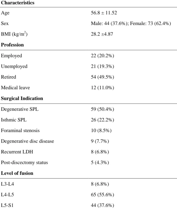
One hundred and seventeen patients (73 women and 44 men) were included in this study. Eighty-three (85.6%) were non-smokers and the mean age at surgery was 56.8 ± 11.52 years old. Mean body mass index (BMI) was 28.2 ± 4.87 kg/m2. The majority of patients (55.6%) were operated at L4-L5 level and the most common indication for surgery was degenerative spondylolisthesis (50.4%). A 32x10mm bullet-shaped cage was used in most of the cases(81%) of the patients., 26x10mm bullet-shaped cages were used in 12%, and other sizes in 7% of the patients (heights from 8 to 12mm and lengths from 26 to 32mm). The demographic data of the included patients are summarized in Table 1. Radiologic evaluation
All patients included had preoperative and postoperative radiologic parameters measured. Since 44 patients were operated at L5-S1 level, the parameters related to disc height and segmental angle of the
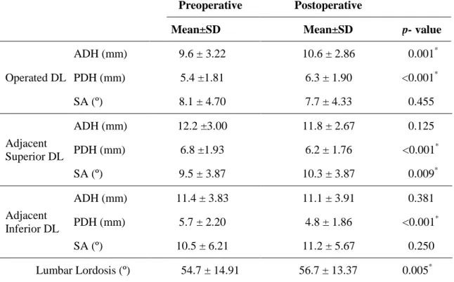
adjacent inferior disc were not accessed in this group. Table 2 summarizes preoperative and postoperative radiologic results. The mean ADH at the operated segment preoperatively and postoperatively was 9.6 ± 3.22 and 10.6 ± 2.86, respectively. Mean PDH at the same level was 5.4 ±1.81 and 6.3 ± 1.90, respectively pre- and postoperatively. These increases in ADH and PDH were both significant. However, the SA at the operated segment did not significantly change from before the surgery to the postoperative follow-uptime of follow-up period. Only PDH showed significant postoperative decrease in the adjacent segments. The mean difference was 0.60 ± 1.79mm (p < 0.001) in the adjacent superior segment and 0.89 ± 2.04mm (p < 0.001) in the adjacent inferior segment. Postoperative significant increase of SA was observed in the adjacent superior segment with a mean gain of 0.89 ± 3.65º (p = 0.009). Preoperative lumbar lordosis mean was 54.7º ± 14.91º and postoperative mean was 56.7º ± 13.37º at 12-month follow up. The difference between preoperative and postoperative periods was statistically significant, denoting a gain of lordosis of 1.97 ± 7.48º (p = 0.005) based on paired analysis. No more significant differences were shown between pre- and postoperative results.
On average, the intersomatic cage was located 2.8 mm from the anterior margin of the vertebral plate and 6.1 mm from the posterior margin, with a slope of 56.7° in relation to the transverse processes line (Table 3). Definite interbody fusion was achieved in 88.9% of patients in the follow-up CT scan of the lumbar spine, obtained 6 to 12 months after surgery. Subsidence of the cage was observed in 21.4% (25) patients. None of the cases required surgical revision.
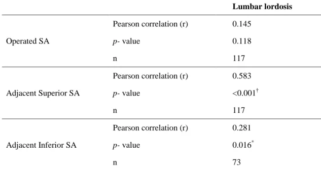
The correlation analysis showed that the postoperative adjacent superior SA has a stronger correlation with the postoperative gain on lumbar lordosis (r: 0.58; p < 0.001). The increase of adjacent inferior SA also correlates with the increment on lumbar lordosis, although to a lesser magnitude (r: 0.28; p = 0.016). No association was observed between operated SA and postoperative lumbar lordosis (Table 4). RegardingRelatively to cage parameters, no correlation was found between anterior-posterior positioning of the cage and postoperative lumbar lordosis or operated SA. Cage subsidence was not associated with absence of fusion or postoperative SA of the operated disc. No significant differences were found in radiological results of patients according to the operated level, Odom´s criteria or surgical indications (t-test and oneway ANOVA were performed).
Clinical evaluation
The median NRS-11 value for low back pain and leg pain was 5 and 4, respectively. InThe mean
ODI questionnaire 23.7% of patient scored 0-20 percentage points, 48.4% between 20-40, 23.7% between 40-60, 4.3% between 60-80 and the mean value was 32.18 ± 1.80. Relatively to Odom´s criteria, the majority of the patients (39.73%) had a “good” result, 38.85% had a “fair” result and
12.98% had an “excellent”, while 8.6% had a “poor” result. Odom’s criteria and ODI classes are summarized in table 5.
Regarding the use of medication for pain relief, the majority of the patients (36.3%) did not require any analgesic medication in the postoperative 6-12 months follow-up. Occasional use of a non-narcotic drug was reported by 13.2% of the patients while 20.9% reported a daily use of those drugs, 19.8% and 9.9% of patients required occasional and daily use of narcotic/opioid drugs, respectively, being tramadol, by far, the most common of these medications.
Seventy-five (78.9%) patients were satisfied with surgical results and 77 patients (78.6%) would redo surgery if they could anticipate the outcome.
The median satisfaction level with treatment and its results was 7 on a scale from 0 to 10, where 0 means not satisfied at all and 10 means completely satisfied.
No significant differences were found in clinical outcomes of patients according to the operated level or surgical indication (t-test and oneway ANOVA were performed).
Correlation between clinical and radiologic evaluation
No significant correlations were found between age and radiological or clinical results. The fusion rate was similar between smokers and non-smokers.
No significant correlations were found between clinical outcomes and radiological results.
Discussion
MIS-TLIF has become a popular method of interbody fusion due to its similarity in terms of effectiveness to the conventional open TLIF with the advantage of minimizing iatrogenic injury and potential for reducing the risk of adjacent segment degeneration7.
Restoration of normal segmental and lumbar sagittal alignment are primary concerns when performing an interbody fusion. There is a paucity of studies relating minimally invasive TLIF with radiological parameters. In our study we focused on the capacity of MIS-TLIF to alter anterior and posterior disc height and segmental disc angle at surgical and adjacent levels and global lumbar lordosis. These parameters might be related to preoperative symptoms and can potentially influence postoperative clinical outcomes19. Failure to restore disc height, disc angle, lumbar lordosis and foraminal height can result in flat back deformity and poor long-term outcomes20,21.
Postoperative improvement of lumbar lordosis is an expected result after an interbody fusion. We observed a slight but significant postoperative mean increase of approximately 2º on lumbar lordosis in our seriescases. Similar to what has been reported with open TLIF13,22–24, improvement in LL has
also been observed in more recent studies involving MIS-TLIF25–29. In contrast, Lee et al.30 only found
a significant increase in segmental lordosis but not in global LL. Previous studies involving TLIF evaluated segmental lordosis and presented variable results. Some reported insufficient ability of the procedure to restore segmental lordosis at the surgical level20,22,31–33, while others showed substantial increases in that parameter19,23,24,30,34. Additionally, Ray et al., reported an increase in the index-level segmental lordosis both using static and expandable interbody devices, but with no effect in overall lumbar lordosis19. Some investigators demonstrated an association between postoperative LL and better clinical scores and recovery of function13,35. However, we did not find a correlation between radiographic results and clinical outcomes in our study.
Disc degeneration causes narrowing of the disc and decrease of foraminal height. Thus procedures improving disc height entail an indirect decompression of spinal nerve roots in the foramina and increase tension in the anterior longitudinal ligament allowing better control of forces affecting the fused levels24. Increase of disc height was demonstrated in previous studies13,20,22,23,25,27,36. In our series, both anterior and posterior disc height were significantly increased at the operated level, however this did not produce a significant change in the segmental angle. In a comparative analysis between ALIF and TLIF performed by Hsieh et al.20, TLIF decreased the segmental angle of the operated segment by 0.1º, which is consistent with our results. However, these authors report an associated reduction in the foraminal height, which does not seem to be the case in our series, since
Formatted: Font color: Red
Formatted: Font color: Red
the PDH was increased. Alternatively, we advocate that the maintenance of the segmental angle results from the compensatory increase of posterior disc height relatively to the anterior gain of height thus producing no alteration of the segmental angle but probably contributing to the increase of foraminal height in addition to the distraction across the disc space at that level.
Several factors related with the surgical technique can be responsible for the lack of improvement of segmental lordosis: the distraction of the disc space is obtained both anteriorly and posteriorly, we use polyaxial screws and we do not perform posterior column osteotomy on the contralateral side of the approach. Regarding the type of cages used (straight, bullet-shaped), it doesn’t seem to be a clear benefit in using other type of cage. In fact, in a recent RCT including 84 patients37 comparing MIS TLIF with straight versus banana cages, the subsidence rate was higher using banana-shaped cages. Moreover, the radiological comparison between preoperative and 12 months follow-up demonstrated unchanged segmental and global lordosis in both groups. Hence, in those cases where lordosis restoration is a major issue, different techniques should be considered, such as anterior/lateral approaches or osteotomies.
On the adjacent superior and inferior segments, we observed a significant postoperative decrease at the posterior disc height. The change in segmental angle from pre- to postoperative was significant only at the adjacent superior segment, with an increase of approximately 1º at final follow-up. These findings are possibly related to the fact that the segmental angle did not change at the level of the surgery. It has been suggested that the postoperative alignment of the fused segment probably influences the biomechanical stresses on the adjacent segments and contributes to adjacent-level degeneration32,38. In our series, the postoperative maintenance of the segmental angle in the operated segment may have caused a compensatory gain of lordosis at the adjacent segments (particularly at the cranial level), as a mechanism to improve the sagittal balance39. The correlation observed between the segmental angles of the adjacent levels and the postoperative lumbar lordosis is also in favor of this hypothesis.
Achievement of a solid fusion is a primary goal of all spinal fusion techniques and is emphasized as prerequisite for a good clinical outcome. Fusion rates after MIS-TLIF have ranged from 92 to 100 %26,27,37,40–42, although some studies reported lower fusion rates11,28,35,43. Our study revealed a fusion
rate that was slightly lower than the majority of the results (approximately 89% at 6 to 12 months after surgery). Therefore, this lower rate observed may be due to an insufficient follow-up time to accurately determine successful fusion. Smoking has been associated to a higher risk of non-fusion after lumbar interbody fusion28, but this was not confirmed in our series.
General practice when performing a TLIF or MIS-TLIF is to place the intersomatic cage as anteriorly as possible to allow a more effective distraction of the intervertebral space and maximize segmental lordosis while decreasing the strain on the rods44. Nonetheless, some studies have reported that the position of the cage had no influence on postoperative lordosis24,45. Like Kepler and his colleagues13, we did not find an association between a more anterior position of the cage and postoperative change on lumbar lordosis. However, they observed that an anterior positioning was associated with increase on disc height of the operated segment and patients with a higher postoperative lumbar lordosis and disc height had better outcomes. This is not consistent with our results since we were not able to find an association between these parameters and clinical outcomes after the intervention. However, it should be taken in account that this being a retrospective study with missing preoperative scores any comparison and correlation with clinical outcomes is difficult.
Assessment of cage subsidence in the endplates after interbody fusion is believed to be important in terms of clinical outcome. Cage subsidence causes reduction of disc and foraminal height, increasing the risk of foraminal stenosis and contributing for the malalignment of the fused segment and lumbar column after surgery correction. In our study, cage subsidence occurred in 21% of the patients. It was not associated with absence of fusion or postoperative SA of the operated disc and none of the patients required revision surgery. Hence this study does not support cage subsidence as a condition related to clinical outcome.
There are some limitations in the present study. Being a retrospective study we were unable to access the postoperative clinical information of all patients. More importantly, preoperative clinical evaluation was not available, making it impossible to compare the postoperative status to the baseline and to adequately assess the clinical result from surgery. We could only obtain an indirect estimate through the clinical scores used and the patients’ satisfaction rate. Another possible limitation is the
duration of follow-up period which may be insufficient to obtain an accurate evaluation of some parameters, such as the fusion rate.
On the other hand, we believe that the main strength of this study is that the population is relatively homogeneous, given that all patients underwent a similar procedure for a degenerative pathology and the population size of 117 patients contributes to the soundness of the results. In addition, this study is intended to be representative of a “real world” population of patients who undergo minimally invasive TLIF for degenerative pathologies.
Conclusion
This study suggests that single-level MIS-TLIF significantly increases disc height but not the segmental angle at the operated level. However, lumbar lordosis seems to get a slight but significant improvement, mostly resulting from an increase of the cranial segmental angle. The clinical significance of these results remains unclear and further studies are necessary to outline it.
Funding sources
This research did not receive any specific grant from funding agencies in the public, commercial, or not-for-profit sectors.
Conflict of interest None.
References
1. Mobbs RJ, Phan K, Malham G, Seex K, Rao PJ. Lumbar interbody fusion: techniques, indications and comparison of interbody fusion options including PLIF, TLIF, MI-TLIF, OLIF/ATP, LLIF and ALIF. J Spine Surg 2015;1:2-18.
Review of Techniques and Outcomes. Spine (Phila. Pa. 1976). 2010;35:S294-S301. 3. Sheehan JM, Shaffrey CI, Jane JA. Degenerative lumbar stenosis: the neurosurgical
perspective. Clin. Orthop. Relat. Res. 2001:61-74.
4. Cole CD, McCall TD, Schmidt MH, Dailey AT. Comparison of low back fusion techniques: Transforaminal lumbar interbody fusion (TLIF) or posterior lumbar interbody fusion (PLIF) approaches. Curr. Rev. Musculoskelet. Med. 2009;2:118-126.
5. Garg J, Woo K, Hirsch J, Bruffey JD, Dilley RB. Vascular complications of exposure for anterior lumbar interbody fusion. J. Vasc. Surg. 2010;51:946-950.
6. Holly LT, Schwender JD, Rouben DP, Foley KT. Minimally invasive transforaminal lumbar interbody fusion: indications, technique, and complications. Neurosurg. Focus 2006;20:E6. 7. Shunwu F, Xing Z, Fengdong Z, Xiangqian F. Minimally Invasive Transforaminal Lumbar
Interbody Fusion for the Treatment of Degenerative Lumbar Diseases. Spine (Phila. Pa. 1976). 2010;35:1615-1620.
8. Lee JC, Jang H-D, Shin B-J. Learning Curve and Clinical Outcomes of Minimally Invasive Transforaminal Lumbar Interbody Fusion. Spine (Phila. Pa. 1976). 2012;37:1548-1557. 9. Ozgur BM, Yoo K, Rodriguez G, Taylor WR. Minimally-invasive technique for
transforaminal lumbar interbody fusion (TLIF). Eur. Spine J. 2005;14:887-894.
10. Pereira P, Buzek D, Franke J, Senker W, Kosmala A, Hubbe U, et al. Surgical Data and Early Postoperative Outcomes after Minimally Invasive Lumbar Interbody Fusion: Results of a Prospective, Multicenter, Observational Data-Monitored Study. Park P, ed. PLoS One 2015;10:e0122312.
11. Franke J, Manson N, Buzek D, Kosmala A, Hubbe U, Rosenberg W, et al. MASTERS-D Study: A Prospective, Multicenter, Pragmatic, Observational, Data-Monitored Trial of Minimally Invasive Fusion to Treat Degenerative Lumbar Disorders, One-Year Follow-Up.
Cureus 2016;8:e640.
12. Price JP, Dawson JM, Schwender JD, Schellhas KP. Clinical and Radiologic Comparison of Minimally Invasive Surgery With Traditional Open Transforaminal Lumbar Interbody Fusion.
Clin. Spine Surg. 2018;31:E121-E126.
13. Kepler CK, Rihn JA, Radcliff KE, Patel AA, Anderson DG, Vaccaro AR, et al. Restoration of lordosis and disk height after single-level transforaminal lumbar interbody fusion. Orthop.
Surg. 2012;4:15-20.
14. Schwender JD, Holly LT, Rouben DP, Foley KT. Minimally invasive transforaminal lumbar interbody fusion (TLIF): technical feasibility and initial results. J. Spinal Disord. Tech. 2005;18 Suppl:S1-6.
15. Hawker GA, Mian S, Kendzerska T, French M. Measures of adult pain: Visual Analog Scale for Pain (VAS Pain), Numeric Rating Scale for Pain (NRS Pain), McGill Pain Questionnaire (MPQ), Short-Form McGill Pain Questionnaire (SF-MPQ), Chronic Pain Grade Scale (CPGS), Short Form-36 Bodily Pain Scale (SF. Arthritis Care Res. (Hoboken). 2011;63:S240-S252. 16. Fairbank JC, Couper J, Davies JB, O’Brien JP. The Oswestry low back pain disability
questionnaire. Physiotherapy 1980;66:271-3.
17. Jensen MP, Karoly P, O’Riordan EF, Bland F, Burns RS. The subjective experience of acute pain. An assessment of the utility of 10 indices. Clin. J. Pain 1989;5:153-9.
18. Calmels P, Béthoux F, Condemine A, Fayolle-Minon I. Outils de mesure des paramètres fonctionnels dans la lombalgie. Ann. Réadaptation Médecine Phys. 2005;48:288-297.
19. Hawasli AH, Khalifeh JM, Chatrath A, Yarbrough CK, Ray WZ. Minimally invasive
transforaminal lumbar interbody fusion with expandable versus static interbody devices: radiographic assessment of sagittal segmental and pelvic parameters. Neurosurg. Focus 2017;43:E10.
20. Hsieh PC, Koski TR, O’Shaughnessy BA, Sugrue P, Salehi S, Ondra S, et al. Anterior lumbar interbody fusion in comparison with transforaminal lumbar interbody fusion: implications for the restoration of foraminal height, local disc angle, lumbar lordosis, and sagittal balance. J.
Neurosurg. Spine 2007;7:379-386.
21. Rice JW, Sedney CL, Daffner SD, Arner JW, Emery SE, France JC. Improvement of Segmental Lordosis in Transforaminal Lumbar Interbody Fusion: A Comparison of Two Techniques. Glob. spine J. 2016;6:229-33.
22. Kim SB, Jeon TS, Heo YM, Lee WS, Yi JW, Kim TK, et al. Radiographic results of single
level transforaminal lumbar interbody fusion in degenerative lumbar spine disease: focusing on changes of segmental lordosis in fusion segment. Clin. Orthop. Surg. 2009;1:207-213. 23. Jagannathan J, Sansur CA, Oskouian RJ, Fu K-M, Shaffrey CI. Radiographic restoration of
lumbar alignment after transforaminal lumbar interbody fusion. Neurosurgery 2009;64:955-964.
24. Ould-Slimane M, Lenoir T, Dauzac C, Rillardon L, Hoffmann E, Guigui P, et al. Influence of transforaminal lumbar interbody fusion procedures on spinal and pelvic parameters of sagittal balance. Eur. Spine J. 2012;21:1200-1206.
25. Kim J-S, Kim D-H, Lee S-H. Comparison between Instrumented Mini-TLIF and Instrumented Circumferential Fusion in Adult Low-Grade Lytic Spondylolisthesis : Can Mini-TLIF with PPF Replace Circumferential Fusion? J. Korean Neurosurg. Soc. 2009;45:74-80.
26. Kim M-C, Chung H-T, Kim D-J, Kim S-H, Jeon S-H. The clinical and radiological outcomes of minimally invasive transforaminal lumbar interbody single level fusion. Asian Spine J. 2011;5:111-6.
27. Lim JK, Kim SM. Radiographic Results of Minimally Invasive (MIS) Lumbar Interbody Fusion (LIF) Compared with Conventional Lumbar Interbody Fusion. Korean J. Spine 2013;10:65-71.
28. Giorgi H, Prébet R, Delhaye M, Aurouer N, Mangione P, Blondel B, et al. Minimally invasive posterior transforaminal lumbar interbody fusion: One-year postoperative morbidity, clinical and radiological results of a prospective multicenter study of 182 cases. Orthop. Traumatol.
Surg. Res. 2015;101:S241-S245.
29. Choi WS, Kim JS, Ryu KS, Hur JW, Seong JH. Minimally Invasive Transforaminal Lumbar Interbody Fusion at L5-S1 through a Unilateral Approach: Technical Feasibility and Outcomes. Biomed Res. Int. 2016;2016.
30. Lee DY, Jung T-G, Lee S-H. Single-level instrumented mini-open transforaminal lumbar interbody fusion in elderly patients. J. Neurosurg. Spine 2008;9:137-144.
31. Kwon BK, Berta S, Daffner SD, Vaccaro AR, Hilibrand AS, Grauer JN, et al. Radiographic analysis of transforaminal lumbar interbody fusion for the treatment of adult isthmic
spondylolisthesis. J. Spinal Disord. Tech. 2003;16:469-76.
32. Watkins RG, Hanna R, Chang D. Sagittal alignment after lumbar interbody fusion: comparing anterior, lateral, and transforaminal approaches. J. spinal Disord. {&} Tech. 2014;27:253-256. 33. Cheng X, Zhang F, Zhang K, Sun X, Zhao C, Li H, et al. Effect of Single-Level
Transforaminal Lumbar Interbody Fusion on Segmental and Overall Lumbar Lordosis in Patients with Lumbar Degenerative Disease. World Neurosurg. 2018;109:e244-e251. 34. Kim JT, Shin MH, Lee HJ, Choi DY. Restoration of lumbopelvic sagittal alignment and its
maintenance following transforaminal lumbar interbody fusion (TLIF): comparison between straight type versus curvilinear type cage. Eur. Spine J. 2015;24:2588-2596.
35. Liang Y, Shi W, Jiang C, Chen Z, Liu F, Feng Z, et al. Clinical outcomes and sagittal alignment of single-level unilateral instrumented transforaminal lumbar interbody fusion with a 4 to 5-year follow-up. Eur. Spine J. 2015;24:2560-2566.
36. Kim M-C, Chung H-T, Kim D-J, Kim S-H, Jeon S-H. The Clinical and Radiological Outcomes of Minimally Invasive Transforaminal Lumbar Interbody Single Level Fusion.
Asian Spine J. 2011;5:111.
37. Choi W-S, Kim J-S, Hur J-W, Seong J-H. Minimally Invasive Transforaminal Lumbar Interbody Fusion Using Banana-Shaped and Straight Cages: Radiological and Clinical Results from a Prospective Randomized Clinical Trial. Neurosurgery 2017;0:1-10.
38. Akamaru T, Kawahara N, Tim Yoon S, Minamide A, Su Kim K, Tomita K, et al. Adjacent segment motion after a simulated lumbar fusion in different sagittal alignments: a biomechanical analysis. Spine (Phila. Pa. 1976). 2003;28:1560-6.
39. Umehara S, Zindrick MR, Patwardhan AG, Havey RM, Vrbos LA, Knight GW, et al. The biomechanical effect of postoperative hypolordosis in instrumented lumbar fusion on instrumented and adjacent spinal segments. Spine (Phila. Pa. 1976). 2000;25:1617-24. 40. Lee CK, Park JY, Zhang HY. Minimally invasive transforaminal lumbar interbody fusion
using a single interbody cage and a tubular retraction system : technical tips, and perioperative, radiologic and clinical outcomes. J. Korean Neurosurg. Soc. 2010;48:219-24.
Fusion. Spine (Phila. Pa. 1976). 2010;35:2273-2281.
42. Rouben D, Casnellie M, Ferguson M. Long-term durability of minimal invasive posterior transforaminal lumbar interbody fusion: a clinical and radiographic follow-up. J. Spinal
Disord. Tech. 2011;24:288-96.
43. Deutsch H, Musacchio MJ. Minimally invasive transforaminal lumbar interbody fusion with unilateral pedicle screw fixation. Neurosurg. Focus 2006;20:E10.
44. Polly DW, Klemme WR, Cunningham BW, Burnette JB, Haggerty CJ, Oda I. The biomechanical significance of anterior column support in a simulated single-level spinal fusion. J. Spinal Disord. 2000;13:58-62.
45. Faundez AA, Mehbod AA, Wu C, Wu W, Ploumis A, Transfeldt EE. Position of interbody spacer in transforaminal lumbar interbody fusion: effect on 3-dimensional stability and sagittal lumbar contour. J Spinal Disord Tech 2008;21:175-180.
Figure captions
Figure 1 - Radiologic parameters measured on preoperative and postoperative radiographs; Anterior disc height (ADH), posterior disc height (PDH), segmental angle (SA) and lumbar lordosis (LL)
Figure 2 – Position o the cage. Cage distance to the anterior (a) and posterior (d) adjacent vertebral endplates was taken from metallic markers within the cage that outline its anterior (b) and posterior (c) borders.
Figure 3 - Cage obliquity. Defined as the angle subtended by the cage length line (CL) and the line connecting the transverse processes (TPL) of the underlying vertebra, on the axial plane of CT scan.
Formatted: Font: Bold Formatted: Font: Bold
Pereira
Highlights
1. ADH and PDH of the operated level were significantly increased by MIS-TLIF.
2. There was no significant change in the SA of the operated level after surgery.
3. Postoperative PDH was significantly decreased at the adjacent levels.
Pereira
Title
How does minimally invasive TLIF (transforaminal lumbar interbody fusion) influence lumbar
radiological parameters?
Author names and affiliations
Cláudia Pereiraa; Pedro Santos Silvaa,b,c; Marisa Cunhaa,b; Rui Vaza,b,c; Paulo Pereiraa,b,c
Affiliations:
a – Faculty of Medicine, University of Porto, Porto, Portugal;
b – Department of Neurosurgery, Centro Hospitalar São João, Porto, Portugal;
c – Neurosciences Center CUF Porto, Portugal
Corresponding author: Cláudia Pereira
Address: Department of Neurosurgery, Centro Hospitalar São João, Alameda Professor Hernâni
Monteiro,4200-319 Porto, Portugal
Email address: [email protected]
Highest academic degrees for all authors: Cláudia Pereira, MB; Pedro Santos Silva, MD; Marisa
Cunha, MD, MSc; Rui Vaz, MD, PhD; Paulo Pereira, MD, PhD
Key words: Lumbar lordosis; Minimally Invasive Surgery; Lumbar radiological outcomes; Single
level fusion; Transforaminal Lumbar Interbody Fusion
Abbreviations list:
ADH: Anterior disc height; BMI: Body mass index; LDH: Lumbar disc herniation; LL: Lumbar
lordosis; MIS-TLIF: Minimally Invasive Transforaminal Lumbar Interbody Fusion; NRS-11: Low
back and Leg pain numeric rating scale; ODI: Oswestry Disability Index; PDH: Posterior disc height;
SA: Segmental angle; SPL: Spondylolisthesis
Abstract
Background: Minimally Invasive Transforaminal Lumbar Interbody Fusion (MIS-TLIF) has become
a popular method of interbody fusion. Clinical outcomes after MIS-TLIF have been reported but few studies have focused on the radiological changes in the segmental parameters of the operated and
adjacent segments and in lumbar lordosis, for single level MIS-TLIF.
Methods: From March 2009 to September 2016, 117 patients who underwent a single-level
MIS-TLIF surgery for lumbar degenerative disease were enrolled in this retrospective study.
Anterior disc height (ADH), posterior disc height (PDH) and segmental angle (SA) of the operated
and adjacent levels and lumbar lordosis (LL) were evaluated on X-rays obtained preoperatively and
postoperatively at 6-12 months follow-up visits. Cage related parameters including fusion and
subsidence rates were analyzed on postoperative CT scans. Clinical assessment used validated outcome scores such as Oswestry Disability Index (ODI) questionnaire and Odom’s criteria.
Results: ADH and PDH of the operated segment increased significantly after surgery, but no
significant changes were seen in SA of that level. Statistically significant decreases were observed in
PDH of both adjacent segments and increases in adjacent superior SA. Lumbar lordosis showed a
slight but statistically significant improvement after surgery that was mostly correlated with the
postoperative increase in adjacent superior SA (r: 0.58; p < 0.001). No significant correlations were
found between clinical and radiological results.
Conclusions: Single-level MIS-TLIF increased disc height but not the segmental angle at the
operated level. Lumbar lordosis improvement after surgery was mainly associated with the increase of the cranial segmental angle.
Introduction
The surgical approach to spinal disorders that require lumbar arthrodesis has evolved significantly
over the years and includes a variety of surgical techniques that range from posterolateral fusion (PLF) to lumbar interbody fusion techniques (LIF).1
Lumbar interbody fusion has been reported to have higher fusion rates, improved deformity
correction, and capability for indirect decompression and increasing of foraminal height.1,2 It can be
performed using five main approaches: posterior lumbar interbody fusion (PLIF), transforaminal
lumbar interbody fusion (TLIF), anterior lumbar interbody fusion (ALIF), oblique lumbar interbody
fusion/anterior to psoas (OLIF/ATP) and lateral lumbar interbody fusion (LLIF).1
Posterior approaches, such as PLIF and TLIF, are frequent options for the treatment of
degenerative lumbar disorders, allowing for complete decompression of the spinal canal and nerve
roots, restoration of intervertebral height, near-total discectomy and, expectably, restoration of
segmental lordosis at the fused level. Additionally, the posterior approaches have minimal risk of
damaging retroperitoneal structures as opposed to ALIF.3–5
However, in the PLIF procedure, significant retraction of the thecal sac and nerve roots is required,
in order to provide adequate access to the posterior disc space. Hence the risk of damage to nerve
roots or conus medullaris, dural tears, epidural fibrosis and neuropathic pain, that usually limit the
technique to the lower spine (L3-S1). The TLIF procedure was developed to overcome this limitation
by providing a more lateral approach and unilateral exposure of the disc space that involves less
neural retraction and decreases the risk of neurological or dural injury. TLIF enables placement of the
graft and the cage within the anterior or middle third of the disc space, aiming to restore lumbar
lordosis and allows preservation of the contralateral lamina, facet, and pars interarticularis. 1,4
As in other open posterior procedures, the iatrogenic injury of soft tissues and paraspinal muscles
is an important cause of postoperative low back pain and can adversely affect short- and long term
patient outcomes. Minimally invasive transforaminal lumbar interbody fusion (MIS-TLIF) was introduced to minimize the morbidity related to muscle trauma without compromising operative and
clinical outcomes and is increasingly being used for lumbar arthrodesis. Reported benefits include less
faster recovery.1,6–12 However, there is a limited number of reports evaluating radiological changes
and their influence on outcomes after MIS-TLIF procedure. The restoration and maintenance of
lumbar and segmental sagittal plane alignment are major concerns when performing intersomatic fusion. Factors related to the intersomatic cage influence the anterior and posterior disc space height
and therefore the alignment and structural stability of the operated segment.13
There is indeed a claim of TLIF to restore segmental lordosis. However, this potentiality of the
procedure is not well documented in the literature, in particular with the use of straight cages in
minimally invasive surgery.
In this study, we reviewed our experience with this type of cages in single level minimally invasive
TLIF and report the differences observed after the surgery in the segmental parameters, either of the
operated disc or the adjacent ones and in lumbar lordosis. In addition, possible correlations of these
variables with the clinical outcomes were investigated.
Methods
Study and inclusion criteria
This study retrospectively identified patients who underwent a single-level MIS-TLIF surgery in
the Department of Neurosurgery of Centro Hospitalar S. João, Porto from March of 2009 to
September of 2016. The surgery was performed by the same team of neurosurgeons affiliated to the
institution and experienced in the procedure. The hospital’s ethics committee approved the study
protocol.
Patients over 18 years, who underwent single-level MIS-TLIF to treat a symptomatic lumbar
degenerative disease, whose preoperative and postoperative standing radiographs, and postoperative
computed tomography (CT) where available, and with a minimum of 6-month follow up in the
outpatients clinic were eligible for the study. Exclusion criteria included: multilevel procedure,
MIS-TLIF performed on levels above L3-L4, previous open lumbar fusion surgery or non-MIS-TLIF MI fusions, adjacent level decompressions or additional procedures such as vertebral augmentation or
received a previous lumbar decompressive surgery at any lumbar level by open or MI procedure were
not excluded from the study. A total of 117 patients accomplished these criteria.
Functional and clinical data were obtained from retrospective review of the outpatient and inpatient medical records, radiographs and CT scans.
Surgical technique
Details of minimally invasive-TLIF technique are well described by previous literature. 7,9,14
Briefly, the patient is positioned prone on a radiolucent operating table following the induction of
general anesthesia. Routine preparation and draping are performed. Under fluoroscopic guidance,
Jamshidi needles are inserted percutaneously in the pedicles of the vertebrae to be fused and K-wires
are passed through the trocars of the needles into the vertebral bodies. On the less symptomatic side of
the patient, pedicle screws are introduced around the K-wires and a rod is inserted and secured to the
screws using a MI spinal fixation system. On the contralateral side, a 4 cm incision is made on the
skin and the fascia, between the entry points of the K-wires, (approximately 4.5 to 5 cm from the
midline), and several muscle-splitting tubular dilators are inserted to create the surgical working
channel and an appropriate-length tubular retractor (22mm diameter) is passed around the dilators and
docked on the facet joint complex. The procedure is then performed by illuminating the tube and
using an operating microscope. Ipsilateral facetectomy is performed and exiting and traversing nerve
roots are decompressed if needed. A standard microdiscectomy followed by endplates preparation is
performed. Then a bullet-shaped PEEK cage filled with local bone chips is inserted obliquely in the
disc space and the remaining bone chips are placed in front and around the cage. Ipsilateral
percutaneous screws and a rod are then placed through the same incision and the screw-rod construct
is compressed in an attempt to create lordosis and provide compression for the interbody graft/cage.
After sufficient irrigation and hemostasis, the surgical wounds are sutured layer by layer.
Clinical Outcome
Personal data such as anthropometric parameters, smoking and professional activity was collected from patients’ clinical records, and postoperative clinical outcome was assessed through validated
using Low back and Leg pain numeric rating scale (NRS-11), Oswestry Disability Index (ODI) questionnaire, Odom’s criteria and Stanford’s score.15–18
Radiologic Outcome
The radiologic parameters were measured on preoperative and postoperative radiographs (lateral
full-length spine or lumbar X-rays). These included anterior and posterior disc height as well as
segmental angle of the operated and adjacent levels and lumbar lordosis. The cage distance to the
anterior and posterior margins of the vertebral endplates, cage obliquity, degree of fusion and cage
subsidence in the vertebral endplates were measured on postoperative CT scans. The postoperative
measurements were performed using radiographs and CT scans obtained at the 6-month and/or
12-month follow-up visits.
The anterior disc height (ADH) was defined as the distance between the inferior endplate to the
superior endplate at the anterior vertebral body line, whereas the posterior disc height (PDH) was the
corresponding measurement at the posterior vertebral body line. The segmental angle (SA) is the
angle between the superior endplate and inferior endplate of the index segment. Lumbar lordosis (LL)
was measured between the superior endplate of L1 and the superior endplate of S1 (Figure 1).
Cage distance to the anterior and posterior adjacent vertebral plate was taken from metallic
markers within the cage that outline its anterior and posterior borders (Figure 2). Cage obliquity was
defined as the angle subtended by the cage length line and the line connecting the transverse processes
of the underlying vertebra, on the axial plane of CT scan (Figure 3).
Fusion was defined as continuous trabeculae of bone bridging the superior and inferior vertebral
endplates on the sagittal and coronal planes of CT scan. Cage subsidence was defined as a sinking of
the cage in the lower vertebral endplate greater than 3mm.
Primary endpoint
In this study, the primary endpoint was defined as a difference in PDH and SA between pre and
postoperative images at the operated level.
Secondary endpoints were: 1) the difference in LL between pre and postoperative images, 2)
correlation between the distance of the cage to the anterior limit of the vertebral endplates and
postoperative changes in LL and SA at the operated segment, and 3) correlation between the difference in pre and postoperative radiological parameters and clinical outcomes.
Statistical analyses
Data analysis was performed using SPSS software. Paired and independent Student t-tests were
used for mean comparison of preoperative and postoperative radiologic and clinical continuous
variables with normal distribution, whereas Mann-Whitney U-test was used for comparison of those
with non-normal distribution. Comparison of means between more than two groups was performed
using oneway ANOVA. Association between variables was assessed using Pearson´s correlation
coefficient for continuous, normal variables and Spearman´s correlation coefficient for
non-parametric variables (both correlation coefficients denoted by r). Statistical significance was defined
as a p-value <0.05.
Results
Sample description and surgical data
One hundred and seventeen patients (73 women and 44 men) were included in this study.
Eighty-three (85.6%) were non-smokers and the mean age at surgery was 56.8 ± 11.52 years old. Mean body
mass index (BMI) was 28.2 ± 4.87 kg/m2. The majority of patients (55.6%) were operated at L4-L5
level and the most common indication for surgery was degenerative spondylolisthesis (50.4%). A
32x10mm bullet-shaped cage was used in most of the cases (81%), 26x10mm bullet-shaped cages
were used in 12%, and other sizes in 7% of the patients (heights from 8 to 12mm and lengths from 26
to 32mm). The demographic data of the included patients are summarized in Table 1.
Radiologic evaluation
All patients included had preoperative and postoperative radiologic parameters measured. Since 44 patients were operated at L5-S1 level, the parameters related to disc height and segmental angle of the
adjacent inferior disc were not accessed in this group. Table 2 summarizes preoperative and
postoperative radiologic results. The mean ADH at the operated segment preoperatively and
postoperatively was 9.6 ± 3.22 and 10.6 ± 2.86, respectively. Mean PDH at the same level was 5.4 ±1.81 and 6.3 ± 1.90, respectively pre- and postoperatively. These increases in ADH and PDH were
both significant. However, the SA at the operated segment did not significantly change from before
the surgery to the postoperative follow-up. Only PDH showed significant postoperative decrease in
the adjacent segments. The mean difference was 0.60 ± 1.79mm (p < 0.001) in the adjacent superior
segment and 0.89 ± 2.04mm (p < 0.001) in the adjacent inferior segment. Postoperative significant
increase of SA was observed in the adjacent superior segment with a mean gain of 0.89 ± 3.65º (p =
0.009). Preoperative lumbar lordosis mean was 54.7º ± 14.91º and postoperative mean was 56.7º ±
13.37º at 12-month follow up. The difference between preoperative and postoperative periods was
statistically significant, denoting a gain of lordosis of 1.97 ± 7.48º (p = 0.005) based on paired
analysis. No more significant differences were shown between pre- and postoperative results.
On average, the intersomatic cage was located 2.8 mm from the anterior margin of the vertebral
plate and 6.1 mm from the posterior margin, with a slope of 56.7° in relation to the transverse
processes line (Table 3). Definite interbody fusion was achieved in 88.9% of patients in the follow-up
CT scan, obtained 6 to 12 months after surgery. Subsidence of the cage was observed in 21.4% (25)
patients. None of the cases required surgical revision.
The correlation analysis showed that the postoperative adjacent superior SA has a stronger
correlation with the postoperative gain on lumbar lordosis (r: 0.58; p < 0.001). The increase of
adjacent inferior SA also correlates with the increment on lumbar lordosis, although to a lesser
magnitude (r: 0.28; p = 0.016). No association was observed between operated SA and postoperative
lumbar lordosis (Table 4). Regarding cage parameters, no correlation was found between
anterior-posterior positioning of the cage and postoperative lumbar lordosis or operated SA. Cage subsidence
was not associated with absence of fusion or postoperative SA of the operated disc. No significant differences were found in radiological results of patients according to the operated level, Odom´s criteria or surgical indications (t-test and oneway ANOVA were performed).