Bone morphogenetic proteins:
from structure to clinical use
1Departamento de Biologia Celular e Molecular, Instituto de Biologia,
Universidade Federal Fluminense, Niterói, RJ, Brasil
2Departamento de Bioquímica, Instituto de Química, Universidade de São Paulo,
São Paulo, SP, Brasil
3Departamento de Ciências Biológicas, Faculdade de Odontologia de Bauru,
Universidade de São Paulo, Bauru, SP, Brasil J.M. Granjeiro1,
R.C. Oliveira3,
J.C. Bustos-Valenzuela2,
M.C. Sogayar2 and R. Taga3
Abstract
Bone morphogenetic proteins (BMPs) are multi-functional growth factors belonging to the transforming growth factor ß superfamily. Family members are expressed during limb development, endochon-dral ossification, early fracture, and cartilage repair. The activity of BMPs was first identified in the 1960s but the proteins responsible for bone induction were unknown until the purification and cloning of human BMPs in the 1980s. To date, about 15 BMP family members have been identified and characterized. The signal triggered by BMPs is transduced through serine/threonine kinase receptors, type I and II subtypes. Three type I receptors have been shown to bind BMP ligands, namely: type IA and IB BMP receptors and type IA activin receptors. BMPs seem to be involved in the regulation of cell prolif-eration, survival, differentiation and apoptosis, but their hallmark is their ability to induce bone, cartilage, ligament, and tendon formation at both heterotopic and orthotopic sites. This suggests that, in the future, they may play a major role in the treatment of bone diseases. Several animal studies have illustrated the potential of BMPs to enhance spinal fusion, repair critical-size defects, accelerate union, and heal articular cartilage lesions. Difficulties in producing and purifying BMPs from bone tissue have prompted the attempts made by several laboratories, including ours, to express these proteins in the recombinant form in heterologous systems. This review focuses on BMP structure, molecular mechanisms of action and significance and potential applications in medical, dental and veterinary practice for the treatment of cartilage and bone-related diseases.
Correspondence
J.M. Granjeiro
Departamento de Biologia Celular e Molecular
Instituto de Biologia, UFF Outeiro de São João Baptista, s/n 24020-150 Niterói, RJ Brasil
Fax: +55-21-3701-1617 E-mail: [email protected]
Presented at SIMEC 2004 (International Symposium on Extracellular Matrix), Angra dos Reis, RJ, Brazil, September 27-30, 2004.
Publication supported by FAPESP.
Received February 23, 2005 Accepted June 16, 2005
Key words
•Bone morphogenetic proteins
•Osteogenic protein •Protein structure •Meta-analysis •Clinical trial
Introduction
Since the discovery of bone morphoge-netic proteins (BMPs) as bone inductive pro-teins by Urist (1), many investigators have shown that BMPs induce stem and mesen-chymal cell differentiation into osteogenic
In response to this stimulus, the cells prolif-erate and differentiate following a pre-de-fined pattern and spatial arrangement. From a physical and chemical point of view, BMPs are proteins secreted by cells, which act as ligands for receptors present on the plasma membrane of different types of cells (auto-crine and para(auto-crine effects), thus establish-ing cell and tissue organization.
The general role of BMPs in the process of bone formation during the development and repair of fractures has been well estab-lished. BMPs are capable of inducing the formation of bone tissue in ectopic sites and in critical-sized bone defects in several ani-mal models. Until recently, however, little was known about the cellular and molecular mechanisms involved in the ectopic ossifi-cation induced by BMPs.
Therefore, the objective of the present review was to clarify some of the basic aspects of the cellular and molecular mechan-isms of action of BMPs, presenting some potential clinical applications and perspec-tives for their utilization.
Structure of bone morphogenetic proteins
The study of BMPs began in the 1960s, with the observation that demineralized bone matrix had the capacity to induce endochon-dral bone formation in subcutaneous and intramuscular pockets in rodents (1,2). This research group subsequently isolated a low-molecular weight glycoprotein from bone and demonstrated that it promoted bone for-mation when ectopically located (2).
Demineralized bone matrix is the bioma-terial of choice for isolation and purification of BMPs, even though these molecules are also present in dentin (3). At the end of the 19th century it was demonstrated that decal-cified bovine bone could be used for the treatment of osteomyelitis. In the middle of the 20th century Lacroix raised the hypo-thesis of the inductive role of bone, naming
it osteogenin. Some years later, Urist (1) transformed the concept of bone repair after accidentally finding that rabbit deminera-lized lyophideminera-lized bone matrix was able to promote new bone formation when implanted into the musculature.
Induction of ectopic bone formation has been consistently demonstrated with the use of rabbit, canine, bovine, and native human xenogenic demineralized bone matrix im-planted in mice. However, rat allogenic ma-trix and native BMPs induce ectopic ossifi-cation in rats but not in other species (4). Therefore, restrictions concerning animal species and their responses to BMPs, prob-ably of an immunological nature, must be considered. Another method of demonstrat-ing the inductive ability of BMPs or demin-eralized bone matrix is their implantation in intramembranous bone defects. Evidence for repair through endochondral ossification confirms the inductive effect of these mate-rials.
Bone morphogenetic proteins belong to the transforming growth factor ß (TGF-ß) superfamily. These proteins are synthesized as large precursor molecules. After dimer-ization, these proteins are cleaved proteolyti-cally at a consensus Arg-X-X-Arg site to generate mature dimers. It has been shown that the N-terminal region controls the sta-bility of the processed mature protein and that the downstream sequence adjacent to the cleavage site determines the efficiency of cleavage (5).
disulfide bond, and a primary structure 40 to 50% similar to that of TGF-ß. In fact, con-firmed aligned segments of common se-quences include BMPs as part of the TGF-ß superfamily (6). However, TGF-ß are found in the bone matrix in higher amounts than BMPs, being classified as cytokines rather than morphogenes. Hence, while BMP-2 in-duce or enhance the expression of alkaline phosphatase and osteocalcin (osteoblast dif-ferentiation markers), TGF-ß1 dramatically inhibit the expression of osteocalcin and the activity of alkaline phosphatase (7).
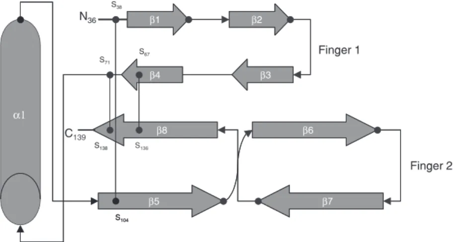
The three-dimensional structure of BMP-7 osteogenic protein 1 (OP-1) was solved (7,8), showing that, although there is limited sequence identity between OP-1 and TGF-ß2, they share a common polypeptide fold. The identity of OP-1 and TGF-ß is less than 38%, but it is 60 and 85%, respectively, relative to BMP-2 and BMP-5. The mono-mer presents three disulfide bonds, the cys-teine knot constituting the monomer core, and four strands of antiparallel ß-sheet, which emanate from the knot forming two finger-like projections (Figure 1). Tabas et al. (9) confirmed the assignment of the BMP-2A gene to chromosome 20.
The signaling cascade of bone morphogenetic proteins
Osteogenesis comprises a sequential cas-cade with three critical phases: migration and mitosis of mesenchymal cells, differen-tiation of mesenchymal cells into chondro-blasts, cartilage formation and, finally, sub-stitution of cartilage by bone. These sequen-tial events are triggered by the binding of plasma fibronectin to the demineralized bone matrix, enhancing adhesion and prolifera-tion of mesenchymal cells at 3 days after implantation. Chondrogenesis is observed after 5 days, reaching its peak at 7-8 days. Cartilage hypertrophy and mineralization are observed after 9 days. Osteoblast differen-tiation depends on angiogenesis and the
high-est level occurs after 10-11 days (10). Se-quentially, the newly formed endochondral bone is remodeled and becomes a hemato-poietic site. The sequence of morphogenetic events in response to the demineralized bone matrix mimics the initial events of skeletal morphogenesis in embryos and of bone re-pair in adults.
Accordingly, the key signals for bone morphogenesis have been identified (Figure 2). The BMP, as a signaling molecule of the TGF-ß superfamily, binds to a type II specif-ic receptor present on the cell membrane and recruits a type I receptor, forming a com-plex. These receptors are transmembrane serine/threonine kinase proteins that self phosphorylate after the formation of the BMP-receptor II-receptor I complex and ac-quire the ability to phosphorylate Smad pro-teins, a family of TGF-ß transducers. Smads are a family of signaling mediators of BMP receptors in vertebrates homologous of Mad (mothers against decapentaplegic,in Droso-phila)and Sma (related to Mad in C. elegans) and can be classified into three subtypes by structure and function, i.e., receptor-regu-lated Smads (R-Smads), common-mediator Smads, and inhibitory Smads. R-Smads are phosphorylated by activated serine/threonine
Figure 1. Schematic diagram of bone morphogenetic protein 7 (OP-1/BMP-7). The mono-mer is stabilized by three disulfide bonds, i.e., Cys-67-Cys-136 and Cys-71-Cys-138 forming a ring through which the third, Cys-38-Cys-104, passes. The cysteine knot consti-tutes the monomer core from which four strands of antiparallel ß-sheets emanate, forming two finger-like projections. Adapted from Ref. 8, with permission.
kinase receptors (BMP-receptor II-receptor I complex). R-Smads interact with common-mediator Smads to form hetero-oligomeric complexes, which then translocate into the nucleus and regulate the transcription of vari-ous target genes (for a review, see Ref. 11). It is not clear whether Smads can recognize specific binding sites and bind to DNA by themselves.
Transient co-expression of BMP-2 with BMP-5, BMP-6 or BMP-7, or BMP-4 tran-siently co-expressed with BMP-7, resulted in more BMP activity than expression of any single BMP, with heterodimeric BMP-2/7
presenting about 20-fold higher specific ac-tivity than BMP homodimers (in vitro alka-line phosphatase induction assay) (12).
Recent studies have identified specific BMP antagonists (i.e., noggin and chordin) and members of the DAN family (i.e., grem-lin). Such antagonists bind to BMP with the same affinity as their specific receptors, blocking signal transduction and thus de-creasing bone formation. Therefore, these antagonists may be used therapeutically in pathological conditions characterized by excessive bone formation (13).
Bahamonde and Lyons (14) demonstrated that BMP-3 has an inhibitory effect on os-teogenesis, presenting a signaling pathway similar to TGF-ß/activin. The ability of BMP-3 to inhibit the activity of BMP-2 seems to result from competition for common signal-ing components of the TGF-ß/activin and BMPs pathways. Since BMP-3 is by far the most abundant BMP in demineralized bone, it probably plays a fundamental role as a modulator of the osteogenic activity of other BMPs in vivo.
These findings are of great clinical rel-evance because of the need to quantitate the amount of BMP-3 when products composed of exogenous BMPs are used to accelerate bone regeneration. The osteogenic potential of BMPs is increased when the antagonists are eliminated. Nevertheless, BMP-3 could be used in the treatment of diseases charac-terized by bone hypermineralization, such as osteopetrosis.
Immune response to bone morphogenetic proteins
The immune mechanisms activated upon implantation of BMPs are not fully under-stood or well defined due to controversies in the literature, which are raising some confu-sion. It seems that the single application of allogeneic BMPs and non-collagenic pro-teins provides a moderate immune response through the production of immunoglobulins
G, but does not decrease the osteoinductive capacity of BMP. On the other hand, a single dose of BMP-non-collagenic protein stimu-lates a high concentration of BMP anti-body, which could inhibit the osteoinductive potential of BMP (15).
Implantation of allogenic or xenogenic BMPs appears to promote recruitment of macrophages, lymphocytes and plasma cells, and production of antibodies which may inhibit osteogenesis. Whereas some studies demonstrate a species-specific effect of BMPs (16), others demonstrate that a single dose of up to 100 mg xenogenic BMP would be safe and would not stimulate a detectable immune response (17). However, a later study by Urist and collaborators (18) showed that a second implantation of BMPs results in intensification of the immune response and in reduced efficacy of the xenogenic BMP in the treatment of critical-size lesions in dogs. Analysis of the immune response after implantation of recombinant human BMP (rhBMP) has not yet been thoroughly studied. Nevertheless, preliminary studies have reported that anti-rhBMP-2 was not produced after implantation of this recombi-nant protein in defects of the mandibles of dogs.
Further studies are certainly necessary to explain the relationship between bone in-duction and the occurrence of immune re-sponses upon implantation of BMPs, and to ensure safety in their use for orthopedic and dental purposes.
Target cells for bone morphogenetic proteins
Numerous studies have demonstrated the cellular and molecular effects of deminera-lized bone matrix and purified or recombi-nant BMPs on several cell lines. At least some pluripotent mesenchymal cell lines, bone marrow cells, osteoblast precursors, myoblasts, fibroblasts, and neural cells re-spond to BMPs. Numerous markers of bone
metabolism such as alkaline phosphatase, parathyroid hormone receptor, osteocalcin, osteopontin, and osteonectin are modulated by BMPs. In spite of the accumulated evi-dence that the BMP-mediated response in-volves the use of specific receptors during the development of cartilage and bone, their signal transduction mechanisms are still un-clear. Indeed, BMPs play an important role during the initial stages of organogenesis.
In mesenchymal and embryonic cells, the most impressive effect of BMPs is the ability to induce differentiation of these cells into osteoblasts, stimulating cartilage for-mation and alkaline phosphatase activity. Other hormones or cytokines cannot modu-late the level of these markers of bone me-tabolism. Noteworthy is the fact that in in
vitro experiments low concentrations of
BMPs promote differentiation of mesenchy-mal cells into adipocytes, whereas high con-centrations of these proteins promote osteo-blast differentiation. This emphasizes the need to specify the doses of BMP in order to predict its effect (19). Osteoblasts treated with rhBMP-2 present rapid differentiation, similarly to mesenchymal cells, with an in-crease in the levels of alkaline phosphatase, osteocalcin, osteopontin, and bone sialopro-tein (20).
Since many types of BMPs can induce endochondral ossification, chondroblasts should also be natural targets of these proteins. In fact, many BMPs have been shown to induce cell proliferation and synthesis and activity of alkaline phosphatase of chondro-blasts and chondrocytes of the growth plate. The nature of the chondrocytes for in vitro
culture has a significant role in the effect of BMPs, showing that the stimuli for these pro-teins are tissue-specific (23).
Bovine BMPs induced an increase of DNA and protein synthesis and also of alka-line phosphatase activity in NIH-3T3 fibro-blasts in a dose-dependent manner (24). In contrast, rhBMP did not induce an increase in alkaline phosphatase in these cells. BMP-2 promoted differentiation of BALB/c-3T3, Swiss-3T3 and 3T3-L1 fibroblasts into adi-pocytes and osteoblasts (20).
Usually, target cells for BMPs differenti-ate into osteoblast-like cells and produce alkaline phosphatase and mineralized tissue. On the other hand, Kaneko et al. (25) exam-ined the direct effect of BMPs on osteoclas-tic bone-resorbing activity in a culture of highly purified rabbit mature osteoclasts. BMP-2 and -4 increase in bone resorption pits excavated by the isolated osteoclasts. BMP-2 also elevated the messenger RNA expression by cathepsin K and carbonic an-hydrase II, which are key enzymes for the degradation of organic and inorganic bone matrices, respectively.
Purified versus recombinant bone morphogenetic proteins
As with every growth factor, BMPs act at very low doses in the tissues, existing in nanograms or micrograms. However, in or-der to isolate a couple of micrograms of BMPs, kilograms of demineralized bone matrix are needed (26). Once isolated, dif-ferent BMPs may be identified by their amino acid sequence. Purification of BMPs from the demineralized bone matrix can be
car-ried out by four distinct methods: 1) enzy-matic digestion, since they resist collagen-ase; 2) ethylene glycol extraction, due to the hydrophobic nature of the BMP molecule; 3) 6 M urea plus 0.5 M CaCl2, since BMPs
can be dissociated from other non-collagen proteins in chaotropic solvents; 4) concanava-lin A affinity chromatography due to their hydrophobic nature and to carbohydrates present in their structure.
In spite of these methods, purification of BMPs is an extremely laborious process and the yields are low. Preparations must be initiated with a minimum of 100 kg of washed fresh cortical bone free of bone marrow (26). Since their molecular weight ranges from 15 to 30 kDa, partial purification of BMPs yields 57 mg of the pool of BMPs per kg of fresh bone (26). Isolation of a particular native BMP yields even smaller amounts of the order of µg/kg tissue. The small amounts of BMPs resulting from such a laborious puri-fication process have stimulated the applica-tion of molecular biology techniques for the cloning and expression of these proteins.
The molecular cloning of the first genes encoding BMPs took place at the end of the 1980’s and more than 30 members of the BMP family have been described (27). The study of different BMPs revealed that their expression pattern and their biological func-tions are not restricted to skeletal develop-ment. Other functions have been identified, such as cell proliferation and differentiation, apoptosis, morphogenesis of various organs, including the skeleton, and organogenesis.
proteins are members of the TGF-ß super-family, except for BMP-1, which has been identified as procollagen C proteinase (30). The molecular cloning of BMP-encoding genes and their identification as TGF-ß rela-tives has enhanced the interest in these pro-teins and has permitted expression and func-tional studies.
In view of the osteoinductive properties of BMP-2 and BMP-7, we set out to isolate the human cDNA counterparts of these molecules to clone them into appropriate transducing vectors in order to produce and purify these recombinant proteins using heterologous bac-terial, mammalian and baculovirus expression systems (Bustos-Valenzuela JC and Sogayar MC, unpublished data). The recombinant pro-teins obtained are being used in blind cDNA cloning strategies to identify and characterize novel potential regulators of the osteoblast differentiation process, to better understand the molecular mechanisms involved in bone formation and to gain new therapeutic insights (drug design and gene therapy).
The osteoinductive properties of recom-binant BMPs are reduced compared to puri-fied BMPs and require the characterization of BMPs by genetic engineering techniques. Bessho et al. (31) have analyzed in detail the effects of purified versus recombinant bo-vine BMP. On the basis of Ca2+ content and
radiographic aspects, they observed that maturation of bone tissue ectopically formed in rat muscle was as much as 10 times greater when bovine BMP was used.
Several hypotheses have been proposed and tested to explain such discrepancies. There are reports about differences in the amino acid sequence of recombinant and purified bovine BMP. In addition, it has been suggested that these cytokines operate coordinately in bone repair, emphasizing the need for several recombinant BMPs simul-taneously to further facilitate the repair. An-other consideration is the type of carrier used to transport the BMP into the defect, with collagen-derived materials being
ex-cellent candidates (31).
Osteogenic protein-1/bone morphogenetic protein-7
OP-1, or BMP-7, was cloned in the 1990’s. The gene encoding OP-1 was found in placenta, hippocampus and osteosarcoma cDNA libraries, using a consensus probe based on the polypeptide chain of bovine BMP-enriched preparations. OP-1 promotes an increase in chondrocyte proliferation and induces chondrogenic differentiation. OP-2 (BMP-8), however, is only expressed during embryogenesis. Like BMP-2 to 6, OP-1 is a homodimeric glycoprotein stabilized by di-sulfide bonds, with molecular weight rang-ing from 32 to 36 kDa. The amino acid sequence of OP-1, initially purified from bovine bone matrix, is approximately 60% similar to that of BMP-2.
Recombinant human OP-1 produced by the expression of the entire cDNA in mam-malian cells was able to induce bone forma-tion in vitro, in a manner similar to prepara-tions of highly purified bovine OP-1 (22). During the initial stages of treatment, OP-1 elicits an intense chondrogenic response and stimulates an osteoblastic response during the course of treatment.
Investigations regarding the biological effects of OP-1 have been carried out, mainly concerning its potential for osteogenesis. It has been demonstrated that OP-1 stimulates cell proliferation, collagen synthesis and os-teoblast differentiation. Studies on animal models have shown that OP-1 is capable of inducing endochondral ossification in seg-mental osteoperiosteal defects (32).
The FDA of the United States has re-cently approved the use of OP-1 for the treatment of spinal fusion, emphasizing its use in orthopedic and dental surgeries (33).
Clinical applications
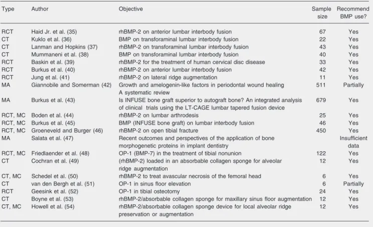
for the descriptor “bone morphogenetic pro-tein”, but only 20 citations for Clinical Trials in Humans. The highest level of scientific evidence arises from meta-analysis and ran-domized controlled trials. Systematic reviews synthesize studies related to the side effects and benefit of treatments. They are based on good quality papers obtained from correct databases and focusing on a specific ques-tion. Avoiding or minimizing bias, system-atic reviews are very helpful in decision making. In meta-analysis, the quantitative combination of results from different studies yields an estimate of the expected effect of a treatment (34). Table 1 summarizes the pa-pers found in PubMed related to clinical trials, randomized clinical trials and meta-analysis, emphasizing the clinical applica-tions proposed for BMPs.
The first studies were clinical trials, one of which a multi-center study, analyzing the effect of BMPs (rhBMP-2) for local alveolar ridge preservation or augmentation (54) or maxillary sinus floor augmentation (53). The great majority of studies concerned spinal fusion (Table 1), with all of them agreeing about the benefit of BMPs. Burkus et al. (43) conducted an integrated analysis of multiple clinical studies involving 679 patients and concluded that rhBMP-2-treated subjects had statistically significant outcomes regarding duration of surgery, blood loss, hospital stay, reoperation rate, median time to return to work, and fusion rates at 6, 12, and 24 months. The safety and effectiveness of 1.5 mg/mL rhBMP-2 applied to an absorbable collagen sponge was demonstrated for the treatment of 450 patients with open tibial fractures.
Table 1. List of publications related to human clinical application of bone morphogenetic proteins (BMP), classified as clinical trial (CT), randomized clinical trial (RCT), multi-center (MC), and meta-analyses (MA).
Type Author Objective Sample Recommend
size BMP use?
RCT Haid Jr. et al. (35) rhBMP-2 on anterior lumbar interbody fusion 67 Yes
CT Kuklo et al. (36) BMP on transforaminal lumbar interbody fusion 22 Yes
CT Lanman and Hopkins (37) rhBMP-2 on transforaminal lumbar interbody fusion 43 Yes
CT Mummaneni et al. (38) BMP on transforaminal lumbar interbody fusion 40 Yes
RCT Baskin et al. (39) rhBMP-2 for the treatment of human cervical disc disease 33 Yes
RCT Burkus et al. (40) rhBMP-2 on anterior lumbar interbody fusion 42 Yes
RCT Jung et al. (41) rhBMP-2 on lateral ridge augmentation 11 Yes
MA Giannobile and Somerman (42) Growth and amelogenin-like factors in periodontal wound healing 511 Partially A systematic review
MA Burkus et al. (43) Is INFUSE bone graft superior to autograft bone? An integrated analysis 679 Yes of clinical trials using the LT-CAGE lumbar tapered fusion device
RCT, MC Boden et al. (44) rhBMP-2 on lumbar arthrodesis 25 Yes
RCT, MC Burkus et al. (45) BMP (INFUSE bone graft) on lumbar interbody fusion 46 Yes
RCT, MC Groeneveld and Burger (46) rhBMP-2 on open tibial fracture 450 Yes
MA Salata et al. (47) Recent outcomes and perspectives of the application of bone Insufficient
morphogenetic proteins in implant dentistry data
RCT, MC Friedlaender et al. (48) OP-1 (BMP-7) in the treatment of tibial nonunion 122 Yes
CT Cochran et al. (49) (rhBMP-2) loaded in an absorbable collagen sponge for alveolar 12 Yes
ridge augmentation
CT, MC Schedel et al. (50) rhBMP-2 to treat avascular necrosis of the femoral head 6 Yes
CT van den Bergh et al. (51) OP-1 in sinus floor elevation 6 Partially
RCT Geesink et al. (52) OP-1 in tibial osteotomy 24 Yes
CT Boyne et al. (53) rhBMP-2/absorbable collagen sponge for maxillary sinus floor augmentation 12 Yes CT, MC Howell et al. (54) rhBMP-2/absorbable collagen sponge device for local alveolar ridge 12 Yes
preservation or augmentation
Significantly superior results compared to standard care were observed in terms of reducing the frequency of secondary inter-ventions and the overall invasiveness of the procedures, with accelerated fracture and wound-healing, and a reduced rate of infec-tion.
In dentistry, BMPs have been tested in periodontal (regeneration of lost bone tissue due to periodontal disease), implant (increase in bone volume for placement of implants, maxillary sinus augmentation) and restor-ative-endodontic (pulpotomies) procedures. Several animal studies have been carried out to evaluate the efficacy of BMPs for maxillary sinus augmentation, and studies in both animals and humans have demonstrated similar, but still unsatisfactory, results, when compared to other procedures. Animal as-says using rhBMP-2 associated with a car-rier (collagen foam) in 3-sided intrabone defects in dogs have demonstrated an in-crease in the rate of bone formation without side effects such as ankylosis or apical bone resorption (55). A clinical trial studying 6 patients (3 of them used as control) indicated that the OP-1 (2.5 mg in 1 g collagen carrier) has the potential to initiate bone formation in the human maxillary sinus within 6 months after a sinus floor elevation operation (51). However, the behavior of this material is not fully predictable.
Barboza et al. (56) have used BMPs as an aid to increase bone crest height prior to the placement of implants. However, Salata et al. (47), in a meta-analysis of 379 scientific reports concerning the use of BMPs on im-plant dentistry, concluded that the number of studies is too small to establish clinical protocols for the improvement of a recipient bone bed prior to implant placement or to enhance the integration process of an im-plant.
Giannobile et al. (57)have shown prom-ising and encouraging results with rhBMP-7 implanted in type III furcation defects in dogs, with no signs of side effects such as ankylosis of the affected roots. However, in a systematic review published later (42) these investigators observed that the majority of the reports available had a low-quality evi-dence rating and that most reports were case studies or case series without controls. They concluded that there were insufficient data to conduct a meta-analysis and that pre-clinical and initial pre-clinical data for growth factors appear promising but are insufficient to draw definitive conclusions, mainly for long-term evaluation.
BMPs have been tested in pulp capping procedures for more than a decade, and have presented enhanced potential as effective agents in the induction of a mineralized bar-rier in the pulp (58). Jepsen et al. (59) used recombinant BMP-7 as a capping agent in minipigs and detected the formation of a thicker dentin barrier in the group treated with recombinant BMP-7 than in the group treated with Ca(OH)2. Ren et al. (60) tested
recombinant BMP-2 associated with fibrin as a capping agent in the pulps of dogs (molars and premolars) and observed the formation of a dentin barrier after one week, this being a better result than for the control group treated with Ca(OH)2 or the
References
1. Urist MR (1965). Bone: Formation by autoinduction. Science, 150: 893-899.
2. Urist MR, Nogami H & Mikulski A (1976). A bone morphogenetic polypeptide. Calcified Tissue Research, 21 (Suppl): 81-87. 3. Bessho K, Tanaka N, Matsumoto J et al. (1991). Human
dentin-matrix-derived bone morphogenetic protein. Journal of Dental Re-search, 70: 171-175.
4. Urist MR, Grant TT, Lindholm TS et al. (1979). Induction of new-bone formation in the host bed by human new-bone-tumor transplants in athymic nude mice. Journal of Bone and Joint Surgery, 61: 1207-1216.
5. Constam DB & Robertson EJ (1999). Regulation of bone morphoge-netic protein activity by prodomains and proprotein convertases.
Journal of Cell Biology, 144: 139-149.
6. Wozney JM, Rosen V, Celeste AJ et al. (1988). Novel regulators of bone formation: molecular clones and activities. Science, 242: 1528-1534.
7. Spinella-Jaegle S, Roman-Roman S, Faucheu C et al. (2001). Op-posite effects of bone morphogenetic protein-2 and transforming growth factor-beta1 on osteoblast differentiation. Bone, 29: 323-330.
8. Griffith DL, Keck PC, Sampath TK et al. (1996). Three-dimensional structure of recombinant human osteogenic protein 1: structural paradigm for the transforming growth factor beta superfamily. Pro-ceedings of the National Academy of Sciences, USA, 93: 878-883. 9. Tabas JA, Zasloff M, Wasmuth JJ et al. (1991). Bone morphogenetic
protein: chromosomal localization of human genes for BMP1, BMP2A, and BMP3. Genomics, 9: 283-289.
10. Pacicca DM, Patel N, Lee C et al. (2003). Expression of angiogenic factors during distraction osteogenesis. Bone, 33: 889-898. 11. Sakou T (1998). Bone morphogenetic proteins: from basic studies to
clinical approaches. Bone, 22: 591-603.
12. Israel DI, Nove J, Kerns KM et al. (1996). Heterodimeric bone morphogenetic proteins show enhanced activity in vitro and in vivo.
Growth Factors, 13: 291-300.
13. Groppe J, Greenwald J, Wiater E et al. (2002). Structural basis of BMP signalling inhibition by the cystine knot protein noggin. Nature, 420: 636-642.
14. Bahamonde ME & Lyons KM (2001). BMP3: to be or not to be a BMP. Journal of Bone and Joint Surgery, 83-A (Suppl 1): S56-S62. 15. Urist MR, Nillsson OS, Hudak R et al. (1985). Immunologic evidence of a bone morphogenetic protein in the milieu interieur. Annales de Biologie Clinique, 43: 755-766.
16. Johnson EE, Urist MR, Schmalzried TP et al. (1989). Autogeneic cancellous bone grafts in extensive segmental ulnar defects in dogs. Effects of xenogeneic bovine bone morphogenetic protein without and with interposition of soft tissues and interruption of blood supply. Clinical Orthopaedics and Related Research, 243: 254-265. 17. Nilsson OS, Urist MR, Dawson EG et al. (1986). Bone repair in-duced by bone morphogenetic protein in ulnar defects in dogs.
Journal of Bone and Joint Surgery. British Volume, 68: 635-642. 18. Nilsson OS & Urist MR (1991). Immune inhibition of repair of canine
skull trephine defects implanted with partially purified bovine mor-phogenetic protein. International Orthopaedics, 15: 257-263. 19. Carrington JL, Chen P, Yanagishita M et al. (1991). Osteogenin
(bone morphogenetic protein-3) stimulates cartilage formation by chick limb bud cells in vitro. Developmental Biology, 146: 406-415. 20. Wang EA, Israel DI, Kelly S et al. (1993). Bone morphogenetic
protein-2 causes commitment and differentiation in C3H10T1/2 and 3T3 cells. Growth Factors, 9: 57-71.
21. Chen TL, Bates RL, Dudley A et al. (1991). Bone morphogenetic protein-2b stimulation of growth and osteogenic phenotypes in rat osteoblast-like cells: comparison with TGF-beta 1. Journal of Bone and Mineral Research, 6: 1387-1393.
22. Sampath TK, Maliakal JC, Hauschka PV et al. (1992). Recombinant human osteogenic protein-1 (HOP-1) induces new bone formation
in vivo with a specific activity comparable with natural bovine osteo-genic protein and stimulates osteoblast proliferation and differentia-tion in vitro. Journal of Biological Chemistry, 267: 20352-20362. 23. Chen P, Vukicevic S, Sampath TK et al. (1993). Bovine articular
chondrocytes do not undergo hypertrophy when cultured in the presence of serum and osteogenic protein-1. Biochemical and Bio-physical Research Communications, 197: 1253-1259.
24. Vukicevic S, Latin V, Chen P et al. (1994). Localization of osteo-genic protein-1 (bone morphogenetic protein-7) during human em-bryonic development: high affinity binding to basement membranes.
Biochemical and Biophysical Research Communications, 198: 693-700.
25. Kaneko H, Arakawa T, Mano H et al. (2000). Direct stimulation of osteoclastic bone resorption by bone morphogenetic protein (BMP)-2 and expression of BMP receptors in mature osteoclasts. Bone, 27: 479-486.
26. Gao T, Lindholm TS, Marttinen A et al. (1996). Composites of bone morphogenetic protein (BMP) and type IV collagen, coral-derived coral hydroxyapatite, and tricalcium phosphate ceramics. Interna-tional Orthopaedics, 20: 321-325.
27. Reddi AH (1997). Bone morphogenetic proteins: an unconventional approach to isolation of first mammalian morphogens. Cytokine and Growth Factor Reviews, 8: 11-20.
28. Wang EA, Rosen V, Cordes P et al. (1988). Purification and charac-terization of other distinct bone-inducing factors. Proceedings of the National Academy of Sciences, USA, 85: 9484-9488.
29. Kirker-Head CA (2000). Potential applications and delivery strate-gies for bone morphogenetic proteins. Advanced Drug Delivery Reviews, 43: 65-92.
30. Hofbauer LC & Heufelder AE (1996). Updating the metalloprotease nomenclature: bone morphogenetic protein 1 identified as procolla-gen C proteinase. European Journal of Endocrinology, 135: 35-36. 31. Bessho K, Kusumoto K, Fujimura K et al. (1999). Comparison of
recombinant and purified human bone morphogenetic protein. Brit-ish Journal of Oral and Maxillofacial Surgery, 37: 2-5.
32. Sampath TK, Rashka KE, Doctor JS et al. (1993). Drosophila trans-forming growth factor beta superfamily proteins induce endochon-dral bone formation in mammals. Proceedings of the National Acad-emy of Sciences, USA, 90: 6004-6008.
33. Vaccaro AR, Patel T, Fischgrund J et al. (2003). A pilot safety and efficacy study of OP-1 putty (RhBMP-7) as an adjunct to iliac crest autograft in posterolateral lumbar fusions. European Spine Journal, 12: 495-500.
34. Sacks HS, Berrier J, Reitman D et al. (1987). Meta-analyses of randomized controlled trials. New England Journal of Medicine, 316: 450-455.
36. Kuklo TR, Rosner MK & Polly Jr DW (2004). Computerized tomogra-phy evaluation of a resorbable implant after transforaminal lumbar interbody fusion. Neurosurgical Focus, 16: E10.
37. Lanman TH & Hopkins TJ (2004). Lumbar interbody fusion after treatment with recombinant human bone morphogenetic protein-2 added to poly(L-lactide-co-D,L-lactide) bioresorbable implants. Neu-rosurgical Focus, 16: E9.
38. Mummaneni PV, Pan J, Haid RW et al. (2004). Contribution of recombinant human bone morphogenetic protein-2 to the rapid cre-ation of interbody fusion when used in transforaminal lumbar interbody fusion: a preliminary report. Invited submission from the Joint Section Meeting on Disorders of the Spine and Peripheral Nerves, March 2004. Journal of Neurosurgery. Spine, 1: 19-23. 39. Baskin DS, Ryan P, Sonntag V et al. (2003). A prospective,
random-ized, controlled cervical fusion study using recombinant human bone morphogenetic protein-2 with the CORNERSTONE-SR al-lograft ring and the ATLANTIS anterior cervical plate. Spine, 28: 1219-1225.
40. Burkus JK, Dorchak JD & Sanders DL (2003). Radiographic assess-ment of interbody fusion using recombinant human bone morphoge-netic protein type 2. Spine, 28: 372-377.
41. Jung RE, Glauser R, Scharer P et al. (2003). Effect of RhBMP-2 on guided bone regeneration in humans. Clinical Oral Implants Re-search, 14: 556-568.
42. Giannobile WV & Somerman MJ (2003). Growth and amelogenin-like factors in periodontal wound healing. A systematic review. An-nals of Periodontology, 8: 193-204.
43. Burkus JK, Heim SE, Gornet MF et al. (2003). Is INFUSE bone graft superior to autograft bone? An integrated analysis of clinical trials using the LT-CAGE lumbar tapered fusion device. Journal of Spinal Disorders and Techniques, 16: 113-122.
44. Boden SD, Kang J, Sandhu H et al. (2002). Use of recombinant human bone morphogenetic protein-2 to achieve posterolateral lum-bar spine fusion in humans: a prospective, randomized clinical pilot trial: 2002 Volvo Award in Clinical Studies. Spine, 27: 2662-2673. 45. Burkus JK, Transfeldt EE, Kitchel SH et al. (2002). Clinical and
radiographic outcomes of anterior lumbar interbody fusion using recombinant human bone morphogenetic protein-2. Spine, 27: 2396-2408.
46. Groeneveld EH & Burger EH (2000). Bone morphogenetic proteins in human bone regeneration. European Journal of Endocrinology, 142: 9-21.
47. Salata LA, Franke-Stenport V & Rasmusson L (2002). Recent out-comes and perspectives of the application of bone morphogenetic proteins in implant dentistry. Clinical Implant Dentistry and Related Research, 4: 27-32.
48. Friedlaender GE, Perry CR, Cole JD et al. (2001). Osteogenic protein-1 (bone morphogenetic protein-7) in the treatment of tibial
nonunions. Journal of Bone and Joint Surgery, 83-A (Suppl 1): S151-S158.
49. Cochran DL, Jones AA, Lilly LC et al. (2000). Evaluation of recombi-nant human bone morphogenetic prote2 in oral applications in-cluding the use of endosseous implants: 3-year results of a pilot study in humans. Journal of Periodontology, 71: 1241-1257. 50. Schedel H, Schneller A, Vogl T et al. (2000). Dynamic magnetic
resonance tomography (MRI): a follow-up study after femur core decompression and instillation of recombinant human bone morpho-genetic protein-2 (RhBMP-2) in avascular femur head necrosis.
Rontgenpraxis, 53: 16-24.
51. van den Bergh JP, ten Bruggenkate CM, Groeneveld HH et al. (2000). Recombinant human bone morphogenetic protein-7 in max-illary sinus floor elevation surgery in 3 patients compared to autog-enous bone grafts. A clinical pilot study. Journal of Clinical Perio-dontology, 27: 627-636.
52. Geesink RG, Hoefnagels NH & Bulstra SK (1999). Osteogenic activity of OP-1 bone morphogenetic protein (BMP-7) in a human fibular defect. Journal of Bone and Joint Surgery. British Volume, 81: 710-718.
53. Boyne PJ, Marx RE, Nevins M et al. (1997). A feasibility study evaluating RhBMP-2/absorbable collagen sponge for maxillary si-nus floor augmentation. International Journal of Periodontics and Restorative Dentistry, 17: 11-25.
54. Howell TH, Fiorellini J, Jones A et al. (1997). A feasibility study evaluating RhBMP-2/absorbable collagen sponge device for local alveolar ridge preservation or augmentation. International Journal of Periodontics and Restorative Dentistry, 17: 124-139.
55. Choi SH, Kim CK, Cho KS et al. (2002). Effect of recombinant human bone morphogenetic protein-2/absorbable collagen sponge (RhBMP-2/ACS) on healing in 3-wall intrabony defects in dogs.
Journal of Periodontology, 73: 63-72.
56. Barboza EP, Duarte ME, Geolas L et al. (2000). Ridge augmentation following implantation of recombinant human bone morphogenetic protein-2 in the dog. Journal of Periodontology, 71: 488-496. 57. Giannobile WV, Ryan S, Shih MS et al. (1998). Recombinant human
osteogenic protein-1 (OP-1) stimulates periodontal wound healing in class III furcation defects. Journal of Periodontology, 69: 129-137. 58. Nakashima M (1994). Induction of dentin formation on canine ampu-tated pulp by recombinant human bone morphogenetic proteins (BMP)-2 and -4. Journal of Dental Research, 73: 1515-1522. 59. Jepsen S, Albers HK, Fleiner B et al. (1997). Recombinant human
osteogenic protein-1 induces dentin formation: an experimental study in miniature swine. Journal of Endodontics, 23: 378-382. 60. Ren WH, Yang LJ & Dong SZ (1999). Induction of reparative dentin