Faculdade de Medicina de Lisboa
R
EVISITING GERM CELL DYNAMICS AND OVARIAN
FOLLICLE ASSEMBLY IN THE MOUSE MODEL
Patrícia Carla Coelho Rodrigues
Orientadores: Prof. Doutor Carlos E. Plancha
Prof. Doutor David F. Albertini
Tese especialmente elaborada para obtenção do grau de Doutor em Ciência Biomédicas, especialidade de Ciências Morfológicas
Faculdade de Medicina de Lisboa
R
EVISITING GERM CELL DYNAMICS AND OVARIAN FOLLICLE
ASSEMBLY IN THE MOUSE MODEL
Patricia Carla Coelho Rodrigues
Orientadores: Prof. Doutor Carlos E. Plancha
Prof. Doutor David F. Albertini
Tese especialmente elaborada para obtenção do grau de Doutor em Ciência Biomédicas, especialidade de Ciências Morfológicas
Júri:
Presidente: Doutor José Luis Bliebernicht Ducla Soares, Professor Catedrático em regime
de tenure e Vice-presidente do Conselho Cientifico da Faculdade de Medicina da Universidade de Lisboa
Vogais:
Doutor David F. Albertini, Professor no University of Kansas Medical Center, Estados Unidos da América (Orientador)
Doutor João Ramalho de Sousa Santos, Professor Associado com Agregação da Faculdade de Ciências e Tecnologia da Universidade de Coimbra
Doutor Vasco Manuel Leal Martins de Almeida, Professor Auxiliar da Faculdade de Ciências da Universidade do Porto
Doutor Manuel Diamantino Pires Bicho, Professor Catedrático da Faculdade de Medicina da Universidade de Lisboa
Doutor António José Saraiva da Cunha Cidadão Professor Associado com Agregação da Faculdade de Medicina de Lisboa
Doutor Carlos Calhaz Jorge, Professor Associado com Agregação da Faculdade de Medicina da Universidade de Lisboa
Todas as opiniões expressas no presente documento são da exclusiva responsabilidade do seu autor, não cabendo qualquer responsabilidade à Faculdade de Medicina de Lisboa pelos conteúdos nele apresentados. All statements made in this document are the sole responsibility of the author; the Faculty of Medicine of Lisbon has no responsibility to the
A impressão desta tese foi aprovada pelo
Conselho Cientifico da Faculdade de Medicina
de Lisboa em reunião de 20 de Outubro de 2015.
PREFACE
This thesis investigates the mouse ovarian follicle reserve establishment, focusing on the mechanisms involved in the perinatal extensive germ cell loss and what leads to the retention of a subset of germ cells forming the primordial follicle reserve. Using imaging techniques and animal models we studied the cytoskeleton, extracellular matrix and junctions involvement in this event. The chapters of this dissertation include two published journal articles, (Rodrigues et al., 2008 J. Cell. Physiol. 216:355-365; Rodrigues et al., 2009 Reproduction 137:709-720).
The present dissertation is outlined as followed:
Chapter 1 – General Introduction divided in two major sections:
1. Oogenesis and folliculogenesis: the beginning, here the developmental trajectory of germ cells until primordial follicle assembly is described.
2. Oogenesis: prospects and challenges for the future. [Rodrigues, P.; Limback, D.; McGinnis, L. K.; Plancha, C. E.; Albertini, D. F. (2008) J. Cell. Physiol. 216: 355-365]. This review describes follicle growth and the factors involved in its growth.
Chapter 2 - Multiple mechanisms of gem cell loss in the perinatal mouse
ovary. [Rodrigues, P. et al., (2009) Reproduction 137: 709-720]. This chapter describes multiple mechanisms involved in germ cell loss, around birth, in the mouse model.
Chapter 3 - Perinatal ovarian remodeling and establishment of the
definitive follicle reserve: the somatic-germ cell interface. [Rodrigues, P.; Limback, D.; McGinnis, L. K.; Albertini, D. F.; Plancha, C. E.]. Here is
described how germ-somatic-extracellular matrix interactions contribute to ovarian follicle establishment in the mouse.
Chapter 4 - Oocyte and hormonal regulation of the ovarian follicle reserve
in the mouse. [Rodrigues, P.; Limback, D.; McGinnis, L. K.; Albertini, D. F.; Plancha, C. E.]. In this chapter both germ cell and hormonal involvement in the ovarian remodeling and ovarian follicle reserve establishment are addressed, using specific genetically manipulated mouse models.
Chapter 5 – Concluding remarks. Here the results are consolidated and
further discussed reinforcing the importance of germ-somatic cell communication in the establishment of definitive ovarian follicular reserve in the mouse.
ABBREVIATIONS ActA ADAMTS-1 AMH AVE bFGF Bcl-2 Bax BBC3 BMP BMP4 BMP7 BMP8b BMP-15 Casp4 Cx Cx37 Cx43 CL CXCR4 cAMP COX-2 dpc Dax Dhh DVE E2 eEF2K ERα or β ECM ExE Figα FGFs FGF9 FSH Activin A
A disintegrin and metalloproteinase with thrombospondin-like motives-1 Anti-Müllerian hormone
Anterior visceral endoderm Basic fibroblast growth factor B-cell lymphoma/leukemia-2 Bcl-2 associated X protein BCL2-binding component 3 Bone morphogenetic protein Bone morphogenetic protein 4 Bone morphogenetic protein 7 Bone morphogenetic protein 8b Bone morphogenetic protein-15 Caspase 4 (also known as Casp11) Connexin
Connexin 37 Connexin 43 Corpora lutea
CXC chemokine receptor 4
Cyclic adenosine monophosphate Cyclooxygenase-2
Days post-coitum
Dosage-sensitive sex reversal (DSS)-Adrenal hypoplasia congenital critical region on the X chromosome, gene 1
Desert hedgehog
Dorsal visceral endoderm Estradiol-17β
Elongation factor 2 kinase Estrogen receptor α or β Extra cellular matrix
Extra-embryonic ectoderm Factor in the germline-alpha Fibroblast growth factors Fibroblast growth factor 9 Follicle-stimulating hormone
FSHr Foxl2 Foxo3 GREL GV GVBD GCNA-1 GDF9 Gja1 Gja4 Ihh ICM KGF KL KO Lhx8 Lhx9 LIF LH LHr MI MII MT MIS Nobox NSN Oct-4 PDK1 PDGF PGCs PG Pin1 PKA POI PR PTEN
Follicle-stimulating hormone receptor Forkhead transcription factor 2 Forkhead transcription factor O3 Gonadal Ridge Epithilial-Like Germinal vesicle
Germinal vesicle breakdown Germ cell nuclear antigen-1 Growth differentiation factor-9 Connexin 43
Connexin 37 Indian hedgehog Inner cell mass
Keratinocyte growth factor Kit ligand
Knockout
LIM homeobox protein 8 LIM homeobox protein 9 Leukemia inhibitory factor Luteinizing hormone
Luteinizing hormone receptor Metaphase I
Metaphase II Microtubules
Meiosis-inducing substance
Newborn ovary homeobox-encoding gene Non-surrounded nucleolus
Octamer-binding transcription factor 4 (also known POU5f1) Phosphatidylinositol-dependent kinase 1
Platelet-derived growth factor Primordial germ cells
Prostaglandin
Peptidyl-prolyl isomerase 1 Protein kinase A
Primary Ovarian Insufficiency Progesterone receptor
RA Sry SSEA-1 Sf1 SDF1 SN Scp3 SI TNAP TGFβ TZP Act-TZP MT-TZP TNFα TNFR2 VE W Wt1 Wnt4 ZP ZP1 ZP2 ZP3 Retinoic acid
Sex-determining gene on the Y chromosome Stage specific embryonic antigen-1
Steroidogenic factor-1 Stromal cell-derived factor Surrounded nucleolus
Synaptonemal complex protein 3 Still
Tissue non-specific alkaline phosphatase Transforming Growth Factors-β
Transzonal projections
Transzonal projections with Actin-filaments Transzonal projections with Microtubules Tumour necrosis factor-α
TNF receptor type 2 Visceral endoderm Dominant white spotting Wilms tumour 1
Wingless-related MMTV integration site 4 Zona pellucida
Zona pellucida 1 Zona pellucida 2 Zona pellucida 3
ACKNOWLEDGEMENTS
This Ph.D. thesis is a joint collaboration between the laboratories of Professor Carlos Plancha in Portugal and Professor David F. Albertini, in the USA. The majority of the lab work was done in the USA. In Portugal the work was preformed at Instituto de Medicina Molecular and Instituto de Histologia e Biologia do Desenvolvimento, Faculdade de Medicina da Universidade de Lisboa. In the USA, it started at Department of Anatomy and Cellular Biology at Tufts University Schools of Medicine, Boston, Massachusetts, and ended at Department of Molecular and Integrative Physiology, University of Kansas Medical Center, Kansas City, Kansas.
This work was only possible due to the generous funding of Fundação para a Ciência e Tecnologia, Portugal (SFRH/BD/6439/2001), Fundação Calouste Gulbenkian, Portugal (nr 78591), and NIH grant HD042076 (USA). I’m very thankful to both my advisors for their mentoring, friendship and allowing me to embrace this journey full of great experiences. I’ve learnt many lessons with you both. Professor Carlos your comments and suggestions were always so assertive and your “coolness” made it all seem so much easier! Thank you for introducing me to Dra. MB! Professor David your ability to simplify, correlate subjects and making me see the “big picture” is extraordinary! I’m forever GRATEFUL!
To my beloved husband and dearest friend Gonçalo. I’m blessed for having met you, and everyday since you are my strength and motivation! AMO-TE!
I wish to thank all my family for their support, understanding and motivation, to my dearest parents and parents’ in-law! My sister, grandparents, sister and brothers’ in-law, hunts, uncles, cousins, nephews, which one way or another were there for me. You are all forever in my heart! Amo-vos!
To all my friends, without them this journey would have been very difficult, for listening, jogging, pushing me to finish this journey, to the extent of making weekly objectives lists so that I could end writing or sharing recipes and keeping me company via skype to cheer me up! Thank you all!
Kansas City
To all Albertini lab members – the A’ team rocks!
To dearest Darlene for your help, friendship, and teachings, I will never forget you, and “I know that life is hard” but I believe it gets better! Also for showing me that Kansas is not “cowboy land”! Love you! To Lynda, we met in Boston, but it was in Kansas City that we were labmates! You are an extraordinary friend, and wonderful person to work and be with, I’ve learned so much with you. I miss our dinners. You are forever in my heart! To Susan, my labmate since Boston! Thank you for your scope teachings and all of your support. With you and Mike we had great fun! Love you always! To Stephanie that taught me to be calm and organized in the lab! You and your husband, Todd, are a wonderful couple that introduce me to “s’mores” and offered me the ”tumbleweed” that I was hoping to find in Kansas! Thank you for your support and friendship! You are in my heart! To Karla that taught me ISH and so much more in the lab, your “coolness” felt so good in the lab! You and your husband, Ollie, are my New Zealander/Australian friends with whom I’ve discovered the Great Plains. I’ll never forget you! To Elena, even though we’ve met in Boston, we only really met in Kansas. Driving through Kansas and New England was so much fun because of you! Gràcies, no t'oblidaré!
Thank you to all Kumar Lab members
Thank you Dr. Kumar for your help with the knockout mouse colony and kind support. Thank you Aparna my wonderful friend for your guidance and help. I love you and your wonderful family!
Thank you to all Christenson Lab members
Thank you Dr. Lane for your support and help submitting a grant, without you I would have been out of funding!
Boston
Thank you all Albertini lab members:
To Gloria for your help and teachings, I’ll always remember you! To Alex (Xana) the other Portuguese in the lab before me, with whom I’ve shared great experiences. Thank you for all your help and understanding. Your smile was the sunshine of many grey days! To Catherine, which left the lab before I arrive, but whom I had the pleasure to met and spent great times! Je t’adore! To Stephan Teilmann for the great scientific discussions during dinner! Tak!
To Subrhadip for the burger lunches, such a great treat! To Peter Geck for the long conversations!
To Raquel, my wonderful Brazilian friend, you made my life so much better! I loved to discover New England and New York with you! You’ll always in my heart! Adoro-te!
To Adriana, the other Brazilian, for many great moments together, and for teaching me how to “sambar”!
Lisbon
Thank you to all Reproductive Biology lab members at IMM
To Catarina M, you are such a great friend and good to work with. If it weren’t for you I would have run from “Catacumba’s”! To Mafalda, dear
friend, I love our afternoon teas! I’ve learned a lot from you! To Paulo, loved your writing and good taste in music! To Sara, which I didn’t overlap much in the lab but our paths crossed somewhere else and I’m happy for it! To Catarina R, Viviana and Rita inters that give a new life to the lab! Thank you
To Dra. Madalena Barata for believing in me, pushing and motivating me to finish my thesis, and for introducing me to Dr. PB. I’m very grateful that our paths crossed!
To João, my embryology colleague, for your endless patience and for being my friend and my mirror! You are the less judgmental person I know and I love you for it!
To Dr. Pierre Boyer, from Hôpital Saint-Joseph - Marseille, who not knowing me accepted me in his lab and taught me all I know of reproductive embryology laboratory. Your support and always-kind way of pushing me to finish the PhD were very important! You and Marie, become my friends, Merci!
To Debbie, for your help reviewing and editing the manuscript. It is always a pleasure talking to you. Merci!
To Dr. Miguel Oliveira e Silva for support and the office space to write!
To Dra. João Mendonça for friendship and support, the room always shines when you come in!
INDEX Preface i Abbreviations iii Acknowledgments vii Index xi Abstract 1 Chapter 1 General introduction
1.1 Oogenesis and folliculogenesis: the beginning
1.2 Ooogenesis: prospects and challenges for the future 1.3 Ovulation 1.4 Objectives 3 3 16 40 42 Chapter 2
Multiple mechanisms of germ cell loss in the perinatal mouse
ovary 65
Chapter 3
Perinatal ovarian remodelling and establishment of the
definitive follicle reserve: the somatic-germ cell interface 97 Chapter 4
Oocyte and hormonal regulation of the ovarian follicle reserve
in the mouse 123
Chapter 5
Concluding remarks 151
Portuguese extended abstract 161
Annexes
ABSTRACT
Development of the mammalian ovary is characterized by extensive germ cell loss and the retention of a subset of germ cells within the primordial follicle reserve. Here we aimed to identify the mechanisms whereby some germ cells are retained while others are lost using novel imaging techniques and animal models.
We observed that multiple mechanisms contributed to germ cell loss at birth. Our results confirmed germ cell loss around birth of approximately 44%, but our apoptotic numbers did not account to all that loss. Besides non-apoptotic germ cell loss through the ovarian surface, the use of serum cultured ovaries, and selective autophagy and apoptosis inhibitors, allowed us to suggest other cell death mechanism besides apoptosis may be involved in follicular reserve establishment around birth.
To investigate germ-somatic cell interactions during ovarian follicle assembly, antibodies against cytoskeleton, extracellular matrix (ECM), and intercellular junctions were used to track expression patterns by immunofluorescence. We could confirm that a true ovarian epithelium is not formed until day 20 after birth. The persistence of gap junctions throughout ovary development implies that ovarian follicle reserve establishment requires an ongoing dialogue between somatic and germ cells.
To better understand this cellular dialogue, we used four mouse models with specific gene deletion: GDF9 (growth differentiation factor 9), Nobox (newborn ovary homeobox gene), Sohlh1 (sperma-and oogenesis basic helix-loop-helix1), and FSHβ (follicle stimulating hormone). We found deficient gap junction communication between germ-somatic cells in the FSHβ and GDF9 models, as well as specific cytoskeletal and ECM modifications in all models. In conclusion both oocyte-intrinsic (GDF9, Nobox, Sohlh1) and ovary extrinsic (FSHβ) factors differentially modulate germ-somatic cell interactions during ovarian development, influencing follicle assembly and ovarian follicle reserve establishment.
Key words: ovarian follicle reserve; germ cells; primordial follicles;
C
HAPTER
# 1
GENERAL INTRODUCTION
1.1 Oogenesis and folliculogenesis: the beginning
The process of germ cell formation is known as gametogenesis. In animal groups that reproduce sexually two different forms of gametogenesis exist:
oogenesis, differentiation of an ovum (females); and spermatogenesis,
sperm differentiation (male) (Wassarman and Albertini, 1994; Gilbert, 1997). In both, female and male, germ cells differentiate from primordial germ
cells (PGCs), thus the first event in gametogenesis is the formation of PGCs.
In females, PGCs go through several transformations, as they progress from PGCs to oogonia to oocytes. In mammals, these events normally take place during fetal life, when oogonia enter meiosis and arrest at prophase of meiosis I remaining arrested for months or years depending on the species (Peters and McNatty, 1980; Wassarman and Albertini, 1994). To grow and mature the oocyte must be enclosed by specialized somatic cells: the granulosa cells. The complex of oocyte - granulosa cells forms the initial ovarian follicle (Buccione et al., 1990; Eppig, 2001). Follicle growth accompanies oocyte growth and maturation, a process usually designated as folliculogenesis (see box 1) (Peters and McNatty, 1980; Eppig, 2001). A theca cell layer differentiates later, in a particularly well-coordinated process, with participation of paracrine and/or autocrine factors (see box 1). The latter stages of follicular growth become dependent on pituitary hormones (Eppig, 2001; Vanderhyden, 2002; Albertini and Barrett, 2003). In the mammalian ovary, the majority of follicles remain dormant and are progressively recruited to the growing pool until the supply of developmentally competent oocytes is exhausted (Hirshfield, 1992). The ultimate goal of oogenesis and folliculogenesis is the production of female sex hormones and healthy oocytes to ensure the development
Box 1 Stages of follicle development and oocyte growth
In the mouse follicle assembly occurs shortly after birth, when few flattened pre-granulosa cells associates with the primary oocyte – primordial follicle (type 2). Then the flattened cells become cuboidal (granulosa cells) and completely surround the oocyte with a single layer of cells – primary follicle (type 3). Multiple layers of granulosa cells surround the oocyte, zona pellucida begins to form, and theca cells start surrounding the follicle – secondary follicle (type 4-5). Small patches of fluid become evident within the granulosa cells, which continue to proliferate – early-antral follicle (type 5-6). The patches of fluid become a single cavity, the antrum – antral follicle (type 6). Continuing to grow, the follicle presents now two types of granulosa cells depending on their location: on the follicular wall – mural; and surrounding the oocyte – cumulus, granulosa cells, and the antrum cavity is now increased – pre-ovulatory follicle (type 7). See figure 8 for ovarian mouse examples.
References: Pedersen and Peters (1968); Peters and McNatty 1980)
of healthy babies, ensuring species continuity. How this happens has been the subject of many investigations.
Origin of germ cells: historical perspective
Primordial germ cells are the precursors of oocytes and sperm. The origin of PGCs is the matter of many discussions and controversies, before becoming a scientific dogma. More than a century ago, Weismann (Heys, 1931) published his theory on “clear distinction between the soma and germ plasm”. This scientist developed his studies in Hydromedusae, proposing the existence of a “germ plasm” (preformed germ cell determinates), which were transmitted only to the future germ cells to ensure totipotency and germline continuity (Saitou et al., 2003). In some organisms including Caenorhabditis elegans, Drosophila melanogaster and
Xenopus laevis, this has been proven to be true, in fact some embryonic
cells, in these organisms inherited morphologically and functionally distinct cytoplasmic components which will later develop as germ cells (Wylie, 1999; Extavour and Akam, 2003). Previous to Weismann, Waldeyer firstly
addressed the idea of the germ cells uniqueness and early separation from somatic cells in vertebrates (Heys, 1931; Everett, 1945). This author observed germ cells within the “germinal epithelium” of the chicken ovary; furthermore he reported that these germ cells had been produced by the epithelium itself (Heys, 1931; Everett, 1945). Since then many theories were formed and summarized (Heys, 1931; Everett, 1945). An interesting and controversial question derived: “Is the number of germ cells finite or is there
de novo formation of germ cells whenever those are needed?” This
question remains open even today.
Regarding germ cell origin, it was not until the 1950’s when Chiquoine (Chiquoine, 1954) used tissue non-specific alkaline phosphatase (TNAP) as a marker for germ cells that early germ cell segregation was accepted without doubt (Ginsburg et al., 1990). This indicated that, unlike flies and frogs, mammals do not have preformed germ cell determinants (Wylie, 1999; Extavour and Akam, 2003). Hence, mammalian germ cell formation involves epigenesis (progressive development from an undifferentiated cell) and not preformation (Pinto-Correia, 1997; Watson and Tam, 2001; Extavour and Akam, 2003; Saitou et al., 2003).
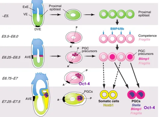
In the mouse, PGCs originate in the base of the developing allontois, between endoderm and mesoderm of the ventral amniotic fold, at 7-7.5 days post-coitum (dpc) (Ginsburg et al., 1990; McLaren, 2003). In humans, these cells first appear around 3 weeks post-fertilization (Motta et al., 2003). After their segregation from somatic cell lineages, PGCs migrate through the embryo to become incorporated in the developing gonads where they finalize their differentiation. During migration PGCs actively proliferate. (Buehr, 1997; McLaren, 2003)
Primordial Germ Cells specification
Lawson and Hage (1994) were able to locate the PGC ancestral population in the most proximal epiblast cells at 6-6.5 dpc (early gastrulation), by injecting single epiblast cells with a lineage marker of
mouse embryos. PGC’s were not yet fully differentiated since some labelled cells contributed to other somatic cell-lineages (Lawson and Hage, 1994). However, this work determined that the founding population of precursor germ cells were allocated early during gastrulation (7.2 dpc) as a group of approximately 40 to 45 cells (Ginsburg et al., 1990; Lawson and Hage, 1994). Studies point to mammalian germ cell-lineages being dependent on external signals rather than being predetermined (Extavour and Akam, 2003; McLaren, 2003; De Felici et al., 2004). Contributing to this conclusion were observations on mutant Bone Morphogenic Protein-4 (Bmp4) mice, which lack PGCs (Lawson et al., 1999) (Fig.1). This observation suggested the involvement of BMP4, an intracellular signalling protein member of the Transforming Growth Factors-β (TGFβ) superfamily, in the specification of germ cells. However, BMP4 signalling was not restricted only to germ cells since these animals also lack allantois (Lawson et al., 1999). Bmp8b mutants also showed a severe, although not as drastic reduction of PGC numbers (Ying et al., 2000). In vitro studies showed that both BMP4 and BMP8b signalling in the epiblast cells have a synergistic action on germline competency acquisition. However, BMP4 is required for epiblast cells to gain germ cell competency before (Ying et al., 2001) (Fig.1). Further studies found that BMP4 induces fragilis expression on the epiblast tissue (Saitou et al., 2002). Fragilis is a putative interferon-inducible gene, which expression in this epiblast cells is thought to induce a transmembrane protein associated with germ cell acquisition of competence (Saitou et al., 2002; Lange et al., 2003). In fact, it has been shown that only the cells with highest expression of fragilis will express stella, the first gene known to be germ cell lineage-restricted (Saitou et al., 2002).
Stella is a nuclear-cytoplasmic protein that seems to be involved in
chromosomal and RNA organization and is predominantly expressed in totipotent and pluripotent cells (Saitou et al., 2003) (Fig.1). Importantly,
stella-positive cells exhibited repressed homeobox genes, suggesting that
homeobox genes repression) and maintenance of pluripontency (Saitou et
al., 2002). The repression of homeobox genes seems to be due to the
expression of Blimp1 (Prdm1), a potent transcriptional repressor of a histone methyltransferase subfamily, in the epiblast at 6.25 dpc and later restricted to the founder germ cell cluster (Ohinata et al., 2005; Saitou et al., 2005) (Fig.1). Another cell pluripontency associated gene is Oct-4, a POU transcription factor encoded by the Pou5f1 gene, which was demonstrated to be expressed in PGCs, unfertilized oocytes and in the inner cell mass (ICM) of blastocysts (Scholer et al., 1990b; Watson and Tam, 2001). Like alkaline phosphatase activity, at 7.0 dpc Oct 4 is still expressed in the epiblast cell but becomes germ cell restricted at 7.5-8.0dpc (Scholer
et al., 1990a) (Fig.1) and is essential for PGCs survival, since Oct-4 loss leads
to germ cell apoptosis (Kehler et al., 2004). These observations indicate that all cells in the proximal epiblast can be in the founder cluster of PGCs, as long as they are in the right place at the right time for specification (Watson and Tam, 2001).
Figure 1 – Primordial Germ Cell specification in the mouse adapted from (Hayashi et al.,
2007). (VE-visceral endoderm; ExE-extra-embryonic ectoderm; DVE-dorsal visceral endoderm; AVE-anterior visceral endoderm)
Oct-4
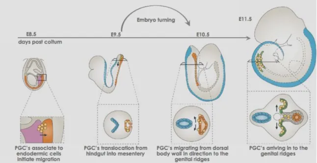
Primordial Germ Cell migration and proliferation: arrival to the genital ridges
At this point in time (7.0-7.5dpc) in the mouse embryo PGC cluster is outside of the embryo proper at the base of the allantois. A day later, 8.5dpc, PGCs are associated with the endodermic cells. As the endodermic cells begin invagination to form the hindgut, PGCs begin migration (Fig. 2) (Byskov, 1982; Motta et al., 1997; McLaren, 2003; Motta et al., 2003). Approximately, one day later (9.5dpc) in the mouse PGCs translocate from the hindgut and actively move dorsally into the mesentery (Fig. 2) (Molyneaux et al., 2001). Next, at 10.5dpc PGCs start migrating directionally from the dorsal body wall to the genital ridges - the future gonads (Fig. 2). Finally at 11.5dpc mouse PGCs populate genital ridges (Fig. 2). (Byskov, 1982; Molyneaux et al., 2001; McLaren, 2003; Molyneaux and Wylie, 2004) During migration PGCs actively proliferate, continuing for 2-3 days after arrival at the genital ridges, allowing the initial 45 cells to become approximately 20 000 at 13.5dpc (Tam and Snow, 1981).
Several mechanisms may be used by the PGCs and surrounding cells for migration: adhesion, where PGCs respond to cell-cell contacts; contact guidance, PGCs move along a preformed molecular pathway in a substrate; and/or chemotaxis, PGCs are attracted by a substance produced elsewhere (Buehr, 1997). It was proposed that PGCs arrive to the hindgut independently but when leaving the gut to the dorsal mesentery (9.5dpc) they form aggregates, extending long process of stage specific embryonic antigen-1 (SSEA-1) forming a network (Gomperts et al., 1994). SSEA-1 has been proposed as a specific cell-surface marker of undifferentiated cells and is often used to identify pluripotent cells (Choi et
al., 1999). More recently it was shown that E-cadherin is essential to the
PGC-PGC connection and motility in the migratory process. Disrupting PGC-PGC adhesion through E-cadherin functional antibodies induced the formation of ectopic germ cells (Bendel-Stenzel et al., 2000; Di Carlo and De Felici, 2000). Interactions between PGCs and the extra cellular matrix
(ECM) in which PGCs translocate, and β1-integrins, cell surface receptors in the ECM, were also shown to be important for PGCs locomotion during
migration (Garcia-Castro et al.,1997; Anderson et al.,1999).
Figure 2 - Primordial germ cells migration into the genital ridges. Adapted from
(Richardson and Lehmann, 2010).
Disruptions in the c-kit/kit-ligand (KL/stem cell factor/mast cell growth factor) leads to germ cell migration problems in addition to alterations in patterns of survival and proliferation (Keshet et al., 1991; Buehr et al., 1993; Pesce et al., 1997). The tyrosine kinase receptor, c-kit and its ligand-KL are important regulators of germ cell development, encoded in the genes White and Steel, respectively and are among the loci most studied in the mouse (Russel et al., 1968; Keshet et al., 1991). In vitro studies lead to the understanding of the importance of these proteins and the surrounding environment in migration, proliferation and survival (Godin et al., 1991; Pesce et al., 1997). Recently, Steel factor was proven to be essential for PGCs migration and proliferation (Runyan et al., 2006). Other important factors on germ cells proliferation are the fibroblast growth factors (FGFs) associated with proliferation of many cell types, including germ cells and mesoderm formation in embryos (Kawase et al., 2004). FGF-4/8/17 are expressed in the neighbouring cells of PGCs, during migration but FGF-4/8 is
expressed by the PGCs when they arrive to the genital ridges. This differential expression of FGF may indicate that during migration proliferation is induced through a paracrine mechanism while within the genital ridges it may switch to an autocrine mechanism (Kawase et al., 2004). These authors also found that without c-kit, FGFs together with KL promote proliferation but the presences of c-kit induced an inhibitory effect on FGF’s-stimulated PGCs proliferation. Peptidyl-prolyl isomerase 1
(Pin1) involvement in PGCs proliferation was demonstrated through Pin1
-/-females, which are born with significantly less oocytes, although PGCs allocation was perfectly normal (Atchison et al., 2003). Further studies showed that PGCs expansion was impaired with no signs of cell cycle arrest or apoptosis, thus indicating prolonged cell cycles, fewer divisions, which in the end results in lower oocyte numbers (Atchison et al., 2003).
Studies in the early nineties suggested that chemotropic substances released by the genital ridges, the target tissue, act as attractants to germ cells - chemotaxis (Godin et al., 1991; Godin and Wylie, 1991; Buehr, 1997). An in vitro study showed that soluble TGFβ1 in the culture media mimicked the signals produced by the genital ridges at 10.5dpc (Godin and Wylie, 1991). More recently it was shown that colonization of genital ridges requires the ligand-receptor interaction – SDF1/CXCR4 (Molyneaux et al., 2003). Stromal cell-derived factor – SDF1 is a member of chemokine CXC subfamily and the only known ligand for the G-protein-coupled receptor CXC chemokine receptor 4 - CXCR4, also a co-receptor for HIV-1 virus in humans (Bleul et al., 1996; Molyneaux et al., 2003; Peng et al., 2004). The ligand, SDF1, is expressed in the dorsal body wall during migration while the receptor, CXCR4, is expressed in the migrating germ cells (Molyneaux et al., 2003). These authors showed that exogenous SDF1 induced abnormal migration, and targeted mutations to CXCR4 lead to genital ridge
colonization problems (Molyneaux et al., 2003). Similarly, SDF1-/- showed
impaired colonization of the genital ridges, indicating that SDF1 is not required for migration but for homing the PGCs (Ara et al., 2003).
The proliferation within the genital ridges is a special program in which PGCs expand in number but also form cysts or clusters, which divide synchronously but without cytokinesis. Thus PGCs develop in a cluster of 4 to 32 cells with cytoplasm connected by an intercellular bridge (Pepling and Spradling, 1998; Pepling et al., 1999). This is a conversed mechanism from invertebrates to vertebrates that occurs before germ cells enter meiosis at the time of sex determination (Pepling et al., 1999). This conservation demonstrates the importance of the intercellular bridge connections between germ cells before meiosis. This mechanism is very well studied in Drosophila, which grow up to 16 germ cells before entering meiosis (de Cuevas and Spradling, 1998). However, striking similarities have been identify in Xenoupus, in which germ cells also divide synchronously to form a cluster of 16 cells entering meiosis afterwards (Pepling et al., 1999) and in the mouse it is predicted for the clusters to grow to 32 germ cells before entering in meiosis (Pepling and Spradling, 1998).
Sex determination
When germ cells reached the genital ridges, they are still undifferentiated. Sex determinant mechanisms vary among the animal kingdom: ratio of X chromosomes to autosomes (worms and flies); or temperature, hormones, environmental cues (reptiles and fishes) (Tilmann and Capel, 2002). In mammals, until the early 2000’s, the ovary was believed to be a default pathway, set to always develop in the absence of “male/testis signals” (Lovell-Badge and Hacker, 1995; Tilmann and Capel, 2002). However, several genes have been implicated in gonad formation in both sexes. Among these are steroidogenic factor-1 (Sf1), Wilms tumour 1 (Wt1), and LIM homeobox protein 9 (Lhx9) (McLaren, 2000; Tilmann and Capel, 2002), which have been implicated by the absence and/or defects in genital ridges of knockout (KO) mouse models (Pangas and Rajkovic, 2006).
In males, the decision to make testes is controlled by the sex-determining gene on the Y chromosome – Sry (Koopman et al., 1991). This gene is
expressed in the mouse embryo from approximately 10.5 to 12.5dpc, only in the supporting cell lineage within the genital ridge (Lovell-Badge and Hacker, 1995; Koopman, 2001; McLaren, 2003). Sry induces the differentiation of Sertoli precursors and its down-regulation coincides with the beginning of testes development (Tilmann and Capel, 2002; Kim and Capel, 2006). FGF9 is involved in the migration of mesonephric cells (Colvin
et al., 2001) and in germ cell survival, hence it is one of the first factors on
the sex-determinant pathway (DiNapoli et al., 2006). This growth factor also “antagonizes” with wingless-related MMTV integration site 4 (Wnt4) and together they form a balanced mechanism of decision between male and female, respectively (Kim et al., 2006).
In female mice, deletion of Wnt4 leads to their masculinization, due to testosterone production, suggesting Wnt4 as a determinant gene for female sex differentiation (Vainio et al., 1999; Jordan et al., 2001; Yao et al., 2004; Heikkila et al., 2005; Kim and Capel, 2006). Wnt4 is expressed in the undifferentiated gonads of mice until 11.5dpc, when it is down regulated in the males but persists in the females (Vainio et al., 1999). Wnt4 controls the expression of follistatin, which is responsible for repressing the formation coelemic vessel (testis formation precursor) and contributes to germ cell survival in the ovary (Yao et al., 2004). Along with Wnt4, Dax1 (Dosage-sensitive sex reversal (DSS)-Adrenal hypoplasia congenital critical region on the X chromosome, gene 1) was also thought to be an ovary regulatory gene (Zanaria et al., 1995). In fact, Dax1, like Wnt4, is down regulated in males when differentiation begins (~12.5dpc) while persisting in the ovary (Swain et al., 1996). Since its overexpression lead to XY sex reversal, antagonizing with Sry (Swain et al., 1998) it was the a perfect candidate for an ovary determinant gene. However, it is not required for ovary development but for testis development instead, working more like an “anti-testis than an ovary determinant” (Yu et al., 1998). Furthermore, Wnt4 and Dax1 have a joint role in controlling female development and testes repression, where Wnt4 up regulates Dax1 (Jordan et al., 2001; Mizusaki et
al., 2003). The finding that female mice lacking the gene forkhead
transcription factor 2 (Foxl2) have oocytes but also have differentiated male somatic cells, lead to the suggestion that sex determination continues throughout ovarian development (Loffler et al., 2003; Baron et al., 2005; Ottolenghi et al., 2005). Foxl2 expression initiates when gonads start differentiating (~12.5dpc) (Loffler et al., 2003), which could implicate it as being a female determinant gene, however the fact that it occurs after oocyte loss, may also be indicative of an oocyte effect (Ottolenghi et al., 2005). Hence Wnt4 continues to be the primary female gene determinant
studied so far (Fig. 3).
Figure 3. Schematic summary of gonad development and sex determination in mice.
Initially to form a bipotential gonad Wt1, Sf1 and Lhx9 are needed. On day 11.5 dpc primordial germ cells arrive into the genital ridge, with no phenotypical difference between sexes. If Sry is present Sox9 is up regulated and in a forward loop induced Fgf9, which inhibits Wnt4, and testis is formed. Müllerian ducts start regression due to Sertoli cells AMH production. However if Wnt4 is dominant, Fgf9 is inhibited and consequently Sox9; Dax1is is up regulated and Foxl2 begins expressing, an ovary is formed. In this case it is the Wolffian duct that regresses.
Germ cell meiosis initiation
Twelve and half days post-coitum, PGCs within the genital ridges initiated differentiation and continue proliferating until approximately 13.5dpc. Around this time, occurs the first germ sex developmental difference between male and female germ cells (Swain, 2006). In the males, germ cells (T-prospermatogonia) arrest in the G1/G0 stage of the mitotic cell
Figure 4. Mouse fetal ovary, 16 days post coitum, cluster delimitation by actin-filaments in red and nuclei in white, detailed image of actin-clustered oogonia on the right. Scale bars 20µm.
cycle around 13dpc (McLaren and Southee 1997). On the other hand, female germ cells (oogonia) enter prophase of the first meiotic division,
arresting in pachytene
stage prior to birth,
resuming meiosis shortly before ovulation (McLaren and Southee, 1997).
Around day 13.5 post-coitum in the mouse,
oogonia are
interconnected with one
another by
inter-cytoplasmic bridges and
surrounded by somatic cells forming the ovigerous cords (Byskov and Lintern-Moore, 1973; Pepling et al., 1999) (Fig. 4). At the same time Stra-8 is expressed and oogonia transit from the mitotic to the meiotic cell cycle, becoming primary oocytes (Hirshfield, 1991; Koubova et al., 2006). They progress through meiotic prophase-I stopping at diplotene. By day 17.5 post-coitum some oocytes are already arrested in diplotene, while others will do so shortly after birth (Hirshfield, 1991).
Germ cell reserve establishment and primordial follicle assembly
In most mammals, female meiosis entry coincides with a major germ cell loss, which in rodents goes up to 80% of the initial oogonia pool (Baker, 1966; Hirshfield, 1991; Tilly, 2001). The reason for the massive loss may be associated with errors in mitosis, or chromosome pairing, (Tilly, 2001; McClellan et al., 2003; Di Giacomo et al., 2005). The proposed mechanisms usually associated with germ cell elimination are: apoptosis (Baker 1966; Peters and McNatty, 1980; Coucouvanis et al., 1993; De Pol et al., 1997; Pepling and Spradling, 2001; Tilly, 2001; Baum et al., 2005) and germ cell extrusion through the ovarian epithelium – or shedding (Byskov and
Rasmussen, 1973; Hirshfield, 1991; Wassarman and Albertini, 1994; Motta et
al., 2003). We will address this subject further in chapter 2. With few
exceptions, (as the lemurs of Madagascar (Gosden, 1995)), the number of germ cells/oocytes is set at birth or shortly after birth and will gradually decrease when ovulation begins (Peters and McNatty, 1980; Byskov, 1982). Recently, this dogma has been challenged in the mouse by Johnson et al., (2004; 2005), Lee et al., (2007), and in human by Bukovsky et al., (2004), generating a very exciting discussion in the recent years among the reproductive scientific community.
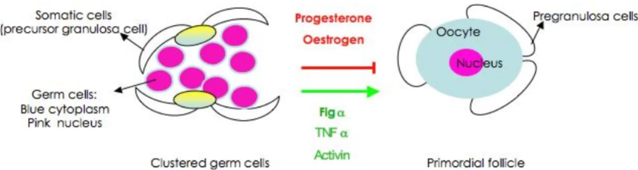
Follicle formation occurs after birth and coincides with cluster breakdown, and major germ cell loss (Pepling and Spradling, 2001). In the embryonic mammalian ovary the oogonia clusters are enclosed within somatic cells structures - the ovigerous cords (Fig 4) (Byskov and Lintern-Moore, 1973). The breakdown of both, ovigerous cords and clusters leads to the formation of primordial follicles; the somatic cells from the cords “invade” the cluster and associate with the surviving primary oocytes (Byskov, 1986; Rajah et al., 1992; Pepling and Spradling, 2001; Sawyer et al., 2002). These invading cells - pre-granulosa cells, are flattened and do not completely enclose the oocyte, this being what characterizes a primordial follicle (Pedersen and Peters, 1968). The assembly of primordial follicles is still not well understood, and will be discussed in chapter 3. Figure 5 summarizes schematically the possible factors involved in cluster breakdown and primordial follicle assembly.
(Marcinkiewicz et al., 2002; Morrison and Marcinkiewicz, 2002) (Bristol-Gould and Woodruff, 2006; Coutts et al., 2007)) factors. Yellow/blue cells are the GREL – gonadal ridge epithelial-like cells (Hummitzsch et al., 2013).
Primordial follicles remain “quiescent” until initial recruitment into the growing pool (McGee and Hsueh, 2000; van den Hurk and Zhao, 2005). Primordial follicles can arrest at this stage up to 1 year (rodents), or to 5 decades (human), waiting for recruitment (van den Hurk and Zhao, 2005). Having overviewed primordial germ cells trajectory to follicle assembly, we will now focus on the central player: the oocyte.
1.2. OOGENESIS: PROSPECTS AND CHALLENGES FOR THE FUTURE
[Publication: Rodrigues, P; Limback, D; McGinnis, L; Plancha, CE; Albertini, DF (2008) J Cell Physiol. 216: 355-365] (numbering of figures has been altered relative to the original article, to maintain the numbering of the previous section)
ABSTRACT
Oogenesis serves a singular role in the reproductive success of plants and animals. Of their remarkable differentiation pathway what stands out is the ability of oocytes to transform from a single cell into the totipotent lineages that seed the early embryo. As our understanding that commonalities between diverse organisms at the genetic, cellular and molecular levels are conserved to achieve successful reproduction, the notion that embryogenesis presupposes oogenesis has entered the day-to-day parlance of regenerative medicine and stem cell biology. With emphasis on the mammalian oocyte, this review will cover 1) current concepts regarding the birth, survival and growth of oocytes that depends on complex patterns of cell communication between germ line and soma, 2) the notion of “maternal inheritance” from a genetic and epigenetic perspective, and 3) the relative value of model systems with reference to current clinical and biotechnology applications.
INTRODUCTION
Oogenesis is a protracted process that encompasses the birth, growth, and maturation of a cell unique in its ability to propagate another generation of organisms. In some sense, it is not a very efficient process when measured in terms of viable offspring. In fact, the relative fecundity of a particular organism varies widely according to the kind of reproductive strategy employed. For example, broadcast spawners like fish and many invertebrates are efficient at oogenesis but the fate of ovulated eggs is left up to the whims of the environment the resultant embryos find themselves in. These kinds of animals make a significant metabolic investment in oogenesis. On the other hand, primates expand oocyte numbers prior to birth and engage in a dramatic course of prolonged attrition with a small fraction of the egg endowment surviving to ovulation throughout the reproductive lifespan. As extreme as organisms may be in both the efficiency of egg production and the size of their “spawn” (hundreds to thousands for invertebrates; one for humans), the goals of oogenesis remain the same: producing a developmentally competent egg capable of generating live offspring.
While developmental biologists have long been fascinated by the mechanisms by which an oocyte acquires and executes its totipotentiality during embryogenesis, a new generation of experimentalists have joined the campaign bringing with them research goals of great global and clinical importance. The emergence of the field of Assisted Reproductive Technologies (ARTs) is dominant amongst these new areas of oocyte biology because of the increasing usage of ARTs to treat human infertility. Since the advent of this technology in the late 1970s, more than 3 million children have been born using ARTs and the prospects for innovations and other modifications in current applications are clear. Oocyte and ovary cryopreservation efforts are ongoing as a means to preserve or restore reproductive function to women who have undergone medical treatments that cause partial or complete sterility. Technology for preservation of
maintain the diversity of living organisms that are rare, endangered, or bred for agricultural or medicinal value. The key point here is that oogenesis is a gradual process by which the properties needed to express developmental competence are acquired at different stages of differentiation and so the timing for intervention strategies and their impact on oocyte quality represent key challenges for the future. In this sense, virtually all clinical applications aimed at female germ line preservation will require identification of appropriate model systems and to unravel the mechanisms that confer and mediate the expression of oocyte developmental competencies.
With this background, the goals of this review are to 1) highlight current mechanistic viewpoints as related to the stages during which mammalian oocytes are born, grow, and mature, 2) revisit the seasoned concept of “maternal inheritance” in light of new data showing that especially in mammals, the relative contributions of the oocytes’ endowment to embryo survival and fetal development have been underestimated, and 3) given the impact that studies of oogenesis will have on biotechnology, stem cell research and animal fecundity, the utility of specific animal models will be discussed in the context of future challenges relevant to human health.
A. OOGENESIS IS A PROTRACTED PROCESS THAT INVOKES FEEDBACK REGULATION AT MANY LEVELS.
1. Primordial to primary follicle transition
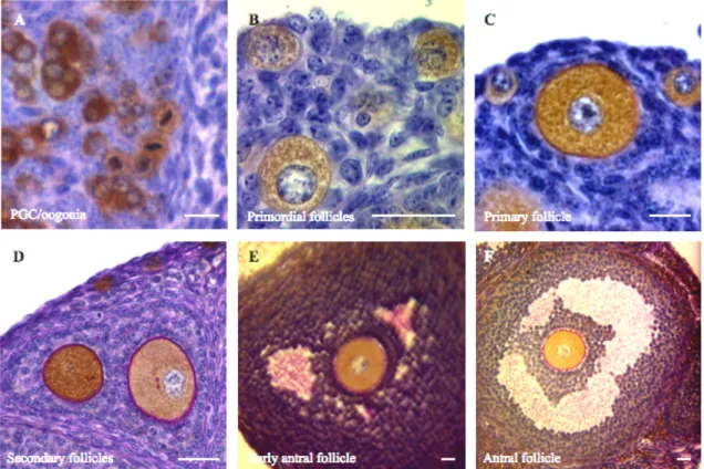
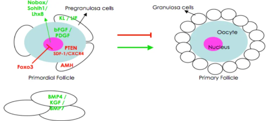
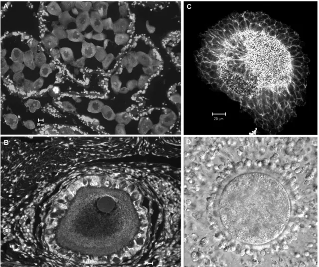
Immediately after primordial follicle assembly some are recruited from the resting pool into the growing population. The mechanism by which primordial follicles are maintained or leave the resting pool is still not well understood. Primordial follicle activation is progressive and initiated with granulosa cell proliferation, and their change in shape from squamous to cuboidal (Hirshfield, 1991; Braw-Tal, 2002). When these cuboidal granulosa cells form a layer that completely surrounds the also enlarging oocyte, the primordial follicle has become a primary follicle (Fig. 6A-C) (Hirshfield, 1991;
Braw-Tal, 2002). Several factors are involved in inhibiting and inducing this transition (Skinner, 2005).
Figure 6. (A-F) Examples of stages in follicle and oocyte growth from oogonia (A) to antral
follicle (F). Oocyte cytoplasm is labeled with mouse vasa homologue (MVH) antibody, a specific oocyte factor; zona pellucida is stained with PAS reaction (pink). Note in B the onset of zona pellucida formation in the primary follicle. Scale bars 20µm.
Anti-Müllerian hormone (AMH), the growth factor that leads to Müllerian duct regression in males, has been sited as responsible for maintaining primordial follicles in the resting pool in females (Durlinger et al., 1999; Durlinger et al., 2002). The observation of early age depletion of primordial follicles in AMH-deficient mice and the fact that AMH is produced in the granulosa cells of developing follicles has lead to the suggestion of AMH as an inhibitor of primordial formation (Durlinger et al., 1999; Durlinger et al., 2002). To further understand the regulatory action of AMH, Nilsson and colleagues (2007) performed a transcriptome analysis of ovaries treated with AMH and known stimulatory factors. Results showed that AMH may decrease the expression of pro-activation factors and increase the
expression of inhibitory factors (Nilsson et al., 2007). Forkhead transcription factor O3 (Foxo3; FOXO subfamily of forkhead transcription factors which are all downstream effectors of the PTEN/PI3K/AKT pathway) was also implicated in inhibition of primordial follicle activation (Castrillon et al., 2003). These scientists generated mice with a null mutation on the Foxo3
locus, and, despite being fairly normal, females exhibited a distinct
phenotype of mass follicle activation, leading to early follicular depletion (Castrillon et al., 2003). In fact, a detailed characterization of the Foxo3 deficient ovary showed that Foxo3 is necessary to suppress primordial follicle activation (John et al., 2007); these animals have viable litters as long as there are follicles available but total follicle depletion happens around 15 weeks of age (John et al., 2007). Accordingly, mice where constitutively active Foxo3a was being expressed in the oocytes exhibited infertile females due to slow follicle development and oocyte growth (John
et al., 2007). Concomitantly, deletion of PTEN (phosphatase and tensin
homolog deleted on chromosome 10; upstream of the Foxo3) gene in mouse oocytes result in activation of the majority of primordial follicles by postnatal day 23, and complete loss by 16 weeks post-birth (Reddy et al., 2008). PTEN is a lipid phosphatase that negatively regulates the PI3K (phosphatidylinositol 3-kinase) signaling pathway of cell proliferation and survival (Cantley and Neel, 1999). It is involved in regulation of primordial follicle activation, since it is this pathway (PI3K-AKT-Foxo3) that kit ligand [KL, growth factor that binds to its cognate receptor – c-kit (Hutt et al., 2006b)] uses to induce oocyte activation (Reddy et al., 2005; Liu et al., 2006). Another signaling pathway, the chemokine SDF-1 and its receptor CXCR4, has been suggested to negatively regulate primordial follicle activation (Holt et al., 2006). Interestingly, in vitro addition of recombinant SDF-1 to neonatal ovaries increased the follicle density but decreased the number of growing follicles when compared to controls, indicating a possible role of this pathway in maintaining primordial follicles in the resting pool (Holt et al., 2006).
On the other hand, KL produced by granulosa cells, is involved in primordial follicle activation (Parrott and Skinner, 1999). Ovaries cultured with and without recombinant KL and/or neutralizing c-kit antibody (ACK-2) demonstrated that KL is necessary and sufficient to promote primordial follicle recruitment into the growing pool (Parrott and Skinner, 1999). Previously, it had been shown that c-kit (produced by the oocyte) and KL are required in ovarian follicle development prior to gonadotropin dependence (Yoshida et al., 1997). Injecting mice with the antibody blocking c-kit function (ACK-2), the authors concluded that KL/c-kit interaction is important for follicle activation in the first 5 days post-birth (Yoshida et al., 1997). Interestingly, leukemia inhibitory factor [LIF; an interleukin 6 class cytokine that affects the growth and development of cells (Taupin et al., 1998)], is also produced by the granulosa cells as another factor shown to promote primordial to primary follicle transition, increasing KL mRNA production in cultured granulosa cells (Nilsson and Skinner, 2002). LIF may act to induce primordial activation through induction of KL expression, as proved by culture of ovaries in the absence or presence of LIF or neutralizing antibody to LIF in a procedure similar to that described for KL (Nilsson et al., 2002). Similarly, applying the same methodology, ovary culture in absence or presence of a factor and its function-blocking antibody, the stimulatory effect on primordial to primary transition was demonstrated for the factors: 1) basic fibroblast growth factor [bFGF; from FGF family with several roles in development: cell proliferation, migration and differentiation (Ornitz and Itoh, 2001)] and is produced in the oocyte (Nilsson and Skinner, 2001); 2) Bmp4 (growth factor from the TGF-β family member), produced by theca and stromal cells, which also was found important for follicle survival (Nilsson and Skinner, 2003); 3) keratinocyte growth factor [KGF; a fibroblast growth factor member that stimulates epithelial cell proliferation (Rubin et al. 1989)] is also produced by precursor-theca, theca and stromal cells (Kezele et al., 2005) and 4) platelet-derived growth factor (PDGF; a growth factor),
interestingly, appears to be produced by the oocyte (Nilsson et al., 2006). Bmp7 (also a growth factor member of the TGF-β superfamily), compared to vehicle, when injected into the ovarian bursa cavity, was also shown to activate primordial follicles and subsequent transition to primary follicle, (Lee et al., 2001).
The presence of three oocyte specific genes was also shown to be essential for primordial to primary transition: newborn ovary homeobox-encoding gene (Nobox) (Suzumori et al., 2002; Pangas et al., 2004),
Sohlh1and Lhx8 (Pangas et al., 2006). Nobox, as the name indicates, is a
homeobox gene consequently involved in regulation of development and is expressed in oocytes of primordial, primary and growing follicles (Suzumori et al., 2002). Studies in deficient mice showed an arrest in follicle growth at primordial stage and a total loss of germ cells by day 14 after birth (Rajkovic et al., 2004). Nobox appears to regulate other important oocyte specific genes such as Oct4 and growth differentiation factor-9 (GDF9; from TGFβ superfamily) (Rajkovic et al., 2004). Both Sohlh1 and Lhx8 are transcriptional factors, which are expressed in germ cell clusters and oocytes of primordial follicles (Pangas et al., 2006). Analysis of female mice lacking the Sohlh1 gene revealed that primary follicles never form and complete depletion of germ cells occurs around 3 weeks post-birth (Pangas et al., 2006). A very similar pattern was encountered for Lhx8
deficient females and because the microarray analysis of Sohlh1-/- ovaries
showed a dramatic downregulation of Lhx8, this may be indicative that Sohlh1 is a major regulator of genes involved in folliculogenesis (Pangas et
al., 2006). Thus, these three genes are crucial for early oogenesis and
folliculogenesis, particularly in primordial follicle activation. Figure 7 summarizes the factors involved in primordial follicle activation.
Figure 7. Schematic representation summarizing the inhibitory and stimulatory factors
involved in the primordial to primary follicle transition.
Even though, FSH and/or LH influences some of these factors later in folliculogenesis, the transition from primordial to primary follicle is independent of gonadotropins (Buccione et al., 1990; Fortune, 2003). The previously described inhibitors and promoters of primordial follicle activation, produced by either oocyte, granulosa or theca cell precursors, reinforce the importance of cell-to-cell communication and illustrate that primordial follicle activation is a highly coordinated process (Albertini and Barrett, 2003; Skinner, 2005; Hutt et al., 2006b). Oocyte and surrounding granulosa cells are coupled to each other throughout folliculogenesis by gap junctions, which are the most important junctions within the ovary (Anderson and Albertini, 1976; Buccione et al., 1990; Albertini and Barrett, 2003). Gap junctions are intracellular membrane channels that allow sharing of small molecules between adjacent cells (Anderson and Albertini, 1976; Kidder and Mhawi, 2002). These are present in the mouse ovary as early as 17 days post coitum (Mitchell and Burghardt, 1986). The gap junction consists of groups of 6 protein subunits – connexins - joined together to form a channel, the connexon which is the functional unit of the gap junction; the end of the connexon from one cell docks with the end of the connexon from the adjacent cell to form the gap junction channel (Bruzzone et al., 1996). In the mouse ovary, several gap junction proteins have been identified, including connexins (Cxs) 37 and 43, which
Mhawi, 2002). Connexin 37 is exclusive to the interface between oocyte and granulosa cell. When Cxs 37 is absent, follicle growth arrests at the preantral stage (Type 4-see box 3) and although there is some growth of the oocyte it cannot initiate meiotic maturation (Simon et al., 1997; Carabatsos et al., 2000b). Connexin 43 is predominantly expressed in granulosa cells. In mice, targeted mutation of cx43 is lethal. Moreover, Cx43 is involved early in development since a reduced number of germ cells arrive to the genital ridges after Cx43 deletion (Juneja et al., 1999; Granot et al., 2002). When neonatal ovaries were cultured and/or grafted into the kidney capsule of adult females, folliculogenesis was arrested at the primary stage (Juneja et al., 1999; Ackert et al., 2001). Together these results underscore the importance of communication between granulosa
cells and the oocyte. Cx37-/- mice support the transition from primordial to
primary follicle, however Cx37 must be expressed for subsequent oocyte maturation and continued folliculogenesis (Liu et al., 2006). On the other hand, Cx43 has an earlier role and is required for connections between granulosa cells in order for these to proliferate, form additional granulosa cell layers and support continued follicle growth (Granot and Dekel, 2002; Gittens and Kidder, 2005; Teilmann, 2005; Simon et al., 2006).
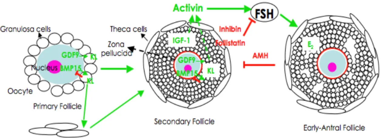
Like females lacking Cx43, females of GDF9 null mice exhibited a block in folliculogenesis at the primary follicle stage (Dong et al., 1996). GDF9 is a member of the TGFβ super family expressed only in the ovary (McPherron and Lee, 1993) and found to be exclusively expressed in oocytes beginning in primary follicles and persisting to later developmental stages (McGrath
et al., 1995). Interestingly, in hamster GDF9 seems to be essential for
primordial follicle formation and granulosa cell differentiation (Wang and Roy, 2006). It was shown that GDF9 stimulates granulosa cell proliferation
(Vitt et al., 2000), which might explain the arrest in folliculogenesis of GDF-9
-/- female mice. Aberrant oocyte-granulosa cell interaction was found in
proliferation, and reinforcing the importance of bidirectional communication between oocyte and granulosa cells and GDF9 paracrine actions in the ovary (Carabatsos et al., 1998; Elvin et al., 1999a; Elvin et al., 1999b). Moreover, it has been demonstrated that the impairment in
folliculogenesis in Cx43-/- ovaries is in part because the granulosa cells
cannot respond adequately to oocyte-derived GDF9 signals (Gittens et al., 2005).
Bidirectional communication is of vital importance to oogenesis and folliculogenesis, as has been shown using GDF9- and Cx43-deficient mice and reviewed by Eppig (2001) and Matzuk and colleagues (2002). The fundamental structures that provide this connection are the transzonal projections (TZPs), which are extensions from granulosa cells to the oocyte surface that establish and maintain the physical contact between these two cell types (Anderson and Albertini, 1976; Albertini and Rider, 1994; Motta et al., 1994; Albertini and Barrett, 2003). At the end of the TZP, where granulosa cells connect to the oocyte surface, are gap junctions (Anderson and Albertini, 1976; Motta et al., 1994). Interestingly, gap junctions at the end of TZPs are heterotypic composites between Cx43 and Cx37 (Kidder and Mhawi, 2002; Albertini and Barrett, 2003; Teilmann, 2005). Disruption of these gap junctions was found to be detrimental to folliculogenesis, as seen in both Cx37 and GDF9 null mouse models although the relationship between these oocyte specific genes has not been fully evaluated (Carabatsos et al., 1998; Carabatsos et al., 2000a). Cytoskeletal components of TZPs include both actin-filaments (Act-TZP) and microtubules (MT-TZP), both of which mediate shape and motility of the TZP (Albertini and Rider, 1994; Albertini et al., 2001; Navarro-Costa et al., 2005). Paracrine and hormonal regulation has been suggested as roles of TZPs for the vectorial secretion and/or uptake of signaling molecules at the oocyte-granulosa interface (Albertini et al., 2001; Combelles et al., 2004). These connections are already present in primordial follicles and continue
throughout folliculogenesis (Motta et al., 1994; Albertini and Barrett, 2003; Teilmann, 2005). The persistence of a coordinated communication network between oocyte and the surrounding somatic cells ensures ovulation of a healthy oocyte that is ready to be fertilized, the ultimate goal of folliculogenesis and oogenesis (Albertini et al., 2001; Eppig, 2001).
2. OOCYTE HYPERTROPHY: FROM PRIMARY TO MULTILAYERED FOLLICLES
The transition from primordial to primary follicle is prolonged to accommodate the growth phase of oogenesis. Hypertrophy of the oocyte is commensurate with a slow rate of granulosa cell proliferation when the follicle forms a second layer around the oocyte in secondary follicles (Fig. 6D). This protracted proliferative phase increases granulosa cells to six or seven layers in the pre-antral stage (Gougeon, 1996; Fortune, 2003). In the mouse, the appearance of a second layer of granulosa cells is accompanied by zona pellucida formation (Braw-Tal, 2002).
Numerous studies have been done in preantral follicles identifying factors responsible for follicle and oocyte growth, but the notion that the oocyte is the driving force for this event has been gaining acceptance (Eppig, 2001; Fair, 2003). In addition to the recognition of GDF9 as an oocyte specific factor, essential for follicular progression further than primary stage (Dong
et al., 1996), another oocyte specific factor was discovered independently
by two laboratories. It is either called bone morphogenetic protein-15 (BMP-15), due to its similarities to the BMP - family (Dube et al., 1998), or GDF9b because of its close homology to GDF9 (Laitinen et al., 1998), (herein referred to as BMP-15). BMP-15 is located on the X-chromosome and has an expression pattern very similar to GDF9 being predominantly in oocytes from primary follicles through ovulation in mouse (Dube et al., 1998), and rat (Otsuka et al., 2000). However, it appears that in some species GDF9 mRNA is present in primordial follicles, particularly in sheep (Galloway et al., 2000), cattle (Bodensteiner et al., 1999) and human (Aaltonen et al., 1999), implying that GDF9 synthesis precedes that of
BMP-15. This difference in species-specific expression patterns is interesting and may reflect local signaling requirements in monovular versus litter bearing (multiovular) animals. For example, studies with BMP-15 mutant mice revealed that null females are subfertile, with decreased ovulation and fertilization rates (Yan et al., 2001), but in sheep null BMP-15 females mimic the GDF9 null mouse phenotype in demonstrating follicular arrest at the primary stage (Galloway et al., 2000). At least in mice, this indicates that BMP-15 is more important in later stages of folliculogenesis, whereas GDF9 is needed earlier (Yan et al., 2001). Both factors were proven essential in sheep (Juengel et al., 2002), and their cooperative effect confirmed when recombinant ovine GDF9 and/or BMP-15 were shown to regulate the proliferation of granulosa cells in rat and ruminants (McNatty et al., 2005a; McNatty et al., 2005b). Furthermore, it was recently suggested that this cooperation is done through BMP – receptor II (Edwards et al., 2007). Interestingly, GDF9 and BMP-15 interact with KL (produced in the granulosa cells) controlling granulosa cell proliferation (Joyce et al., 2000; Otsuka and Shimasaki, 2002; Thomas and Vanderhyden, 2006). These growth factors have antagonistic roles: GDF9 inhibits KL expression in granulosa cells (Joyce et al., 2000; Wu et al., 2004), while BMP-15 acts as an activator of KL expression in granulosa cells, which in turn inhibits BMP-15 in a negative feedback loop (Otsuka and Shimasaki, 2002; Hutt et al., 2006a). At the same time KL from the granulosa cell seems to be the theca cell “organizer”, inducing interstitial cell recruitment to form the theca layer (Parrott and Skinner, 2000). Evidence for GDF9 inhibition of KL comes from GDF9 null mice that demonstrate up-regulation of both KL and inhibin (Elvin
et al., 1999a). Surprisingly, follicles of mice with a double knock-out (KO) for
GDF9 and Inhibin-α developed to multilayered stages before ovarian tumors appeared. This indicates that granulosa cells proliferate without both factors, and that the up-regulation of inhibin in GDF9 null alone is responsible for preventing granulosa cell proliferation since its absence is sufficient to promote proliferation (Wu et al., 2004). Several models have