Comprehensive Identification of
Krüppel-Like
Factor
Family Members Contributing to the
Self-Renewal of Mouse Embryonic Stem Cells
and Cellular Reprogramming
Hyojung Jeon1☯, Tsuyoshi Waku2☯, Takuya Azami1, Le Tran Phuc Khoa3, Jun Yanagisawa4,5, Satoru Takahashi1,6,7, Masatsugu Ema8,9*
1Department of Anatomy and Embryology, Faculty of Medicine, University of Tsukuba, 1-1-1 Tennoudai, Tsukuba, Ibaraki, 305–8577, Japan,2Graduate School of Pharmaceutical Sciences, The University of Tokyo, Hongo, Bunkyo-ku, Tokyo, 113–0033, Japan,3Department of Anatomy and Embryology, Human Biology Program, School of Integrative and Global Majors, University of Tsukuba, 1-1-1 Tennodai, Ibaraki, 305–8575, Japan,4Graduate School of Life and Environmental Sciences, University of Tsukuba, 1-1-1 Tennodai, Tsukuba, Ibaraki, 305–8577, Japan,5Center for Tsukuba Advanced Research Alliance, University of Tsukuba, 1-1-1 Tennodai, Tsukuba, Ibaraki, 305–8577, Japan,6International Institute for Integrative Sleep Medicine, Life Science Center, University of Tsukuba, 1-1-1 Tennodai, Tsukuba, Ibaraki, 305–8577, Japan,7Animal Resource Center, University of Tsukuba, 1-1-1 Tennodai, Tsukuba, Ibaraki, 305–8577, Japan,8Department of Stem Cells and Human Disease Models, Research Center for Animal Life Science, Shiga University of Medical Science, Seta, Tsukinowa-cho, Otsu, Shiga, 520–2192, Japan, 9PRESTO, Japan Science and Technology Agency, 4-1-8 Honcho Kawaguchi, Saitama, 332–0012, Japan
☯These authors contributed equally to this work.
Abstract
Pluripotency is maintained in mouse embryonic stem (ES) cells and is induced from somatic cells by the activation of appropriate transcriptional regulatory networks.Krüppel-like factor gene family members, such asKlf2,Klf4andKlf5, have important roles in maintaining the undifferentiated state of mouse ES cells as well as in cellular reprogramming, yet it is not known whether otherKlffamily members exert self-renewal and reprogramming functions when overexpressed. In this study, we examined whether overexpression of any represen-tativeKlffamily member, such asKlf1–Klf10, would be sufficient for the self-renewal of mouse ES cells. We found that onlyKlf2,Klf4, andKlf5produced leukemia inhibitory factor (LIF)-independent self-renewal, although most KLF proteins, if not all, have the ability to occupy the regulatory regions ofNanog, a critical Klf target gene. We also examined whether overexpression of any ofKlf1-Klf10would be sufficient to convert epiblast stem cells into a naïve pluripotent state and found thatKlf5had such reprogramming ability, in addition toKlf2andKlf4. We also delineated the functional domains of theKlf2protein for LIF-independent self-renewal and reprogramming. Interestingly, we found that both the N-terminal transcriptional activation and C-N-terminal zinc finger domains were indispensable for this activity. Taken together, our comprehensive analysis provides new insight into the contribution ofKlffamily members to mouse ES self-renewal and cellular reprogramming. OPEN ACCESS
Citation:Jeon H, Waku T, Azami T, Khoa LTP, Yanagisawa J, Takahashi S, et al. (2016) Comprehensive Identification ofKrüppel-Like Factor Family Members Contributing to the Self-Renewal of Mouse Embryonic Stem Cells and Cellular Reprogramming. PLoS ONE 11(3): e0150715. doi:10.1371/journal.pone.0150715
Editor:Johnson Rajasingh, University of Kansas Medical Center, UNITED STATES
Received:November 5, 2015
Accepted:February 18, 2016
Published:March 4, 2016
Copyright:© 2016 Jeon et al. This is an open access article distributed under the terms of the
Creative Commons Attribution License, which permits unrestricted use, distribution, and reproduction in any medium, provided the original author and source are credited.
Data Availability Statement:All relevant data are within the paper and its Supporting Information files.
Funding:This work was supported in part by a grant from PRESTO, Japan Science and Technology Agency (JST) (to M. E.). This work was also supported by a Grant-in-Aid for JSPS Fellows (to T. A. and T. W.).
Introduction
Mouse embryonic stem (ES) cells are derived from the inner cell mass of the blastocyst and can be maintained indefinitely in a self-renewing state in culture [1,2]. The ability to direct the dif-ferentiation of ES cells toward a specific cell fate is a highly pursued goal in regenerative medi-cine [3]. However, the utilization of ES cells for therapeutic purposes will require a better understanding of the molecular mechanisms underlying the regulation of pluripotency [4,5]. Previous studies revealed that the pluripotency of ES cells is maintained by multiple soluble factors, such as LIF [6,7], and by nuclear factors [8–15], including putative core transcription factors such asOct3/4,Sox2,Klffamily members andNanog. A reduction in extracellular-sig-nal-regulated kinase (ERK) activity strongly promotes pluripotency. Activation of the fibro-blast growth factor (Fgf)4–Fgf receptor(R)–ERK pathway destabilizes the pluripotent state and promotes a primed state [16–18]. Furthermore, the simultaneous inhibition of ERK and glyco-gen synthase kinase-3 (Gsk3) beta (with the inhibitor 2i) dramatically stabilizes the self-renewal process of mouse ES cells [19]. However, the precise molecular mechanisms of self-renewal remain elusive.
Induced pluripotent stem cells (iPSCs) can be derived from lineage-restricted cells, such as fibroblasts, by the forced expression of defined transcription factors [20,21]. Although previous studies have indicated that Krüppel-like transcription factors (Klfs) are essential for the repro-gramming of somatic cells into a pluripotent state, the molecular mechanisms underlying these processes remain unknown [10,11,22]. The processes involved in cellular reprogramming from somatic cells and the epiblast to generate iPSCs might be similar.
Epiblast stem cells (EpiSCs) are pluripotent stem cells derived from the epiblast of embryos at the egg cylinder stage and retain the ability to differentiate into all three embryonic germ lay-ers [23,24]. The properties and gene expression patterns of EpiSCs are very similar to those of human ES cells derived from embryos at the blastocyst stage. ES cells and EpiSCs are said to be in a naïve or in a primed state of pluripotency, respectively [25]. EpiSCs can be converted into iPSCs by reprogramming factors such asNanog,Esrrb, andKlffamily members includingKlf2
andKlf4[26,27]. The processes of self-renewal and cellular reprogramming share common transcription factors, indicating that both processes might be governed by similar molecular mechanisms.
Previous analysis indicated that expression ofKlf2,Klf4andKlf5is associated with an undif-ferentiated state in mouse ES cells, and loss-of-function gene knockout (KO) studies indicated that a triple KO ofKlf2,Klf4andKlf5resulted in defective self-renewal, and the introduction of
Klf2,Klf4, orKlf5, but notKlf10rescued the defective self-renewal phenotype [11]. Our previ-ous analysis ofKlf5KO ES cells indicated that a lack ofKlf5resulted in the spontaneous differ-entiation of mouse ES cells and that the phenotype was rescued byKlf4expression.
Overexpression ofKlf2,Klf4, orKlf5is sufficient to maintain the undifferentiated state of mouse ES cells in the absence of LIF [10,28,29], yet it is still unknown whether otherKlffamily members have such a function, and the functional domains required for self-renewal and cellu-lar reprogramming are not clear.
reprogramming. This indicated that the ability to reprogram EpiSCs into naïve pluripotent stem cells is correlated with the ability to maintain the pluripotent state. Taken together, our results provide a comprehensive view on the mechanisms involved in self-renewal and repro-gramming byKlffamily members.
Materials and Methods
Plasmids
The coding regions of mouseKlf1–Klf10were amplified from cDNAs derived from mouse ES cells or tissues, sequenced, and then introduced into the multicloning site of a pPB-FLA-G-HA-CAG-ireshygroR vector (Sanger Institute, Cambridge, UK).
Culture of Mouse ES Cells and Overexpression of
Klf
The OCRG9 ES cell line expressing green fluorescent protein (GiFP) (Oct3/4-CFP-irespuroR:: Rex1-GFP) was generated as described previously [30] and a gift from Dr. Hitoshi Niwa (Kumamoto University). All ES cells were maintained in mouse ES medium consisting of Dul-becco’s modified Eagle’s medium supplemented with 10% fetal bovine serum, 1 mM non-essential amino acids, 100μM 2-mercaptoethanol, 1 mM L-glutamine and 1,000 U/ml LIF on gelatin coated dishes as described [10]. In experiments, 1 × 10⁷ES cells were electroporated with 15μg of total DNA (piggyBac transposase and Klf plasmid; pPB-CAG-FLAG-HA-Klf-ire-shygroR) and cultured in the presence of 150μg/ml hygromycin B with or without LIF for 7 days; 1μg/ml puromycin was included in the medium used for the LIF-independent self-renewal assay to select Oct3/4-positive puromycin-resistant colonies.
Generation of EpiSCs with Stable Overexpression of Klfs
MouseΔPE-Oct3/4 EpiSCs were kindly provided by Dr. Austin Smith (Cambridge University) [27]. Transient transfection of mouse EpiSCs was performed on fibronectin-coated dishes seeded with 1 × 10⁶cells using 12μl Lipofectamine 2000 reagent (Invitrogen) with 4μg total DNA (piggyBac transposase and Klf plasmid) per well of a six-well culture plate. The EpiSCs were cultured in N2B27 medium supplemented with human activin A (10 ng/ml; R&D) and basic fibroblast growth factor (bFgf) (5 ng/ml; Wako Pure Chemical Industries) as described previously [27] and then selected with 250μg/ml hygromycin B and 1μg/ml puromycin for 7 days starting from 24 h post-transfection.
For the chimera experiment, 129 EpiSCs were generated as described previously and obtained from Dr. Paul Tesar (Case Western Reserve University) [24] and transfected with a piggyBac transposase and floxed Klf5 plasmid, and cultured in the presence of 200μg/ml G418.
Reprogramming Assays
Wells of six-well plates were coated with fibronectin and EpiSCs were plated at a density of 2–4×10⁴cells per well in EpiSC culture medium. After 24 h, the medium was replaced with NDiff227 medium (StemCells Inc.) supplemented with LIF (1000 U/ml) and 2i [containing a mitogen/extracellular signal-regulated kinase (Mek) inhibitor, PD0325901, 1μM (Cayman) and the Gsk3 inhibitor CHIR99021, 3μM (Cayman)] and changed every 24 h.
Blastocyst Injection
generous gift from Dr. Andras Nagy, Samuel Lunenfeld Research Institute). GFP-positive and Klf5 cassette-negative colonies were screened by polymerase chain reaction (PCR). The GFP-labeled iPSCs were injected into blastocysts that were transferred into surrogate mice. Embryos (9.5 and 13.5 dpc) were recovered and inspected under a fluorescence microscope.
Western Blot Analysis
ES cells and EpiSCs were lysed on ice for 10 min in RIPA buffer (50 mM Tris-HCl pH 7.5, 150 mM NaCl, 0.5% sodium deoxycholate, 1% NP-40, 0.1% sodium dodecyl sulfate (SDS)) supple-mented with a complete protease inhibitor cocktail (Roche). The extracts were clarified by cen-trifugation at 20,000gfor 5 min and suspended in sample buffer. Ten micrograms of cell extract were resolved on 10% SDS polyacrylamide gels and transferred onto polyvinylidene fluoride membranes (Millipore). Anti-hemagglutinin (HA; 1:1000) and anti-β-actin (1:4000) antibodies were used for western blotting. Proteins were detected using Immobilon kits (Millipore).
Chromatin Immunoprecipitation (ChIP) Assay
This assay was conducted as described [31]. The cells were fixed with 1% formaldehyde, and then glycine was added to a final concentration of 0.125 M. The cells were collected in SDS lysis buffer (50 mM Tris-HCl pH 8.1, 1% SDS, 10 mM EDTA, and protease inhibitors from Nacalai Tesque). The samples were sonicated and then centrifuged at 15,800gat 4°C for 15 min. After an aliquot (whole-cell extract) had been removed as an input sample, the supernatants were diluted in ChIP dilution buffer (16.7 mM Tris-HCl pH 8.1, 16.7 mM NaCl, 1.2 mM EDTA, 1.1% Triton X-100, 0.01% SDS). The diluted samples were precleared with 50μl of protein G Sepharose beads (GE Healthcare), and then the supernatants were incubated with 4μg of normal mouse IgG (Santa Cruz) or an anti-FLAG-M2 antibody (Sigma-Aldrich). The immunocomplexes were collected by incubation with 100μl of protein G Sepharose beads (GE Healthcare), and then washed with the following buffers: low salt wash buffer (0.1% SDS, 1% Triton X-100, 2 mM EDTA, 150 mM NaCl, and 20 mM Tris–HCl pH 8.1); high salt wash buffer (0.1% SDS, 1% Triton X-100, 2 mM EDTA, 500 mM NaCl, and 20 mM Tris–HCl pH 8.1); and LiCl wash buffer (0.25 mM LiCl, 1% IGEPAL-C630, 1% sodium deoxycholate, 1 mM EDTA, and 10 mM Tris–HCl pH 8.1). Finally, the beads were washed twice with 1 ml of TE buffer (1 mM EDTA and 10 mM Tris–HCl pH 8.0). The immunocomplexes were then eluted by adding 200μl of elution buffer (10 mM DTT, 1% SDS, 100 mM NaHCO3). After reversal of cross-linking by adding NaCl, the remaining
pro-teins were digested with proteinase K. The purified DNA was analyzed using quantitative (q) PCR to determine which fragments were present in the precipitate. The primers for qPCR were as follows: 50-gaggatgccccctaagctttccctccc-30and 50-cctcctaccctacccaccccctattctccc-30for the
Nanogpromoter; and 50
-tcagcactaaccatacaagttcatc-30
and 50
-agcgaagaggtggctggtag-30
for the
Nanogenhancer.
Immunohistochemistry and Alkaline Phosphatase Assay
Reverse-Transcription (RT) qPCR Analysis
For RT-qPCR analysis, first-strand cDNA was synthesized from total RNA using a QuantiTect Reverse Transcription kit (Qiagen). qPCR was performed with SYBR Premix Ex Taq II (Takara) and analyzed on a Thermal Cycler Dice Real Time System (TP850; Takara). The amount of target RNA was estimated using an appropriate standard curve and divided by the estimated amount ofβ-actin.
Statistical Analysis
Statistical analyses were performed using unpaired Student’sttests with Microsoft Office Excel. Data are expressed as the mean with standard error. Differences between means were considered significant atP<0.05.
Results
Although previous reports indicated that overexpression ofKlf2,Klf4, orKlf5achieves LIF-independent self-renewal of mouse ES cells [10,11,28,29], it is still unknown whether otherKlf
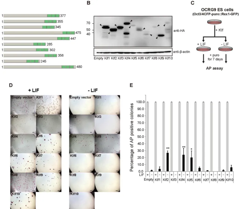
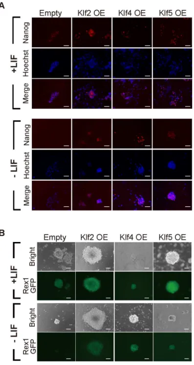
family members might have similar activity. Therefore, we attempted a comprehensive investi-gation into whether overexpression of representativeKlffamily members would be sufficient to maintain the undifferentiated state of mouse ES cells. First, we surveyed the phylogenetic tree of theKlffamily, which is composed of four subclasses, and selectedKlf1–Klf10out of 18 mem-bers, because they represent the four subfamilies (data not shown) [32]. After a FLAG-HA epi-tope tag was introduced into the N-terminus of eachKlfto monitor the protein expression level (Fig 1A), the expression vector was introduced into OCRG9 (Oct3/4-CFP-irespuroR:: Rex1-GFP) ES cells [30], which possesses aRex1-GFP pluripotency marker. Western blot anal-ysis confirmed that the epitope-taggedKlfproteins were overexpressed in the ES cells at vari-able levels (Fig 1B). Evaluation of relative expression levels of epitope-taggedKlfproteins normalized toβ-actin showed that levels of overexpressed Klf6, Klf7, Klf8 and Klf10 protein were low, but not significantly different from that of Klf5 (S1 Fig). The ES cells were cultured in the presence of puromycin to select Oct3/4-positive cells, in the presence or absence of LIF (Fig 1C and 1D). All the samples, including those expressingKlf1–Klf10, showed many undif-ferentiated alkaline phosphatase (AP)-positive colonies in the presence of LIF, yet only cells overexpressingKlf2,Klf4orKlf5led to significant numbers of AP-positive colonies in the absence of LIF (Fig 1D and 1E). We performed immunohistochemistry on colonies cultured in the absence of LIF and confirmed high levels of endogenous Nanog protein (Fig 2A). We also confirmed GFP fluorescence driven by theRex1promoter (Fig 2B), indicating that the undif-ferentiated state of pluripotency had been maintained.
Overexpression experiments clearly indicated thatKlf2,Klf4andKlf5, but not any otherKlf
family member, were able to maintain the undifferentiated state in the absence of LIF. It is not known why only these three members have this ability. To investigate the molecular mecha-nism underlying the pluripotency maintained byKlffamily members, we focused on the
Nanoglocus, because this gene is important for the self-renewal of mouse ES cells [9,14,33] and
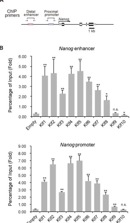
is controlled by the Klf2, Klf4, and Klf5 transcription factors [10,11]. Previous reports indicated that Klf2, Klf4 and Klf5 activateNanogthrough binding to the promoter and its 30enhancer
Mouse EpiSCs can spontaneously reprogram into the naïve state of iPSCs, but at a very low rate [34]. Transient expression of somatic reprogramming factors, such asKlf4,Nanog,Esrrb, andKlf2, cause more efficient and quicker reversion of EpiSCs into iPSCs, implying that this reversion mimics cellular reprogramming from fibroblasts into iPSCs. Previous analysis also indicated thatKlf2andKlf4, but notKlf5, enhance reprogramming of EpiSCs into iPSCs [28]. Fig 1. Comprehensive identification ofKrüppel-like factor(Klf) family members whose overexpression achieves leukemia inhibitory factor (LIF)-independent self-renewal of mouse embryonic stem (ES) cells.(A) A schematic representation of Klfs. These proteins have highly homologous C-terminal DNA binding domains characterized by three C2H2zinc finger motifs, shown in green boxes. The number of amino acids is shown to the left. (B)
Western blot of proteins from mouse ES cells overexpressing FLAG-HA epitope-tagged Klf. Anti-HA and anti-β-actin antibodies were used to detect HA-tagged Klf and endogenousβ-actin, respectively. Arrowheads indicate HA-tagged Klf proteins. (C) A schematic illustration of the experimental outline to assay the ability of a Klf protein to maintain self-renewal. (D) Generation of colonies from ES cells carrying either the empty vector or a Klf expression vector in the presence or absence of LIF. Scale bar: 2 mm. (E) The percentage of AP-positive colonies. Asterisks indicate statistical significance.*P<0.05;
**P<0.01.
We comprehensively examined the reprogramming ability of Klfs (Fig 4A). Expression vectors carrying epitope-taggedKlfgenes were introduced into EpiSCs and protein levels were con-firmed using an anti-HA antibody (Fig 4B). When cells were transferred from EpiSC cultures containing bFGF and activin A into ES medium containing LIF and 2i, only the EpiSCs overex-pressingKlf2,Klf4, orKlf5generated significant numbers of AP-positive colonies (Fig 4C). A Fig 2. Expression of pluripotency-related markers in Klf-overexpressing mouse ES cells.(A) Immunostaining for Nanog in mouse ES cells overexpressing (OE) a Klf protein, cultured in the presence or absence of LIF. (B) Rex1-GFP expression in Klf-overexpressing ES cells grown in the presence or absence of LIF. Scale bar: 100μm.
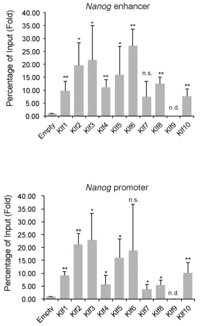
previous report indicated thatKlf5expression could not enhance reprogramming of EpiSCs to iPSCs [28]; therefore, we carefully evaluated whether iPSCs generated byKlf5overexpression Fig 3. ChIP analysis of the binding of epitope-tagged Klf toNanogregulatory regions in mouse ES cells.(A) Schematic representation of theNanogproximal promoter and distal enhancer. Two key regulatory regions (the enhancer and proximal promoter) ofNanogare shown as red and blue bars, respectively. Horizontal arrows indicate the primers used for PCR in site-specific ChIP assays. (B) ChIP analysis of the binding of Klf to the promoter and enhancer ofNanog. Asterisks indicate statistical significance.*P<0.05;
**P<0.01; n.s.; not significant.
were indeed pluripotent. The RT-qPCR analysis showed elevated expression levels of naïve markers, such asNanog,Klf2,Klf4,Esrrb,Stella, andRex1, while the expression levels of primed state markers, such asFgf5andBrachyurywere reduced (Fig 4D). Endogenous Nanog protein was also present in the iPSCs (Fig 4E). TheOct3/4gene contains distal and proximal enhancers; the distal one drives naïve pluripotent stem cells, while both enhancers are required for gener-ating primed pluripotent stem cells [27]. The iPSCs generated by transientKlf5expression showed strong GFP fluorescence without the presence of the proximal enhancer (Fig 4F), also indicating that the iPSCs were indeed naïve pluripotent stem cells. We then performed chimera formation, which is the gold standard assay for pluripotency (Fig 5). The iPSCs reprogrammed by overexpression ofKlf5were injected into blastocysts and the resultant blastocysts were returned into the uterus of a surrogate mother. E9.5 fetuses generated from two independent iPSCs exhibited strong contribution of GFP-positive iPSCs (Fig 5A). Furthermore, E13.5 fetuses exhibited strong contribution of GFP-positive iPSCs throughout the whole embryo Fig 4. Identification of the ability of Klf family members to reprogram EpiSCs into iPSCs.(A) Experimental outline for evaluating the reprogramming activity of a Klf protein. EpiSCs carrying Oct3/4-∆PE-GFP were cultured in N2B27 medium supplemented with LIF, the Mek inhibitor PD0325901, and the Gsk3 inhibitor CHIRON (CHIR99021) for 5–7 days and were then subjected to AP assays. (B) Western blot analysis of EpiSCs expressing epitope-tagged Klf. (C) Percentage of AP-positive colonies generated from EpiSCs overexpressing a Klf protein. (D) RT-qPCR analysis of iPSCs. (E) Immunostaining for Nanog in iPSCs generated by overexpression of Klf2, Klf4 and Klf5. (F) GFP fluorescence in EpiSCs generated by Klf2, Klf4 and Klf5 overexpression. Scale bar: 100μm. Asterisks indicate statistical significance.*P<0.05;**P<0.01; n.s.; not significant.
doi:10.1371/journal.pone.0150715.g004
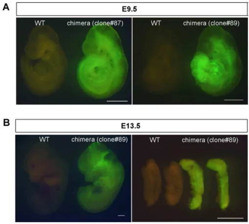
Fig 5. Chimera formation with iPSCs reprogrammed from EpiSCs.(A) Chimeras at E9.5 generated from two independent iPSC lines (#87 and 89). (B) Chimera and its genital ridge at E13.5 generated from iPSC line #89. Scale bar: 1 mm.
including the genital ridge (Fig 5B), indicating that iPSCs have the ability to differentiate into all three germ layers, including the germ cell lineage.
To gain insight into howKlf2,Klf4andKlf5might regulate reprogramming of EpiSCs to iPSCs, we performed ChIP assays to assess the occupation of Klf factors at theNanoglocus under EpiSC culture conditions. Most of the Klf proteins examined, except for Klf9, occupied
theNanogpromoter and enhancer, indicating that occupation itself was not sufficient for
reprogramming (Fig 6). Our analysis indicated that Klf2, Klf4, and Klf5 did not affectNanog
transcription under EpiSC culture conditions (data not shown).
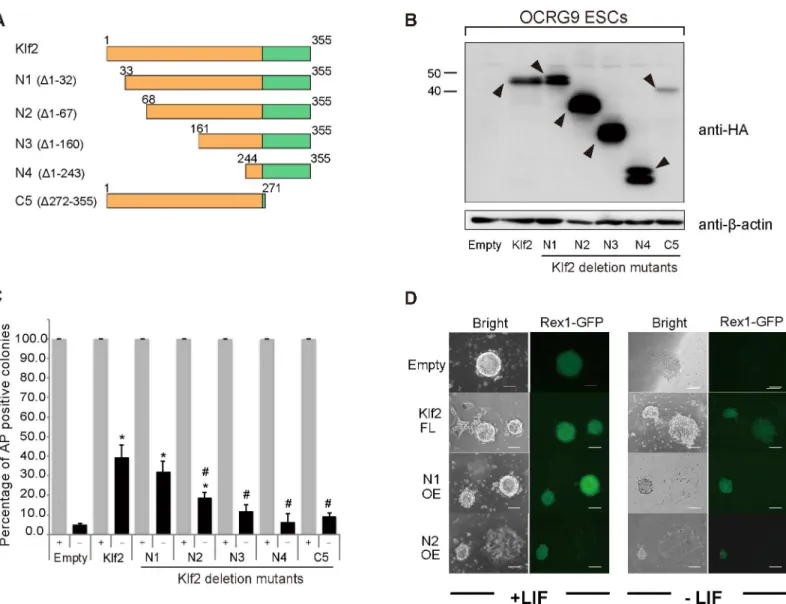
To delineate the functional domains required for LIF-independent self-renewal, we com-pared the amino acid sequences of Klf2, Klf4 and Klf5, and found that these three Klf proteins Fig 6. ChIP analysis of the binding of Klf toNanogregulatory regions in mouse EpiSCs.ChIP experiments were performed with an anti-FLAG antibody to identify the promoter and enhancer ofNanog. Asterisks indicate statistical significance.*P<0.05;**P<0.01; n.s.; not significant; n.d.; not detected.
share homologous regions in the N-terminal half, which acts in transcription, as well as in the C-terminal zinc finger DNA-binding domain (S2 Fig). Because Klf2 is the smallest of these pro-teins, deletion mutants forKlf2were created (Fig 7A). Western blot analysis confirmed that exogenous Klfs were overexpressed in ES cells in the presence of LIF (Fig 7B). When LIF-inde-pendent self-renewal activity was evaluated, N-terminal deletion resulted in a reduction in AP-positive clones (Fig 7C). Deletion of two zinc fingers also abolished the efficiency of this self-renewal activity. Thus, our analysis clearly indicates that both the N-terminal transcriptional modulation domain and the C-terminal zinc finger DNA-binding domain are required for the full activity of Klfs (Fig 7). We also delineated the functional domains required for reprogram-ming of EpiSCs into iPSCs (Fig 8). When variousKlfdeletion mutants were overexpressed in Fig 7. Delineation of the domains of Klf2 required for the self-renewal of mouse ES cells.(A) Schematic illustration of Klf2 deletion (Δ) mutants. Klf2 is highly homologous to Klf4 and belongs to the same subclass. A FLAG-HA tandem epitope tag was fused to the N-terminus of each of the deletion mutants. The zinc finger domain is shown as green boxes. (B) Western blot analyses of Klf2 and Klf2 deletion mutants with an anti-HA antibody. Arrowheads indicate HA-Klf proteins. (C) Percentage of AP-positive colonies in ES cells overexpressing full-length Klf2 or Klf2 deletion mutants, grown in the presence or absence of LIF. (D) Expression of the pluripotency-related marker Rex1. Rex1-GFP expression is seen in full-length Klf2, and N-terminal deletion mutants (N1, N2) in the absence of LIF. Scale bar: 100μm. Asterisks and hash symbols indicate statistical significance.*P<0.05 vs. empty vector; #P<0.05 vs. full-length Klf;*
P<0.01; n.s.; not significant; n.d.; not detected.
EpiSCs in the presence of bFGF and activin A (Fig 8A), we found that both the N-terminal transcription modulation domain and the C-terminal zinc finger DNA-binding domain were required for full activity (Fig 8B). This was similar to the domains required for self-renewal.
Discussion
Our analysis identified that only Klf2, Klf4, and Klf5 have the abilities to both maintain the undifferentiated state of mouse ES cells and to reprogram EpiSCs into iPSCs when overex-pressed. We also delineated functional domains of Klf for LIF-independent self-renewal and reprogramming and found that both the N-terminal transcription modulation domain and the C-terminal zinc finger DNA-binding domain were required for LIF-independent self-renewal and reprogramming. This finding indicates that the ability to reprogram EpiSCs into naïve plu-ripotent stem cells is correlated with the ability to maintain the pluplu-ripotent state.
A previous report by Wang et al. [35] indicated that the reprogramming ability of Oct3/4, Nanog and Sox2 could be boosted by using a chimeric protein including VP16, a potent tran-scriptional activator, in place of the inherent trantran-scriptional modulation domain, indicating that transcriptional activation is important for the reprogramming ability of those core factors. On the other hand, only a Klf4-VP16 chimeric protein provided a similar level of reprogram-ming activity, suggesting that the transcriptional modulation domain exerts inherent activity.
EpiSCs can be converted into iPSCs by naïve-state transcription factors such asNanog,
Esrrb,Klf2andKlf4[26,27]. Overexpression ofKlf2,Klf4orKlf5maintain the undifferentiated
state of mouse ES cells in the absence of LIF [10,28,29]. It is of note that induced expression of Klf4 and Klf5 are associated with the naïve state of rabbit iPS and ES cells [36], indicating a potential conserved role of Klf factors in the naïve state among species.
Previous“omics”studies using mass spectroscopy indicated that Oct3/4 exists in a large complex containing Sall4, Tcfcp2l1, Dax1, Esrrb, and other epigenetic modifiers in mouse ES cells [37]. Similarly, Nanog binds to Sox2, Mta2, Sall4, and PolII [38]. Interestingly, Klf5 is a protein that forms a complex with Oct3/4 and Nanog [37,38]. Our previous study [10] indi-cated that Klf5 is in a complex with Metastasis-associated protein (MTA2), a component of Nucleosome Remodeling Deacetylase (NuRD). Our results here will be useful for the further exploration of functional domains that contribute to self-renewal and reprogramming. It will be interesting to identify possible cofactors that are important for self-renewal and reprogram-ming, leading to elucidation of the molecular mechanisms involved.
Supporting Information
S1 Fig. Relative expression levels of epitope-tagged Klf protein normalized toβ-actin. Sig-nal intensities of epitope-tagged Klf protein were normalized to that ofβ-actin. Three indepen-dent sets of data were used to calculate statistical relevance.
(TIF)
S2 Fig. Multiple sequence alignment of mouse Klf protein using ClustalW.Klf2, Klf4 and Klf5 share highly homologous C-terminal DNA binding domains characterized by three C2H2 zinc finger motifs. An(asterisk) indicates positions with a single, fully conserved residue. A:
(colon) indicates conservation between groups of strongly similar properties, scoring>0.5 in
Fig 8. Delineation of Klf2 domains required for reprogramming EpiSCs to iPSCs.(A) Western blot analysis of EpiSCs expressing epitope-tagged Klf2 and deletion (Δ) mutants. Closed arrowheads indicate epitope-tagged Klf2 proteins. (B) Fluorescence images of a reprogrammed Oct3/4-ΔPE-GFP EpiSC colony with a Klf2 deletion domain on day 7. Open arrowheads indicate GFP fluorescence. OE, overexpression. Scale bar: 100μm.
the Gonnet PAM 250 matrix. A. (period) indicates conservation between groups of weakly sim-ilar properties scoring0.5 in the Gonnet PAM 250 matrix.
(TIF)
S1 Table. Primers used for RT-qPCR analysis. (PDF)
Acknowledgments
We thank Drs Hitoshi Niwa (Kumamoto University) and Tomoyuki Tsukiyama (Shiga Uni-versity of Medical Science) for helpful discussion and reagents. We also thank Drs Austin Smith (Cambridge University) and Paul Tesar (Case Western Reserve University) for provid-ing EpiSC lines. This work was supported in part by a grant from PRESTO, Japan Science and Technology Agency (JST) (to M. E.). This work was also supported by a Grant-in-Aid for JSPS Fellows (to T. A. and T. W.).
Author Contributions
Conceived and designed the experiments: ME. Performed the experiments: HJ TW TA LTPK. Analyzed the data: HJ TW TA LTPK JY ST ME. Wrote the paper: ME.
References
1. Evans MJ, Kaufman MH. Establishment in culture of pluripotential cells from mouse embryos. Nature. 1981; 292(5819): 154–156. PMID:7242681.
2. Martin GR. Isolation of a pluripotent cell line from early mouse embryos cultured in medium conditioned by teratocarcinoma stem cells. Proc Natl Acad Sci U S A. 1981; 78(12): 7634–8. PMID:6950406. 3. Murry CE, Keller G. Differentiation of embryonic stem cells to clinically relevant populations: lessons
from embryonic development. Cell. 2008; 132(4): 661–80. PMID:18295582. doi:10.1016/j.cell.2008. 02.008
4. Niwa H. How is pluripotency determined and maintained? Development. 2007; 134(4): 635–46. PMID: 17215298
5. Posfai E, Tam OH, Rossant J. Mechanisms of pluripotency in vivo and in vitro. Curr Top Dev Biol. 2014; 107: 1–37. PMID:24439801. doi:10.1016/B978-0-12-416022-4.00001-9
6. Smith AG, Heath JK, Donaldson DD, Wong GG, Moreau J, Stahl M, et al. Inhibition of pluripotential embryonic stem cell differentiation by purified polypeptides. Nature. 1988; 336(6200): 688–90. PMID: 3143917.
7. Williams RL, Hilton DJ, Pease S, Willson TA, Stewart CL, Gearing DP, et al. Myeloid leukaemia inhibi-tory factor maintains the developmental potential of embryonic stem cells. Nature. 1988; 336(6200): 684–7. PMID:3143916.
8. Cartwright P, McLean C, Sheppard A, Rivett D, Jones K, Dalton S. LIF/STAT3 controls ES cell self-renewal and pluripotency by a Myc-dependent mechanism. Development. 2005; 132(5): 885–96. PMID:15673569.
9. Chambers I, Colby D, Robertson M, Nichols J, Lee S, Tweedie S, et al. Functional expression cloning of Nanog, a pluripotency sustaining factor in embryonic stem cells. Cell. 2003; 113(5): 643–55. PMID: 12787505.
10. Ema M, Mori D, Niwa H, Hasegawa Y, Yamanaka Y, Hitoshi S, et al. Kruppel-like factor 5 is essential for blastocyst development and the normal self-renewal of mouse ESCs. Cell Stem Cell. 2008; 3(5): 555–67. PMID:18983969. doi:10.1016/j.stem.2008.09.003
11. Jiang J, Chan YS, Loh YH, Cai J, Tong GQ, Lim CA, et al. A core Klf circuitry regulates self-renewal of embryonic stem cells. Nat Cell Biol. 2008; 10(3): 353–60. PMID:18264089. doi:10.1038/ncb1698 12. Masui S, Nakatake Y, Toyooka Y, Shimosato D, Yagi R, Takahashi K, et al. Pluripotency governed by
Sox2 via regulation of Oct3/4 expression in mouse embryonic stem cells. Nat Cell Biol. 2007; 9(6): 625–35. PMID:17515932.
14. Mitsui K, Tokuzawa Y, Itoh H, Segawa K, Murakami M, Takahashi K, et al. The homeoprotein Nanog is required for maintenance of pluripotency in mouse epiblast and ES cells. Cell. 2003; 113(5): 631–42. PMID:12787504.
15. Nichols J, Zevnik B, Anastassiadis K, Niwa H, Klewe-Nebenius D, Chambers I, et al. Formation of plu-ripotent stem cells in the mammalian embryo depends on the POU transcription factor Oct4. Cell. 1998; 95(3): 379–91. PMID:9814708.
16. Burdon T, Stracey C, Chambers I, Nichols J, Smith A. Suppression of SHP-2 and ERK signalling pro-motes self-renewal of mouse embryonic stem cells. Dev Biol. 1999; 210(1): 30–43. PMID:10364425. 17. Hamilton WB, Kaji K, Kunath T. ERK2 suppresses self-renewal capacity of embryonic stem cells, but is
not required for multi-lineage commitment. PLoS One. 2013; 8(4): e60907. PMID:23613754. doi:10. 1371/journal.pone.0060907
18. Kunath T, Saba-El-Leil MK, Almousailleakh M, Wray J, Meloche S, Smith A. FGF stimulation of the Erk1/2 signalling cascade triggers transition of pluripotent embryonic stem cells from self-renewal to lineage commitment. Development. 2007; 134(16): 2895–902. PMID:17660198.
19. Ying QL, Wray J, Nichols J, Batlle-Morera L, Doble B, Woodgett J, et al. The ground state of embryonic stem cell self-renewal. Nature. 2008; 453(7194): 519–23. PMID:18497825. doi:10.1038/nature06968 20. Takahashi K, Tanabe K, Ohnuki M, Narita M, Ichisaka T, Tomoda K, et al. Induction of pluripotent stem
cells from adult human fibroblasts by defined factors. Cell. 2007; 131(5): 861–72. PMID:18035408. 21. Takahashi K, Yamanaka S. Induction of pluripotent stem cells from mouse embryonic and adult
fibro-blast cultures by defined factors. Cell. 2006; 126(4): 663–76. PMID:16904174.
22. Nakagawa M, Koyanagi M, Tanabe K, Takahashi K, Ichisaka T, Aoi T, et al. Generation of induced plu-ripotent stem cells without Myc from mouse and human fibroblasts. Nat Biotechnol. 2008; 26(1): 101–6. PMID:18059259.
23. Brons IG, Smithers LE, Trotter MW, Rugg-Gunn P, Sun B, Chuva de Sousa Lopes SM, et al. Derivation of pluripotent epiblast stem cells from mammalian embryos. Nature. 2007; 448(7150): 191–5. PMID: 17597762.
24. Tesar PJ, Chenoweth JG, Brook FA, Davies TJ, Evans EP, Mack DL, et al. New cell lines from mouse epiblast share defining features with human embryonic stem cells. Nature. 2007; 448(7150): 196–9. PMID:17597760.
25. Nichols J, Smith A. Naive and primed pluripotent states. Cell Stem Cell. 2009; 4(6): 487–92. PMID: 19497275. doi:10.1016/j.stem.2009.05.015
26. Gillich A, Bao S, Grabole N, Hayashi K, Trotter MW, Pasque V, et al. Epiblast stem cell-based system reveals reprogramming synergy of germline factors. Cell Stem Cell. 2012; 10(4): 425–39. PMID: 22482507. doi:10.1016/j.stem.2012.01.020
27. Guo G, Yang J, Nichols J, Hall JS, Eyres I, Mansfield W, et al. Klf4 reverts developmentally pro-grammed restriction of ground state pluripotency. Development. 2009; 136(7): 1063–9. PMID: 19224983. doi:10.1242/dev.030957
28. Hall J, Guo G, Wray J, Eyres I, Nichols J, Grotewold L, et al. Oct4 and LIF/Stat3 additively induce Krup-pel factors to sustain embryonic stem cell self-renewal. Cell Stem Cell. 2009; 5(6): 597–609. PMID: 19951688. doi:10.1016/j.stem.2009.11.003
29. Niwa H, Ogawa K, Shimosato D, Adachi K. A parallel circuit of LIF signalling pathways maintains pluri-potency of mouse ES cells. Nature. 2009; 460(7251): 118–22. PMID:19571885. doi:10.1038/ nature08113
30. Toyooka Y, Shimosato D, Murakami K, Takahashi K, Niwa H. Identification and characterization of sub-populations in undifferentiated ES cell culture. Development. 2008; 135(5): 909–18. PMID:18263842. doi:10.1242/dev.017400
31. Ito I, Waku T, Aoki M, Abe R, Nagai Y, Watanabe T, et al. A nonclassical vitamin D receptor pathway suppresses renal fibrosis. J Clin Invest. 2013; 123(11): 4579–94. PMID:24135137. doi:10.1172/ JCI67804
32. Bieker JJ. Kruppel-like factors: three fingers in many pies. J Biol Chem. 2001; 276(37): 34355–8. PMID: 11443140.
33. Silva J, Nichols J, Theunissen TW, Guo G, van Oosten AL, Barrandon O, et al. Nanog is the gateway to the pluripotent ground state. Cell. 2009; 138(4): 722–37. PMID:19703398. doi:10.1016/j.cell.2009.07. 039
35. Wang Y, Chen J, Hu JL, Wei XX, Qin D, Gao J, et al. Reprogramming of mouse and human somatic cells by high-performance engineered factors. EMBO Rep. 2011; 12(4): 373–8. PMID:21399616. doi: 10.1038/embor.2011.11
36. Honsho K, Hirose M, Hatori M, Yasmin L, Izu H, Matoba S, et al. Naïve-like conversion enhances the
difference in innate in vitro differentiation capacity between rabbit ES cells and iPS cells. J Reprod Dev. 2015; 61(1): 13–9. PMID:25345855doi:10.1262/jrd.2014-098
37. van den Berg DL, Snoek T, Mullin NP, Yates A, Bezstarosti K, Demmers J, et al. An Oct4-centered pro-tein interaction network in embryonic stem cells. Cell Stem Cell. 2010; 6(4): 369–81. PMID:20362541. doi:10.1016/j.stem.2010.02.014