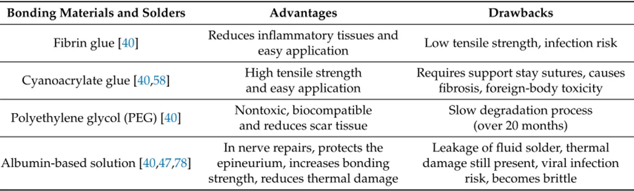
Overview on the evolution of laser welding of vascular and nervous tissues
14
0
0
Texto
Imagem
![Figure 1. Schematics of laser welding of a nervous tissue [40].](https://thumb-eu.123doks.com/thumbv2/123dok_br/15489876.1041324/3.892.352.539.656.810/figure-schematics-laser-welding-nervous-tissue.webp)
+5

![Figure 6. Schematics of the laser-assisted vessel soldering [51].](https://thumb-eu.123doks.com/thumbv2/123dok_br/15489876.1041324/9.892.333.565.662.861/figure-schematics-laser-assisted-vessel-soldering.webp)
Documentos relacionados