Received 25 June, 2014 Accepted 21 Aug., 2014 (006422)
1Center of Biological and Health Sciences, Univates, Lajeado, RS, Brazil, e-mail: ivanbustamante@univates.br 2Center of Science and Technology, Univates, Lajeado, RS, Brazil
*Corresponding author
Molecular screening of bovine raw milk for the presence of Shiga
toxin-producing
Escherichia coli
(STEC) on dairy farms
Tatiane VENDRAMIN1, Débora Mara KICH1, Rachel Dias MOLINA1, Claucia Fernanda Volken de SOUZA2,
Rosangela Uhrig SALVATORI1, Adriane POZZOBON1, Ivan Cunha BUSTAMANTE-FILHO1*
1 Introduction
Escherichia coli is a Gram-negative, nonsporulating, rod shaped, flagellated, and facultative anaerobic bacteria belonging to the Enterobacteriaceae Family. Based on its virulence, this species is classified as enterotoxigenic E. coli (ETEC), enteropathogenic E. coli (EPEC), enterohemorrhagic E. coli
(EHEC), attaching and effacing E. coli (AEEC), and Shiga toxin-producing E. coli (STEC) (Bavaro, 2012).
The Shiga toxin-producing E. coli are an important emerging group of foodborne pathogens. These strains harbor the phage-encoded genes stx1 and stx2, coding for cytotoxins that cause severe tissue damage, specially STX2 that cause a variety of human illnesses ranging from diarrhea to hemorrhagic colitis (HC), thrombotic thrombocytopenia purpurea (TTP), and hemolytic uremic syndrome (HUS) with fatal consequences (Gyles, 2007; Petruzziello-Pellegrini & Marsden, 2012; Walker et al., 2012). In Brazil, in a prospective clinical, observational, multicenter, controlled and non-randomized study, conducted between 2001 and 2005, 92.3% of patients with HUS were infected by STEC. These infected patients’ ages ranged from 8 months to 6 years and 3 months, with an average age of 2 years and 5 months. However, almost half of these patients (46%) were less than 2 years old. These results show a higher risk for pediatric patients, also common victims of foodborne illnesses(Souza et al., 2011).
Ruminants, especially bovines, are the principal reservoir of STEC, and human contamination is often associated with consumption ground beef and direct contact with the animals or their environment (Savoye et al., 2011) STEC strains are
highly pathogenic to human in low infectious doses, causing food-borne diseases through consumption of contaminated water or food(Dweik et al., 2012). Bovine milk and other dairy products such as yogurt and cheese have been associated with diseases caused by STEC (Chapman et al., 1993; Prandel et al., 2008; Martin & Beutin, 2011). Outbreaks of milk-borne diseases associated with STEC, including pasteurized dairy products, have been reported worldwide emphasizing the importance of STEC control for public health(Morgan et al., 1993; Upton & Coia, 1994; Seghal et al., 2008).
The identification of the presence of microorganisms in the food is the gold standard method to determine food poisoning sources. In most clinical laboratories, identification procedures are based on microbiological culture of milk and biochemical tests of the isolated bacteria. There are disadvantages associated with microbiological culture, such as: a milk culture may yield no bacteria from truly contaminated milk due to the presence of very low numbers of bacteria in the samples, and a negative culture may also be due to the presence of residual therapeutic antibiotics that may inhibit bacterial growth in vitro, more likely, sub lethally-injured or stressed bacteria may not grow directly on selective media, unless allowed to recover on selectively first(Riffon et al., 2001). The detection of STEC strains are labor-intensive, and the total time required for strain characterization is typically 72 h, involving methods such as cytotoxicity assays and Sorbitol MacConkey Agar platting. On the other hand, molecular methods are sensitive, specific, and rapid approaches for detection and characterization of microbiological contaminants in food.
Abstract
Milkborne transmission of Shiga toxin- producing Escherichia coli (STEC) has raised considerable concern due to recent outbreaks worldwide and poses a threat to public health. The aim of this study was to develop a sensitive and specific multiplex PCR assay to detect the presence of STEC in bovine raw milk. To identify E. coli (ATCC 25922) contamination, the gene uspA
was used, and PCR sensitivity and specificity were accessed by testing diluted samples ranging from 2 to 2.0 × 10⁶ CFU/mL. To
detect STEC, the stx1 and stx2 genes were selected as targets. After reaction standardization, the multiplex assay was tested in raw milk collected from 101 cows on dairy farms. PCR assay for E. coli detection had a specificity of 100% and sensitivity of 79% (P<0.0001), with a lower detection limit of 2 CFU/mL. Multiplex PCR assay had 100% sensitivity for E. coli positive raw milk samples, and 31.1% were contaminated with STEC, 28.3% of stx2, and 1.9% of stx1. The multiplex PCR assay described in the present study can be employed to identify and screen E. coli harboring stx1 and stx2 genes in raw milk on dairy farms and in industries.
Keywords: shiga-toxin; cattle; multiplex PCR; milk.
OI:
µL of phenol-chloroform-isoamyl alcohol (25:24:1) (Sigma-Aldrich, St Louis, MO, USA) and centrifuged (12000 x g, 5 min). A total volume of 26.5 µL of 2M Sodium acetate (Sigma-Aldrich, St Louis, MO, USA) was added to the supernatant, followed by addition of 400 µL of absolute ethanol (Merck, Darmstadt, GER) and overnight incubation at 4°C. The samples were centrifuged (12000 x g, 20 min) and the pellets with DNA were ressuspended in TE buffer (10 mM Tris-HCL, 1 mM EDTA, pH 8,0), and stored at –20°C until use. The optical density ratio (260/280 nm) of DNA preparations was considered suitable of downstream applications when greater than 1.6.
2.4 Molecular characterization of STEC in milk samples
A multiplex PCR was designed to determine the occurrence of E. coli harboring stx1 and/or stx2 virulence genes in the raw milk samples. Milk DNA samples were used as templates for PCR assays. All primers were from Invitrogen (Carlsbad, CA, USA), and the molecular biology reagents were from Biotools (Madrid, Spain). The names of specific primer sequences, predicted fragment size, and reaction concentrations are presented in Table 1. The uspAgene encoding the universal stress protein was used to detect E. coli DNA (Chen & Griffiths, 1998). To determine the presence of STEC, primers based on stx1 (Genebank accession number AB083043.1) and stx2
(Genebank accession number AB030484.1) genes were designed for this study. Multiplex PCR conditions were 1.75 mM MgCl2, 20 mM Tris-HCl, 50mM KCL (pH 8.4), 500 µM dNTPs, 1.5 U
Taq DNA polymerase, and 100 ng of template DNA; 2% DMSO (Sigma-Aldrich, St Louis, MO, USA) was added to a total volume of 25 µL. Amplification reactions were carried out using Techne® TC-512 termocycler (Techne® Barloworld Scientific, Stone, Staffordshire, UK) as follows: 5 min at 94°C, 35 cycles each consisting of 2 min at 94°C, 1 min at 58°C, 1 min at 72°C, and a final extension step of 10 min at 72°C. Characterization of presence of E. coli producer of Shiga I and/or Shiga II toxins were performed by agarose (Invitrogen, Carlsbad, CA, USA), gel electrophoresis stained with ethidium bromide (Sigma-Aldrich, St Louis, MO, USA). E. coli O157:H7 Edl 933 strain (kindly provided by Dr. Caroline de Nardi from Instituto Federal de São Paulo, Brazil) was used as positive control; PCR without template DNA and/or DNA polymerase was performed to be used as negative control.
To determine PCR sensitivity and specificity, sterile whole milk was artificially contaminated with 2 to 2.0 × 10⁶
CFU/mL of E. coli (ATCC 25922) culture in stationary phase grown in brain heart infusion (BHI) broth (Sigma-Aldrich, St Louis, MO, USA) for 18-24 h at 36°C (Guido, 2003). The The development of PCR-based methods for the detection
of pathogens or virulence factors (Shahi et al., 2013; Branquinho & Ferreira, 2012; Gandra et al., 2011) can be used for the initial screening of the presence of microorganisms in samples of milk and dairy products. Furthermore, the present study, through direct isolation of bacterial DNA in milk samples, aimed to develop the multiplex PCR to assess the prevalence of E. coli and STEC in samples of fresh milk.
2 Materials and methods
2.1 Collection of raw milk samples
This study was carried out on three dairy farms located in Rio Grande do Sul State, Brazil. For the microbiological and molecular biology analyses, 15 mL of raw milk samples of 101 cows were collected. The milk was drawn by hand directly into sterile test tubes and transported to the laboratory in a cooler containing ice.
2.2 Microbiological analysis
To determine contamination of E. coli in milk, Brazilian federal normative (IN62) of the Ministry for Agriculture, Livestock and Supply was followed and used as the standard method (Brasil, 2003). Milk samples were cultured in Violet Red Bile agar (Oxoid Cambridge, UK) at 36°C for 24 h. From the samples that tested positive for E. coli, three colonies were transferred to Brilliant Green medium (Acumedia Lansing, USA) and incubated at 36°C for 48 h. One loopful of cultures presenting gas production was streaked onto a EC broth (Acumedia Lansing, USA) and incubated at 36°C for 48 h. The samples presenting gas formation were cultured at 36°C in Eosin Methylene Blue Agar (EMB, Oxoid Cambridge, UK.). After 24 h,
E. coli colonies were counted with standard technique.
2.3 Preparation of template DNA
For DNA extraction, 200 µL of raw milk sample were used. Briefly, 20 µL of Tween-20 were added to the samples, followed by centrifugation (12000 x g, 15 min). Pellets were suspended in 60 µL extraction buffer (100 mM Tris-HCl, 100 mM EDTA, 250 mM NaCl, pH 8.0) (Invitrogen, Carlsbad, CA, USA), 30 µL 10% SDS (Sigma-Aldrich, St Louis, MO, USA) , 15 µL proteinase K (Ambion®, Austin, TX, USA) (20 mg/mL), and 195 µL ultrapure water, and they were incubated for 1 h at 37°C. Subsequently, 100 µL of buffered phenol (ANRESCO, Solon, OH, USA) were added, and the samples were centrifuged (12000 x g, 5 min); the supernatant was collected and mixed with 100
Table 1. Primer sequences for detection of STEC in raw milk samples.
Primers Sequence (5’- 3’) Product size Target gene Concentration
uspAF CCGATACGCTGCCAATCAGT
884 bp uspA 0.5 µM
uspAR ACGCAGACCGTAGGCCAGAT
stx1F GATTTATCTGCATCCCCGTACG
346 bp stx1 0.5 µM
stx1R CTTACGCTTCAGGCACATACAG
stx2F GGTTAAGCCACTGCCTTTAACTTC
461 bp stx2 1.5 µM
contamination less than 2.0 × 10⁵ CFU/mL since the limit for
100% sensitivity was 2.0 × 10⁶ CFU/mL.
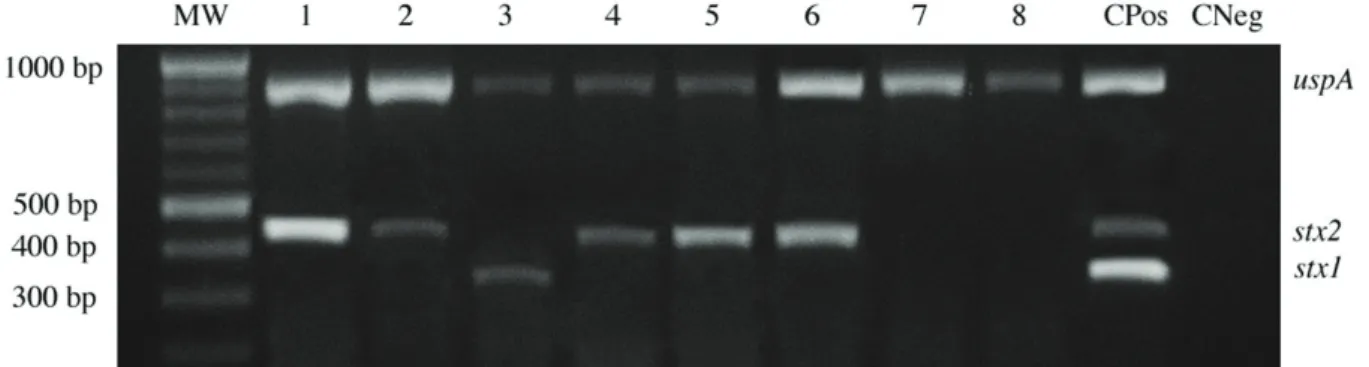
The molecular characterization of STEC was accessed by means of multiplex PCR. The combination of the three sets of primers allowed the detection of the genes of interest as they produce different sized PCR specific products. Hence, after thorough trials (data not shown), the primer pairs to detect the
stx1 and stx2 genes were found to be highly specific (Figure 2). The multiplex assay detected that among all E. coli positive samples, 31.1% were contaminated with STEC. The stx2
fragment was found in 15 samples (28.3%), and stx1 in only one sample (1.9%), which was not stx2 positive. Environmental contaminations and poor herd and milking managements are important causes of reduction of milk quality. The high frequency of stx2 in faecal samples collected from buffalo, cows, and goats described elsewhere (Islam et al., 2008; Stephan et al., 2008; Bandyopadhyay et al., 2011, 2012) might explain the prevalence of stx2 positive STEC in raw milk. In humans, epidemiologic data suggest that E. coli O157 strains that express stx2 are more important than stx1 in the development of HUS, and that strains that express stx2 alone are more likely to be associated with the progression to HUS than strains that produce both stx1 and stx2 (Griffin, 1995)
In this study, the presence of STEC harboring the genes
stx1 and stx2 in raw milk sample was detected. STEC harboring
stx1 and stx2 genes were reported to be typical cattle colonizers (Brett et al., 2003; Vu-Khac & Cornick, 2008). Some authors have recently investigated 593 foodborne STEC strains for their serotypes and for nine virulence genes (stx1, stx1c, stx1d, stx2,
stx2b, stx2e, stx2g, E-hlyand eae), and they observed a significant association of stx1and stx2 genes with bovine meat and milk products. These authors compared the properties of foodborne STEC with published data on faecal STEC from food producing animals, and found that virulence profiles and serotypes of STEC from food showed remarkable similarities to those of faecal STEC that were from the same animal species. Based on these results, it has been pointed out that food-producing animals represent the most important source for the entry of STEC in the food chain (Martin & Beutin, 2011).
It is noteworthy to mention the findings of Kumar et al. (2013), who showed the presence of pathogenic E. coli
O157:H7 in pasteurized milk samples. These authors described a higher detection signal after enrichment, suggesting the presence of viable cells after pasteurization or post-pasteurization contamination. Therefore, fermented dairy products manufactured using raw milk contaminated with
E. coli O157:H7, STEC, or other pathogenic strain can pose a threat to human health, demanding special attention of health and food agencies worldwide. Studies with emphasis quantification was performed at this stage by plating on the
depth of the microorganism suspensions of PCA (Plate Count Agar), followed by incubation at 36°C for 48 h. Dilution curves were repeated 8 times, and each point was analyzed in triplicate. DNA extraction and PCR were performed as described above.
2.5 Statistical analyses
All statistical analyses were performed using the Prism 5 (Graph Pad Software). The prevalence and frequency of contamination and sensitivity and specificity determination were accessed with exact 95% confidence interval (CI), Kruskal-Wallis test, Fisher’s exact test and Chi-square; p < 0.05 was considered statistically significant.
3 Results and discussion
To validate the use of PCR to detect Escherichia coli
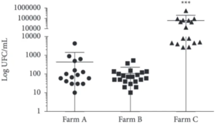
contamination in raw milk, the sensitivity and specificity for the primers designed for the uspA gene were checked. The method was compared to microbiological analysis, which showed a total prevalence of contamination of raw milk by E. coli of 53.5% (against to 51.2% determined by PCR), with no difference (P = 0.3623) between the farms evaluated. However, the contamination level on farm C (60,508.8 ± 31,653.8) was higher (P < 0.0001) than that on farms A and B, 442.8 ± 261.27 and 106.6 ± 27.3, respectively (Figure 1).
100% specificity and 79% sensitivity were observed when PCR was performed with DNA obtained from milk artificially contaminated with 2 CFU/mL (P < 0.0001) (Table 2). When PCR was compared to microbiological analysis, 100% specificity (CI: 0.9328 to 1.000) was observed (P < 0.001); however, lower sensitivity (52.48%; CI: 0.4230 to 0.6251) was found. These results may be related to occurrence of samples with
Table 2. Sensitivity and specificity of PCR assay for contamination of raw milk by E. coli. Each concentration was tested in 24 samples.
E. coli concentration in milk samples (CFU/mL)
2.0 × 10⁶ 2.0 × 10⁵ 2.0 × 10⁴ 2.0 × 10³ 2.0 × 10² 2.0 × 101 2.0
Sensitivity (%) 100 83.3 95.8 83.3 95.8 83.3 79.1
Specificity (%) 100 100 100 100 100 100 100
P < 0.0001 for all values. Fisher Exact test.
14(4), 317-323. PMid:22610457. http://dx.doi.org/10.1007/s11894-012-0264-6
Branquinho, M. R., & Ferreira, R. T. B., & Cardarelli-Leite, P. (2012). Use of real-time PCR to evaluate two DNA extraction methods from
food. Food Science and Technology, 32(1), 112-118.
Brasil. Ministério da Agricultura, Pecuária e Abastecimento. (2003). Oficializa os métodos analíticos oficiais para análises microbiológicas
para controle de produtos de origem animal e água (Instrução
Normativa nº 62, de 26 de agosto 2003). Diário Oficial da República Federativa do Brasil.
Brett, K. N., Ramachandran, V., Hornitzky M. A., Bettelheim K. A., Walker M. J., & Djordjevic S. P. (2003). Stx1c is the most common
Shiga toxin 1 subtype among Shiga toxin-producing Escherichia
coli isolates from sheep but not among isolates from cattle.
Journal of Clinical Microbiology, 41(3), 926-936. PMid:12624011
PMCid:PMC150265. http://dx.doi.org/10.1128/JCM.41.3.926-936.2003
Chapman, P. A., Wright, D. J., & Higgins R. (1993). Untreated milk
as source of verotoxigenic Escherichia coli O157:H7. Veterinary
Record, 133(7), 171-172. PMid:8236710. http://dx.doi.org/10.1136/
vr.133.7.171
Chen, J., & Griffiths, M. W. (1998). PCR differentiation of Escherichia
coli other Gram negative bacteria using primers derived from the
nucleotide sequences flanking the gene encoding the universal
stress protein. Letters in Applied Microbiology, 27(6), 369-371. http://
dx.doi.org/10.1046/j.1472-765X.1998.00445.x
Gandra, E. A., Fernandez, M. A., Silva, J. A., & Silva, W. P. (2011). Standardization of a multiplex PCR for the identification of
coagulase-posivite Staphylococcus. Ciência e Tecnologia de
Alimentos, 31(4), 946-949.
http://dx.doi.org/10.1590/S0101-20612011000400019
Dweik, M., Stringer, R. C., Dastider, S. G., Wu, Y., Almasri, M., & Barizuddin, S. (2012). Specific and targeted detection of viable
Escherichia coli O157:H7 using a sensitive and reusable impedance
biosensor with dose and time response studies. Talanta, 94, 84-89.
PMid:22608418. http://dx.doi.org/10.1016/j.talanta.2012.02.056
Guido, M. C. (2003). Detecção de DNA deBrucella abortuspela PCR
em leite bubalinoexperimentalmente contaminado pela amostra
1119-3 (Tese de doutorado). Universidade de São Paulo, São Paulo.
Griffin, P. M. (1995). Escherichia coli O157:H7 and other
enterohemorrhagic Escherichia coli. In M. J. Blaser, P. D. Smith, J.
I. Rovin, H. B. Greenberg & R. L. Guerrant (Eds.), Infections of the
gastrointestinal tract (pp. 739-761). New York: Raven Press.
on detection of pathogens and their virulence factors in food sources may be useful to prevent infection and development of associated diseases.
Our findings on raw milk contamination are meaningful and should be considered since even one Shiga toxin-producing
E. coli in a food sample may lead to gastrointestinal or urogenital disorder due to their multiplication in the body or the food itself during storage in poor conditions (Gyles, 2007). Therefore, strict hygiene and management practices for dairy herd and milk processing and storage must be adopted to avoid unwanted illness due contaminated milk and milk products consumption.
4 Conclusion
This report presents a quick, sensitive, and specific method for molecular characterization of STEC. In addition, the screening for STEC in three dairy herds showed a significant prevalence of Shiga toxin-producing E. coli contamination in raw milk.
Acknowledgements
The authors are grateful for the financial support provided by FUVATES.
References
Bandyopadhyay, S., Lodh C., Rahaman, H., Bhattacharya, D., Bera, A. K., Ahmed, F. A., Mahanti A., Samanta, I., Mondal, D. K., Bandyopadhyay, S., Sarkar, S., Dutta, T. K., Maity, S., Paul, V., Ghosh, M. K., Sarkar, M., & Baruah K. K. (2012). Characterization of shiga
toxin producing (STEC) and enteropathogenic Escherichia coli
(EPEC) in raw yak (Poephagusgrunniens) milk and milk products.
Research in Veterinary Science, 93(2), 604-610. PMid:22226073.
http://dx.doi.org/10.1016/j.rvsc.2011.12.011
Bandyopadhyay, S., Mahanti, A., Samanta, I., Dutta, T. K., Ghosh, M. K., Bera, A. K., Bandyopadhyay, S., & Bhattacharya, D. (2011).
Enterotoxigenic Escherichia coli (ETEC) from diarrhoeic lambs of
Arunachal Pradesh, India. Tropical Animal Health and Production,
43(3), 705-710. PMid:21104315.
http://dx.doi.org/10.1007/s11250-010-9757-1
Bavaro, M. F. (2012). E. coli O157:H7 and other toxigenic strains: the
curse of global food distribution. Current Gastroenterology Reports,
Figure 2. Specificity of multiplex PCR for detection of STEC in raw milk samples. Fragment identifications are presented on the right – uspA
(884 bp), stx1 (346 bp) and stx2 (461 bp). Lanes: MW: 100 bp ladder; 1 to 8: milk samples; CPos: positive control (E. coli harboring stx1 and stx2
Savoye, F., Feng, P., Rozand, C., Bouvier, M., Gleizal, A., & Thevenot, D. (2011). Comparative evaluation of a phage protein ligand assay with real-time PCR and a reference method for the detection of
Escherichia coli O157:H7 in raw ground beef and trimmings. Journal
of Food Protection, 74(1), 6-12. PMid:21219756. http://dx.doi.
org/10.4315/0362-028X.JFP-10-271
Seghal, R., Kumar, Y., & Kumar, S. (2008). Prevalence and geographical
distribution of Escherichia coli O157 in India a 10 year survey.
Transactions of the Royal Society of Tropical Medicine and Hygiene,
102(4), 380-383. PMid:18321544. http://dx.doi.org/10.1016/j.
trstmh.2008.01.015
Shahi, S. K., Singhm V. K., & Kumar, A. (2013). Detection of Escherichia coli and associated β-lactamases genes from diabetic foot ulcers by multiplex PCR and molecular modeling and docking of SHV-1, TEM-1, and OXA-1 β-lactamases with
clindamycin and piperacillin-tazobactam. PLoS One, 8(7), e68234.
PMid:23861873 PMCid:PMC3701671. http://dx.doi.org/10.1371/ journal.pone.0068234
Souza, R. L., Carvalhaes J. T. A., Nishimura, L. S., Andrade, M. C., & Guth B. E. C. (2011). Hemolytic uremic syndrome in pediatric
intensive care units in São Paulo, Brazil. Open Microbiology Journal,
5, 76-82. PMid:21804902 PMCid:PMC3143539. http://dx.doi.
org/10.2174/1874285801105010076
Stephan, R., Schumacher, S., Corti, S., Krause, G., Danuser, J., & Beutin, L. (2008). Prevalence and characteristics of shiga toxin-producing
Escherichia coli in swiss raw milk cheeses collected at producer level.
Journal ofDairy Science, 91(7), 2561-2565. PMid:18565913. http://
dx.doi.org/10.3168/jds.2008-1055
Upton, P., & Coia, J. E. (1994). Prevalence and geographical distribution of Escherichia coli O157 in India a 10 year survey. Lancet, 344(8928), 1015. http://dx.doi.org/10.1016/S0140-6736(94)91670-5
Vu-Khac, H., & Cornick, N. A. (2008). Prevalence and genetic profiles
of Shiga toxin-producing Escherichia coli strains isolated from
buffaloes, cattle, and goats in central. Veterinary Microbiology,
126(4), 356-363. PMid:17716835. http://dx.doi.org/10.1016/j.
vetmic.2007.07.023
Walker, C. L., Applegate, J. A., & Black, R. E. (2012).
Haemolytic-uraemic syndrome as a sequela of diarrhoeal disease. Journal of
Health, Population and Nutrition, 30(3), 257-261. http://dx.doi.
org/10.3329/jhpn.v30i3.12288
Gyles, C. L. (2007). Shigatoxin-producing Escherichia coli: an overview.
Journal of Animal Science, 85(13), 45-62. PMid:17085726. http://
dx.doi.org/10.2527/jas.2006-508
Islam, M. A., Abdus , S. M., Boer, E., Rijkelt, R. B., Marcel, H. Z., Kaisar , A. T., & Heuvelink, A. E. (2008). Prevalence and genetic
characterization of shiga toxin-producing Escherichia coli
isolates from slaughtered animals in Bangladesh. Applied and
Environmental Microbiology, 74(17), 5414-5421. PMid:18641151
PMCid:PMC2546655. http://dx.doi.org/10.1128/AEM.00854-08 Kumar, A., Grover S., & Batish V. K. (2013). Application of multiplex
PCR assay based on uidR and fliCH7 genes for detection of
Escherichia coli O157:H7 in milk. Journal of General andApplied
Microbiology, 59(1), 11-19. PMid:23518514. http://dx.doi.
org/10.2323/jgam.59.011
Martin, A., & Beutin, L. (2011). Characteristics of shiga
toxin-producing Escherichia coli from meat and milk products of different
origins and association with food producing animals as main
contamination sources. InternationalJournal ofFood Microbiology,
146(1), 99-104. PMid:21371769. http://dx.doi.org/10.1016/j.
ijfoodmicro.2011.01.041
Morgan, D., Newman, C. P., Hutchinson, D. N., Walkar, A. M., Rowe, B., & Majid, F. (1993). Verotoxin producing Escherichia coli O157:H7
infections associated with the consumption of yoghurt. Epidemiology
and Infection, 111(2), 181-187. PMid:8405146 PMCid:PMC2271388.
http://dx.doi.org/10.1017/S0950268800056880
Petruzziello-Pellegrini, T. N., & Marsden P. A. (2012). Shiga toxin-associated hemolytic uremic syndrome: advances in pathogenesis
and therapeutics. Current Opinion in Nephrology and Hypertension,
21(4), 433-440. PMid:22660553. http://dx.doi.org/10.1097/
MNH.0b013e328354a62e
Prandel, N., Bertin Y., Martin C., & Livrelli V. (2008). Molecular analysis of shiga toxin-producing Escherichia coli strains isolated from Hemolytic-Uremic Syndrome patients and dairy samples in
France. Applied and Environmental Microbiology, 74(7), 2118-2128.
PMid:18245246 PMCid:PMC2292610. http://dx.doi.org/10.1128/ AEM.02688-07
Riffon, R., Sayasith, K., Lhalil, H., Dubreuil, P., Drolet, M., & Lagace, J. (2001). Development of a rapid and sensitive test for identification
of major pathogens in bovine mastitis by PCR. Journal of Clinical
Microbiology, 39(7), 2584-2589. PMid:11427573 PMCid:PMC88189.