Vol.55, n. 5: pp. 653-660, September-October 2012
ISSN 1516-8913 Printed in Brazil BRAZILIAN ARCHIVES OF
BIOLOGY AND TECHNOLOGY
A N I N T E R N A T I O N A L J O U R N A L
Endophytic Fungi Occurring in
Ipomoea carnea
Tissues and
their Antimicrobial Potentials
Kumanand Tayung
1*, Meenakshi Sarkar
2and Paran Baruah
31, 2Department of Botany, North Orissa University, Takatpur, Baripada, India. 3Divison of Plant Science, North East
Institute of Science and Technology, Jorhat, India
ABSTRACT
The aim of this work was to study the endophytic fungi associated with the tissues of Ipomoea carnea, a common invasive plant of India. A total of 69 isolates belonging to ten taxa comprising 1.45% Zygomycetes, 10.14% Coelomycetes, 62.32% Hypomycetes, 18.84% sterile mycelia and 7.25% unidentified species were obtained. Species of Curvularia, Aspergillus, Fusarium, Colletotrichum and sterile fungus were isolated as dominant endophytes. Colonization frequency of Curvularia (7.25%) was highest which was isolated from all the tissues. The samples collected during the monsoon harbored more endophytes and showed higher species richness than the samples obtained in summer season. Of the total isolates, 15 isolates (21.74%) displayed antimicrobial activity, inhibiting at least one of the test microorganisms that comprised of pathogenic bacteria (Staphylococcus aureus, Bacillus subtilis, Escherichia coli, Salmonella typhi, Pseudomonas fluorescens, Shigella dysentriae) and fungi (Trichophyton rubrum, Aspergillus fumigatus, Trichophyton sp). The results provided promising baseline information on the endophytic fungal diversity associated with I. carnea tissues and their potential exploitation as antimicrobial agents.
Key words: Ipomoea carnea, Endophytic fungi, Curvularia, antimicrobial activity
*Author for correspondence: [email protected]
INTRODUCTION
Fungi that colonized inner plants tissues without causing any disease symptoms are called endophytic fungi. Over the years, several plants have been investigated for fungal endophytes, both from the temperate (Bills 1996) and tropical plants host (Suryanarayan et al. 2002, 2003). Studies have shown that fungal endophytes are neither incident residents nor merely latent pathogens of plant hosts but also exert several beneficial effect. Their functional roles may be attributed to protect the host from the insect pests (Azevedo et al. 2000), fungal pathogens (Arnold et al. 2003; Dingle and McGee 2003), or increase the host fitness in harsh environments (Redman et al. 2002). Besides, endophytic fungi have been
recognized as repository of biologically active substances (Tan and Zou 2001).
and their derivatives with more bioactivity than those of their respective hosts. This gives paucity of work on endophytes of other plant species. Invasive plants have unique survival features and are less competitive and abundant in their native ranges than in their invaded ranges (Broennimann et al. 2007). It is suggested that associative endophytes of invasive plants could contribute to their greater competitiveness and may also produce novel allelochemicals that might have inhibitory effects (Shipunov et al. 2008). Many invasive species have the natural capability to adapt in wide range of habitats. It is believed that such adaptability might be due to some associated endophytes because recent study indicates that endophytes influence the protection and growth of an invasive plant (Newcombe et al. 2009). However, there is meager information on fungal
endophytes of invasive plant species. Ipomoea
carnea, a native of South America is common invasive plant in all the states of India. The plant has unique survival features, also used as folk medicine for the treatment of skin diseases and leucoderma (Agarwal and Uppadhay 1979). The aim of the this work, therefore, was to study the endophytic fungi communities associated with
Ipomoea carnea tissues and their antimicrobial potentials.
MATERIALS AND METHODS
Collection of plant materials
Ipomoea carnea growing in and around North Orissa University campus (21o93’ N and 86o76’ E), Orissa, India was identified and two sites were selected. Samplings were done for a period of six months, representing two season, summer (April-June) and monsoon (July-September). The samples were collected from five individual healthy and symptomless plants from each site. The collection of leaves, stems and seeds were made from each selected plant 122 cm above the ground with a help of ethanol-disinfected knife and placed in sterile poly ethylene bags separately. The samples were brought to the laboratory, washed thoroughly in running water and shade dried under fan before isolation procedure was undertaken.
Isolation of endophytes
For the isolation of endophytes, samples (stems, leaves and seeds) were surface sterilized by immersing them sequentially in 70% ethanol for 3min and 0.5% sodium hypochloride (NaOCl) for 1min and rinsed thoroughly with sterile distilled water (Bills, 1996). The excess water was dried under laminar airflow chamber. For stem samples, outer barks were removed and inner tissues of 1 x 0.5 cm (approx.) segments were carefully dissected out with a sterile scalpel. The seeds were dissected in two equal halves after removing the seeds coat and leaves were cut into 0.5 cm2 sizes (approx.) without midrib under aseptic conditions. In each Petri dish, 5-6 segments were placed on
Potato Dextrose Agar medium (PDA),
supplemented with penicillin-G (60mg L-1) and streptomycin sulphate (80 mg L -1) to inhibit the bacterial contaminant. For each sample, 200 segments were plated and the plates were incubated at room temperature (30° C approx.) for 4-6 weeks in dark. The plated segments were observed once a day for the growth of endophytic fungi. Hyphal tips growing out the plated segments were immediately transferred into PDA slant, purified and maintained at 4°C. The efficiency of surface sterilization procedure was tested as per the method described by Schultz et al. (1998). The fungal isolates were identified based on their morphological and reproductive characters using the standard identification manuals (Barnett and Hunter 1998; Subramanian 1971). The fungal cultures that failed to sporulate were categorized as sterile mycelia. All the isolates were maintained in Potato dextrose agar slant.
Data analysis
The relative frequency of colonization (% CF) was calculated as the number of segments colonized by a specific fungus divided by the total number of segments plated X 100 and dominant endophytes were calculated as percentage colony frequency divided by the sum of percentage of colony frequency of all endophytes X 100.
Simpson’s index of Diversity was calculated using
the formula: 1-D
Σ n (n-1) Where D = ---
N (N-1)
N = the total number of organisms of all species
Shannon-Wiener diversity index was calculated using the following formula:
S
Hs = - (Pi) (ln Pi), where i =1
Hs – symbol for the diversity in a sample of S species or kinds
S – the number of species in the sample Pi – relative abundance of ith species or kinds measures, = ni/N
N – total number of individuals of all kinds ni – number of individuals of i
th
species ln – log to base 2
Fungal cultivation and metabolites extraction
The fungal endophytes were cultivated on Potato dextrose broth by placing the agar blocks of pure culture (3mm in diameter) of actively growing culture in 250ml Erlenmeyer flasks containing 100ml of the medium. The flasks were incubated in BOD shaking incubator for three weeks at 27±10C with periodic shaking at 150 rpm. The culture was filtered through sterile cheese cloth to remove the mycelial mats. The liquid broth was extracted with equal volume of ethyl acetate in a separating funnel by vigorous shaking for 10 min. The cell mass was separated and the from the filtrate, ethyl acetate was evaporated. The resultant
compound was dried with MgSO4 and
concentrated to yield the crude extract. The crude extract was then dissolved in Dimethyl sulphoxide (DMSO) for antimicrobial bioassay.
Determination of Antimicrobial activity
The antimicrobial activity of the crude metabolites was determined by the agar cup diffusion method against six bacteria (Escherichia coli, Bacillus subtilis, Staphylococcus aureus, Salmonella typhi, Pseudomonas fluorescens, Shigella dysentriae)
and three fungi (Trichophyton rubrum, Aspergillus
fumigatus, Trichophyton sp.) as test pathogens. These test microbes were obtained from the Institute of Microbial Technology (IMTECH), Chandigarh, India. Nutrient agar plates were inoculated with overnight culture of each bacterial suspension. Similarly, for the fungal pathogens, Sabouraud's agar plates were inoculated with each fungal suspension. The plates with the inoculated organisms were evenly spread out with sterile
cotton swabs. Agar cups were prepared by scooping out the medium with a sterile cork borer (7mm in diameter). The cups were then filled with 100µL of the crude extract dissolved in DMSO and incubated at 35±1°C for 24 h for bacterial and 48 h for fungal pathogens. The plates were observed for zone of inhibition and compared with the control (i.e., a cup filled with DMSO solution only). Minimum inhibitory concentration (MIC) was determined only for the bacterial pathogens (B. subtilis and E. coli) by microbroth dilution assay on 96-wells microplates according to the National Committee for Clinical Laboratory Standard (NCCLS, 2003). All the determinations were conducted in duplicate.
Estimation of crude metabolites
The λ-max in ethyl acetate of the crude
metabolites of endophytic fungi was determined by using a UV spectrophotometer (SPECORD 210). A standard curve of the bioactive metabolites was calibrated and the concentrations of the bioactive metabolites produced were determined by comparing the optical density at 230 nm.
RESULTS
A total of 69 isolates belonging to ten taxa including three isolates of sterile mycelia and two unidentified species were obtained from the leaves, barks and seeds of Ipomea carnea (Table 1). Of the total species isolated, 1.45% was Zygomycetes, 10.14% Coelomycetes, 62.32% Hypomycetes, 18.84% sterile mycelia and 7.25% unidentified species. Among the endophytes,
species of Curvularia, Aspergillus, Fusarium,
Table 1 - Occurrence of endophytic fungi in different tissues of Ipomea carnea in two different seasons.
Endophytic fungi
Colonization frequency (% CF)
Total
Frequency of dominant
endophytes
Summer Monsoon
Stem Total Seed Stem Leaf Seed
Acremonium sp. -- -- -- -- 1.0 0.5 03 4.35
Aspergillus sp. -- 1.5 -- -- 1.0 -- 05 7.25
Aspergillus flavus -- -- -- 1.0 -- -- 02 2.90
Aspergillus fumigatus -- -- -- 0.5 -- 1.0 03 4.35
Cladosporium sp. -- -- -- -- -- 0.5 01 1.45
Colletotrichum sp. 1 -- 2.0 -- -- -- -- 04 5.80
Colletotrichum sp. 2 -- -- 1.5 -- -- -- 03 4.35
Curvularia sp. 1 -- 3.5 -- -- -- 1.5 10 14.50
Curvularia sp. 2 1.5 -- -- -- 1.0 0.5 06 8.70
Fusarium sp. 1 -- -- 1.5 -- -- -- 03 4.35
Fusarium sp. 2 -- -- -- -- -- 1.0 02 2.90
Sterile fungus sp. 1 4.0 -- -- -- -- -- 08 11.60
Sterile fungus sp. 2 1.0 -- -- -- -- -- 02 2.90
Sterile fungus sp. 3 -- -- -- -- 1.0 0.5 03 4.35
Rhizopus sp. -- -- -- -- -- 0.5 01 1.45
Trichoderma sp. 1.0 -- -- -- -- -- 02 2.90
Trichoderma viridae -- -- -- 1.5 -- -- 03 4.35
Trichoderma harzianum -- -- -- 1.0 -- 0.5 03 4.35
Unidentified sp. 1 -- -- -- -- -- 2.0 04 5.80
Unidentified sp. 2 -- -- -- -- 0.5 -- 01 1.45
No. of isolates recovered 15 14 06 08 09 17 69 --
*Colonization frequency 7.5 7.0 3.0 4.0 4.5 8.5 -- --
Among the isolates, Acremonium, Cladosporium
and Rhizopus were represented by the single species and were isolated only during the monsoon season. More isolates of sterile mycelia were obtained from the barks during the summer season. Species of Fusarium were isolated only from the seeds in both the seasons. High
colonization of Collectotrichum was observed
from the leaves and bark samples collected during the summer. In general, more endophytes were isolated during the monsoon than in the summer
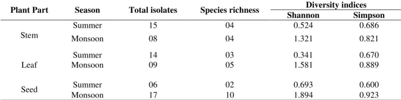
season. The Shannon-Wiener and Simpson diversity indices for fungi were highest in the seeds obtained during the monsoon season but were lower in the leaves sample collected during the summer (Table 2). It indicated higher number of isolates but low in species richness. However, monsoon isolates showed higher species richness, although the number of isolates was less than that of summer. In general, the samples obtained during the monsoon showed higher species richness than from those obtained in the summer.
Table 2 - Species richness and diversity of endophytic fungi in tissues of Ipomoea carnea.
Plant Part Season Total isolates Species richness Diversity indices Shannon Simpson
Stem
Summer 15 04 0.524 0.686
Monsoon 08 04 1.321 0.821
Leaf
Summer 14 03 0.341 0.670
Monsoon 09 05 1.581 0.889
Seed Summer 06 02 0.693 0.600
Of the total isolates, 15 strains (21.74%) showed
activity against the tested microorganisms
inhibiting either bacterial or fungal pathogens. Almost all the isolates exhibited antibacterial activity inhibiting at least one of the test bacterial pathogens but 46.6% of the isolates displayed both antibacterial and antifungal activity (Table 3). Out of the total isolates displaying antimicrobial activity, two isolates inhibited all the test
pathogens. Among the bacterial pathogens, E. coli
was found to be most susceptible to all the crude extracts. Almost all the fungal pathogens showed resistant to the crude extracts, except the
metabolites of Curvularia which exhibited
antifungal activity against all the tested fungal pathogens, i.e., Trichophyton rubrum, Aspergillus fumigatus and Trichophyton sp.
Table 3- Antimicrobial activity of metabolites of some endophytic fungi Zone of inhibition (mm) Endophytic fungi
Sa Bs Ec St Pf Sd Tr Af Tp
Aspergillus sp + -- + + -- + -- -- --
Collectotrichum sp. 1 + + + + + ++ -- -- --
Collectotrichum sp. 2 -- + ++ -- + + -- -- --
Cladosporium sp. + + + + -- + + -- --
Curvularia sp. 1 + + ++ + + + + + +
Curvularia sp.2 + ++ ++ ++ ++ ++ + + +
Mycelia sterilia sp. 1 + + + ++ + + -- -- --
Mycelia sterilia sp. 2 -- ++ ++ -- + + -- -- --
Mycelia sterilia sp.3 -- -- + -- + ++ + -- --
Fusarium sp. 1 ++ +++ +++ ++ ++ ++ -- -- --
Fusarium sp. 2 + + ++ ++ + ++ + -- --
Trichoderma sp -- -- + -- + + -- -- --
Trichoderma viridae + -- + -- -- + -- -- --
Unidentified sp. 1 ++ +++ +++ ++ +++ +++ ++ -- --
Unidentified sp. 2 -- + + -- + -- -- + --
Negative control -- -- -- -- -- -- -- -- --
Negative control: dimethly sulphoxide (the medium to dissolve crude metabolites) 100 µl
Sa- Staphylococcus aureus; Bs- Bacillus subtilis; Ec- Escherichia coli; St- Salmonella typhi ; Pf- Pseudomonas fluorescens;
Sd- Shigella dysentriae; Tr- Trichophyton rubrum ; Af- Aspergillus fumigatus; Tp- Trichophyton sp.
--: no antimicrobial activity; +: the inhibition zone is less than 10 mm;
++: the inhibition zone is from 10 mm to 20 mm; +++: the inhibition zone is above 20 mm
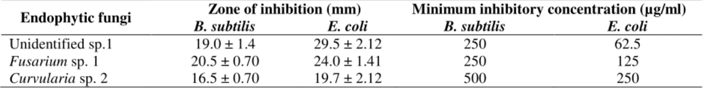
Considerable antimicrobial activity was observed in three endophytic fungi, Curvularia sp. 2,
Fusarium sp. 1 and an unidentified strain. The metabolites of these three endophytic fungi were studied for their minimum inhibition concentration (MIC) considering one Gram positive and Gram negative bacteria, B. subtilis and E. coli, respectively. The crude extract of Curvularia sp. 2
showed highest MIC value of 500µg ml-1 against
B. subtilis while that of the unidentified strain showed lowest MIC of 62.5µg ml-1 against E. coli. All the three crude extracts exhibited lower MIC values against E. coli indicating higher efficacy of these metabolites in inhibiting this test bacterium. The zone of inhibition and MIC values are presented in Table 4.
Table 4- Inhibition zone and MIC values of some potent endophytic fungi
Endophytic fungi Zone of inhibition (mm) Minimum inhibitory concentration (µg/ml)
B. subtilis E. coli B. subtilis E. coli
Unidentified sp.1 19.0 ± 1.4 29.5 ± 2.12 250 62.5
Fusarium sp. 1 20.5 ± 0.70 24.0 ± 1.41 250 125
Curvularia sp. 2 16.5 ± 0.70 19.7 ± 2.12 500 250
The crude metabolites of these fungi were also determined for their λ-max by calibrating the concentration of the metabolites dissolved in ethyl acetate using UV spectrophotometer at 230 nm. Three different standard curves were obtained corresponding to their optical density. The λ-max (peak wavelength) were 1.346, 1.503 and1.093 for
Curvularia sp. 2, Fusarium sp. 1 and an unidentified strain, respectively.
DISCUSSION
Endophytic fungi have been found associated with every plant species investigated so far from tropical and temperate hosts, yet they are poorly investigated because of their cryptic and ephemeral nature (Rodrigues and Petrini 1997; Strobel 2002). There are few reports on the endophytic fungi of invasive plant species, despite their abundance in certain invasive hosts (Shipunov et al. 2008). In the present study, rich
endophytic fungal diversity was obtained
comprising Zygomycetes, Coelomycetes,
Hypomycetes, sterile mycelia and unidentified genera from the tissues of Ipomoea carnea, a common invasive species. Among the endophytes,
class Hyphomycetes was dominant. Such
dominance of Hyphomycetes as endophytes has also been reported from several plants such as
Azadirachta indica and Terminalia indica
(Mahesh et al. 2004; Tejesvi et al. 2005), indicating their ubiquity among the plant kingdom. Environmental conditions may also play an important role in assemblages and diversity of endophytic fungi. It is generally believed that plants growing in lush tropical rainforests, where competition for light and nutrients might be severe, could be most likely to host the greatest number of endophytes than the temperate parts of the world. The dominant endophytic isolates were species of Curvularia, Aspergillus, Fusarium and
Colletotrichum. These fungi are also found commonly as the plant pathogens and they might have evolved to endophytic lifestyle due to loss of virulence (Freeman and Rodrigues 1993). This can be exemplified from the fact that species of
Fusarium is regarded as destructive plant pathogens with important economic impacts and also cause severe human infections (Guarro and Gene 1995). But, at present, they have been isolated as endophytes from many plant species
with some of them displaying biological activity (Chakravarthi et al. 2008; Kour et al. 2008; Deng et al. 2009). Among the dominant endophytes, the colonization frequency of Curvularia was highest indicating their systemic colonization in the host tissues. Such systemic colonization of Curvularia
has also been reported from Dichanthelium
lanuginosum, a plant growing in geothermal sites of Lassen Volcanic National Park (LVNP) and Yellowstone National Park (YNP), where average annual temperature varied between 20 and 50°C. Artificially inoculated plants with the spores of this Curvularia sp. could resist constant soil temperatures of 50°C for three days and intermittent temperatures as high as 65°C for ten days (Redman et al. 2002). It could also be speculated that the colonization of Curvularia in the tissues of I. carnea might have similar role, as this plant grew in extreme environments.
The isolation of Fusarium from the seeds and
environmental conditions, invasive species might harbor interesting endophytic microbes with multiple applications. The present study on endophytes of I. carnea with antimicrobial activity could be an endeavor in this direction.
ACKNOWLEDGEMENTS
The authors are grateful to Vice-Chancellor, North Orissa University for constant support and Head of the Department, Botany for providing necessary facilities to carry out the work. This work is a part of thesis submitted by M. Sarkar to University of North Orissa, India.
REFERENCES
Agarwal RK, Uppadhay RK. Antimicrobial activity of metal complexes prepared from the leaf proteins of I. carnea Jacq. Indian Drugs Phar. Ind. 1979; 14: 23-25.
Arnold AE, Mejía LC, Kyllo D, Rojas EI, Maynard Z, Robbins N, Herre EA. Fungal endophytes limit pathogen damage in a tropical tree. Proc of National Academy Sciences of the United States of American. 2003; 100: 15649-15654.
Azevedo JL, Jr Maccheroni W, Pereira JO, Araujo WL. Endophytic microorganisms: a review on insect control and recent advances on tropical plants. Electron J of Biotechnol. 2000; 3: 41-65.
Barnett HL, Hunter BB. Illustrated Genera of Imperfect Fungi. APS Press, St. Paul, Minnesota, USA; 1996. Bills GF. Isolation and analysis of endophytic fungal
communities from woody plants. In: Redlin, S.C. and Carris, L.M. (eds). Endophytic Fungi in Grasses and Woody Plants: systematics, ecology, and evolution. American Phytopathological Society Press, St Paul; 1996.
Broennimann O, Treier UA, Muller-Scharer H, Thuiller W, Peterson AT, Guisan A. Evidence of climatic niche shift during biological invasion. Ecol Lett. 2007; 10: 701- 709.
Cannon PF, Simmons CM. Diversity and host preference of leaf endophytic fungi in the Iwokrama Forest Reserve, Guyana. Mycologia. 2002; 94: 210-220.
Chakravarthi BVSK, Das P, Surendranath K, Karande AA, Jayabaskaran C. Production of paclitaxel by Fusarium solani isolated from Taxus celebica. J of Bioscience. 2008; 32: 1-9.
Deng BV, Liu K H, Chen W Q, Ding XW, Xie XC. Fusarium solani, Tax-3, a new endophytic taxol-producing fungus from Taxus chinensis. World J of Microbiol and Biotechnol. 2009; 25: 139-143.
Dingle J, Mcgee DA. Some endophytic fungi reduce the density of pustules of Puccinia recondita f. sp. tritici in wheat. Mycol Res. 2003; 107: 310-316.
Freeman S, Rodrigues RJ. Genetic conversion of a fungal plant pathogen to a nonpathogenic, endophytic mutualist. Science. 1993; 260: 75–78.
Guarro J, Gene J. Opportunistic Fusarial infections in humans. European J of Clinical Microbiol and Infect Diseases. 1995; 14: 741–754.
Kour A, Shawl AS, Rehman S, Sultan P, Qazi PH, Suden P, Khajuria RK, Verma V. Isolation and identification of an endophytic strain of Fusarium oxysporum producing podophyllotoxin from Juniperus recurva. World J of Microbiol and Biotechnol. 2008; 24: 1115–1121.
Mahesh B, Tejesvi MV, Nalini MS, Prakash HS, Kini KR, Subbiah V, Shetty HS. Endophytic microflora of inner bark of Azadirachta indica A. Juss. Current Science. 2005; 88: 218-219.
Mohanta J, Tayung K, Mohapatra UB. Antimicrobial potentials of endophytic fungi inhabiting three Ethno-medicinal plants of Similipal Biosphere Reserve, India. Internet J of Microbiol. 2008; 5.
Newcombe G, Shipunov A, Eigenbrode SD, Raghavendra AKH, Ding H, Anderson CL, Menjivar R, Crawford M, Schwarzländer M. Endophytes influence protection and growth of an invasive plant. Communicative and Integrat Biol. 2009; 2: 29-31. Redman RS, Sheehan KB, Stout RG, Rodriguez RJ,
Henson JM. Thermotolerance generated by plant/fungal symbiosis. Science. 2002; 298: 1581. Rodrigues KF. The foliar fungal endophytes of the
Amazonian palm Euterpeoleracea. Mycologia. 1994; 86: 376-385.
Rodrigues KF, Petrini O. Biodiversity of endophytic fungi in tropical regions. In: Hyde KD. (eds). Biodiver of Tropical Micro Fungi. Hong Kong, Hong Kong University Press. 1997.
Schultz B, Guske S, Dammam U, Boyle C. Endophyte-host interactions II. Defining symbiosis of the endophyte-host interaction. Symbiosis. 1998; 25: 213-227.
Shipunov A, Newcombe G, Raghavendra AKH, Anderson CL. Hidden diversity of endophytic fungi in an invasive plant. American J of Bot. 2008; 95: 1096-1108.
Strobel GA. Microbial gifts from the rainforest. Can J of Phytopathol. 2002; 24: 14-20.
Strobel GA, Daisy B. Bioprospecting for Microbial Endophytes and Their Natural Products. Microbiol and Mol Biol Rev. 2003; 67: 491-502.
Subramanian CV. Hypomycetes an account of Indian species except Cercospora. Indian Council of Agricultural Research Publication, New Delhi. 1971. Suryanarayanan TS, Murali TS, Venkatesan G.
Occurrence and distribution of fungal endophytes in tropical forests across a rainfall gradient. Can J of Bot. 2002; 80: 818–826.
Suryanarayanan TS, Venkatesan G; Murali TS. Endophytic fungal communities in leaves of tropical forest trees: diversity and distribution patterns. Current Science. 2003; 85: 489– 493.
Suryanarayanan TS, Kumaresan V, Johnson JAFoliar fungal endophytes from two species of the mangrove Rhizophora. Can J of Microbiol. 1998; 44: 1003- 1006.
Tan RX, Zou WX. Endophytes: a rich source of functional metabolites. Nat. Prod. Rep. 2001; 18: 448-459.
Tayung K, Jha DK. Antimicrobial evaluation of some fungal endophytes isolated from bark of Himalayan yew. World J of Agric Sciences. 2006; 2: 489-494. Tejesvi MV, Mahesh B, Nalini MS, Prakash HS, Kini
KR, Subbiah V, Shetty HS. Endophytic fungal assemblages from inner bark and twig of Terminalia arjuna W. & A. (Combretaceae). World J of Microbiol and Biotechnol. 2005; 2: 1535-1540. Wilson D. Ecology of woody plant endophytes. In:
Bacon CW and White JF. (eds). Microbial Endophytes. Marcel Dekker, Inc., New York; 2000.