Analysis of the Intranuclear Life of
Nonsense Transcripts
Dissertação para obtenção do Grau de Doutor em
Biologia, Especialidade de Biologia Molecular
Orientadora: Doutora Luísa Romão Loison, Investigadora Principal,
Instituto Nacional de Saúde Dr. Ricardo Jorge
Co-orientador (Elo de ligação): Prof. Doutor Pedro Viana Baptista,
Professor Auxiliar com Agregação, Faculdade de Ciências e
Tecnologia da Universidade Nova de Lisboa
Júri:
Presidente: Prof. Doutor Miguel de Oliveira Correia
Arguentes: Doutora Cecília Maria Pais de Faria de Andrade Arraiano Doutor João Manuel Lopes Borges Lavinha
Vogais: Prof.ª Doutora Isabel Maria Godinho de Sá Nogueira Prof.ª Doutora Margarida Henriques da Gama Carvalho
Doutora Ana Luísa Ribeiro da Silva
Ana Sofia João Morgado
Mestre
Analysis of the Intranuclear Life of
Nonsense Transcripts
© Ana Morgado ISBN:
Preface
This thesis is the outgrowth of my four year research training, carried out at Departamento de Genética Humana of the Instituto Nacional de Saúde Dr. Ricardo Jorge, seeking a Ph.D. degree in the Doctoral Program in Biology at Faculdade de Ciências e Tecnologia, Universidade Nova de Lisboa.
This Ph.D. thesis provides new insights to a eukaryotic cellular surveillance mechanism, the nonsense-mediated mRNA decay (NMD), which detects and rapidly degrades messenger RNAs harbouring premature translation-termination codons (PTCs). Although mammalian NMD depends on translation, and requires recognition of the PTC by the cytoplasmic ribosomes, the work presented here shows that the presence of a PTC can also affect the nuclear metabolism of the abnormal transcripts. Therefore, this study poses as an additional support to the emerging view that elimination of transcripts harbouring PTCs might comprise several NMD-related surveillance pathways, acting both in the nucleus and in the cytoplasm of mammalian cells.
Part of the results from this Ph.D. thesis were published in a peer-reviewed international scientific journal:
Ana Morgado, Fátima Almeida, Alexandre Teixeira, Ana Luísa Silva, Luísa Romão (2012) Unspliced precursors of NMD-sensitive β-globin transcripts exhibit decreased steady-state levels in erythroid cells. PLoS ONE 7(6): e38505. (doi: 10.1371/journal.pone.0038505)
Many individuals and institutions helped me to bring my Ph.D. project to fruition.
I would like to thank Dr. Luísa Romão for allowing me to work in her lab and for all her guidance, unconditional support and friendship over the past four years. I am also grateful for her patience, supervision and for her help during the elaboration of my manuscript and this thesis.
I am also very grateful to Prof. Isabel Sá-Nogueira and Prof. Pedro Baptista from Faculdade de Ciências e Tecnologia, who greatly contributed to the accomplishment of my Ph.D. studies.
My acknowledgement to Dr. Maria do Carmo-Fonseca and her lab at Instituto de Medicina Molecular in Lisbon for all the support. Special thanks to Dr. Noélia Custódio for kindly teaching me the fluorescence in situ hybridization technique and for providing cell lines and plasmids.
I would also like to thank Dr. Andre Furger and Dr. Nuno Nunes from the Genes and Development Laboratory at the University of Oxford for providing me the opportunity to receive scientific training at their lab. I greatly appreciated your hospitality and support.
I also acknowledge Dr. João Lavinha for allowing me to carry my Ph.D. work at Departamento de Genética Humana and I wish to thank all my colleagues from the department, whose endless kindness and assistance were crucial for the completion of my Ph.D. project.
Thank you to all my lab colleagues for the encouragement, companionship and support. Alexandre Teixeira, Ana Ramos, Andreia Coelho, Angela Inácio, Bruno Silva, Cristina Barbosa, Isabel Peixeiro, Filipa Tomé, Francisco Pereira, and Rafaela Lacerda, it was a great pleasure working with all of you. A special thanks to Ana Luísa Silva for her invaluable friendship, help and advice throughout the execution of my work and preparation of this thesis. To Rute Martins and Paulo Matos, thank you for sharing with me your laboratory expertise and good spirits.
Abstract
Nonsense-mediated mRNA decay (NMD) is a quality control mechanism that detects and rapidly degrades mRNAs carrying premature translation-termination codons (PTCs). Mammalian NMD depends on both splicing and translation, and requires recognition of the premature stop codon by the cytoplasmic ribosomes. Surprisingly, some published data have suggested that nonsense codons may also affect the nuclear metabolism of the nonsense-mutated transcripts. Therefore,
we hypothesized that human β-globin transcripts sensitive to NMD could have a singular subcellular localization and processing state in mammalian cells nuclei. To determine if PTCs could influence nuclear events, we have established mouse erythroleukemia (MEL) cell lines stably transfected with wild-type or PTC-containing human β-globin genes. Subsequently, we analyzed the accumulation of NMD-competent β-globin transcripts versus wild-type counterparts
using two different approaches: visualization of transcripts localization by fluorescence in situ
hybridization (FISH); and quantification of pre-mRNA steady-state levels by ribonuclease protection assays (RPA) and reverse transcription-coupled quantitative polymerase chain reaction (RT-qPCR).
FISH analysis shows that MEL cells stably expressing PTC-containing β-globin transcripts present a marked tendency to display an abnormal speckled-like pattern of localization in the nucleus. However, in addition to the presence of the PTC, other effectors may act on the β-globin transcripts localization, as some wild-type β-globin MEL cells presented this abnormal FISH phenotype as well. On the other hand, our analyses by RPA and RT-qPCR clearly show that β- -globin pre-mRNAs carrying NMD-competent PTCs, but not those containing a NMD-resistant PTC, exhibit a significant decrease in their steady-state levels relatively to the wild-type or to a missense-mutated β-globin pre-mRNA. Conversely, in non-erythroid HeLa cells, human β-globin pre-mRNAs carrying NMD-competent PTCs accumulate at normal levels. Half-life analysis of these pre-mRNAs in MEL cells demonstrate that their low steady-state levels do not reflect significantly lower pre-mRNA stabilities when compared to the normal control. Furthermore, our results also provide evidence that the relative splicing efficiencies of intron 1 and 2 are unaffected.
In conclusion, our set of data highlights potential nuclear pathways that induce a selective downregulation of PTC-containing β-globin pre-mRNA in MEL cells, albeit not affecting their stability or splicing effectiveness. These specialized nuclear pathways, which may act in concert with the general NMD mechanism, might discriminate the NMD-sensitive transcripts as abnormal in a promoter- and/or cell line-specific manner, probably to obtain optimal NMD activity.
Resumo
O mecanismo de decaimento do mRNA mediado por codões nonsense (nonsense-mediated mRNA decay, NMD) constitui uma via de controlo de qualidade celular que permite a detecção e
rápida degradação de transcritos portadores de codões de terminação da tradução prematuros (CTPs). Nas células de mamíferos, o mecanismo de NMD depende dos processos de splicing e
tradução, ocorrendo o reconhecimento do CTP no citoplasma durante a tradução dos mRNAs processados. Trabalhos publicados por vários autores sugerem que os codões nonsense podem
afectar igualmente o metabolismo nuclear dos transcritos portadores destas alterações. Desta forma, postulámos a hipótese de que transcritos do gene da β-globina humana sensíveis ao NMD podem apresentar uma localização subcelular e um estado de processamento característicos, no núcleo de células de mamíferos. Para determinar se os CTPs podem influenciar eventos nucleares, foram estabelecidas linhas celulares eritroleucémicas de ratinho (MEL) estavelmente transfectadas com o gene normal da β-globina humana ou com variantes portadoras de CTPs. A localização celular dos respectivos transcritos da β-globina foi analisada através de FISH (fluorescence in situ hybridization), e os níveis do pre-mRNA foram quantificados através de
ensaios de RPA (ribonuclease protection assays) e RT-qPCR (reverse transcription-coupled quantitative polymerase chain reaction).
A análise por FISH mostrou que linhas celulares MEL que expressam transcritos da β-globina contendo CTPs apresentam frequentemente um padrão de distribuição anómalo no núcleo. No entanto, para além da presença do codão nonsense, outros factores podem afectar a localização
dos transcritos da β-globina, visto que este fenótipo foi igualmente observado em alguns clones de células MEL expressando o gene normal da β-globina humana. Por outro lado, a análise quantitativa efectuada nestas linhas demonstra claramente que os níveis de pre-mRNA da β- -globina portador de CTPs, sensíveis ao NMD, são significativamente inferiores aos dos transcritos normais, não sendo, no entanto, este efeito observado na linha celular não eritróide HeLa. Curiosamente, a análise dos tempos de meia-vida destes pre-mRNAs demonstram que os níveis reduzidos destes transcritos não reflectem uma estabilidade significativamente reduzida em comparação com o controlo normal. Adicionalmente, os resultados obtidos fornecem evidências de que as eficiências de splicing dos intrões não são afectadas.
Em conclusão, o trabalho aqui apresentado evidencia potenciais mecanismos que induzem uma redução selectiva do pre-mRNA da β-globina humana portador de mutações nonsense em
células eritroleucémicas, sem no entanto, afectar a sua estabilidade ou eficiência de splicing.
Estes mecanismos nucleares poderão funcionar combinadamente com o mecanismo geral de NMD, actuando especificamente em determinados tecidos e/ou genes, visando um controlo de qualidade celular mais eficaz dos transcritos sensíveis ao NMD.
Palavras-chave: mecanismo de decaimento do mRNA mediado por mutações nonsense,
Contents
Preface ... vii
Abstract ... ix
Resumo ... xi
Figure Index ... xvii
Table Index ... xix
List of Abbreviations, Acronyms and Symbols ... xxi
CHAPTER I. General Introduction... 1
I.1. mRNA Biogenesis and Quality Control ... 3
I.1.1. Overview of the gene expression steps ... 3
I.1.2. mRNA quality control ... 6
I.1.2.1. mRNA quality control in the nucleus ... 7
I.1.2.1.1. Nuclear degradation ... 7
I.1.2.1.2. Export to the cytoplasm for degradation ... 9
I.1.2.1.3. Retention in transcription site-associated foci ... 10
I.1.2.1.4. Retention at the nuclear pore complex ... 11
I.1.2.1.5. Transcriptional downregulation ... 11
I.1.2.1.6. Nuclear mRNA quality control in mammals ... 12
I.1.2.2. mRNA quality control in the cytoplasm ... 14
I.2. Nonsense-mediated mRNA Decay ... 16
I.2.1. NMD targets and functions ... 16
I.2.1.1. NMD implications in disease ... 17
I.2.2. Molecular basis of the NMD pathway in mammals ... 18
I.2.2.1. Overview of the translation mechanism ... 19
I.2.2.2. Essential NMD factors ... 22
I.2.2.2.1. UPF proteins ... 22
I.2.2.2.2. SMG proteins ... 23
I.2.2.3. Splicing and the NMD rule ... 24
I.2.3. Models for PTC definition... 25
I.2.3.1. The role of the pioneer round of translation ... 26
I.2.3.2. Surveillance complex assembly and NMD triggering ... 27
I.2.3.3. Alternative NMD pathways in mammals ... 30
I.2.3.4. An evolutionary conserved 3’ UTR model for PTC recognition ... 30
I.2.4. Pathways of mRNA decay associated with mammalian NMD ... 32
I.2.5. Subcellular localization of NMD ... 34
I.2.5.1. Nuclear aspects of NMD ... 37
I.2.5.1.1. Nonsense-codon induced partitioning shift ... 37
I.2.5.1.2. Nonsense-associated altered splicing ... 38
I.2.5.1.3. Nonsense-mediated upregulation of pre-mRNA ... 39
I.3. Human β-globin as a Model System for Studying NMD ... 42
I.4. Aim of Study ... 44
CHAPTER II. Materials and Methods ... 45
II.1. Gene Constructs ... 47
II.2. Cell culture, Stable Transfection and Drug Treatments... 47
II.2.1. MEL cells ... 47
II.2.2. HeLa cells ... 48
II.3. Transgene Integrity and Copy Number Analysis ... 48
II.4. RNA Fluorescence In Situ Hybridization ... 49
II.4.1. RNA probes preparation ... 49
II.4.2. Preparation, fixation and pre-treatment of MEL cells ... 49
II.4.3. Hybridization ... 50
II.4.4. Posthybridization washes ... 50
II.4.5. Immunodetection ... 50
II.4.6. Microscopy ... 51
II.5. RNA Isolation ... 51
II.6. Ribonuclease Protection Assays ... 51
II.7. Reverse Transcription-coupled Quantitative PCR ... 52
II.8. 3’-Rapid Amplification of cDNA Ends ... 52
II.9. Statistical Analysis ... 53
CHAPTER III. Results ... 55
III.1.Establishment of Mouse Erythroleukemia Cell Lines with Stably Integrated Human β-globin Genes ... 57
III.2.Subcellular Localization of Human β-globin Transcripts Bearing a Nonsense Codon in Erythroid Cells ... 59
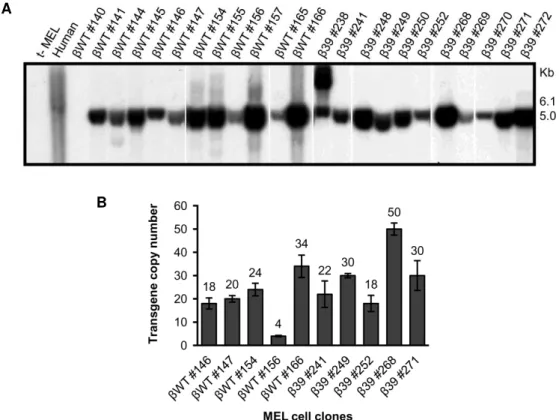
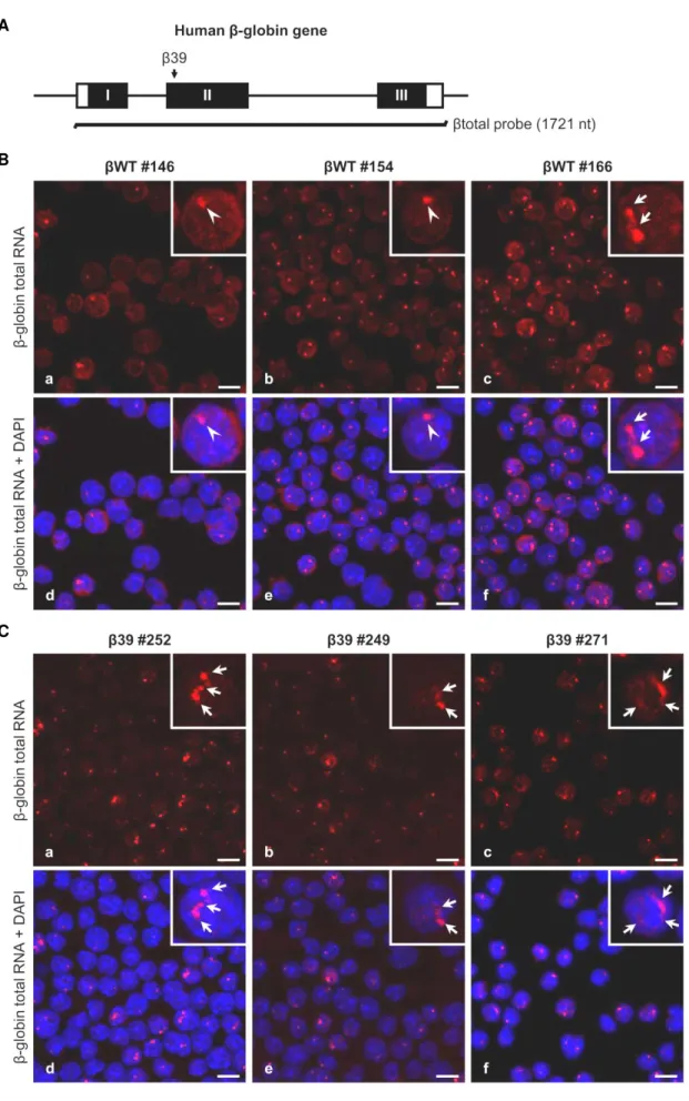
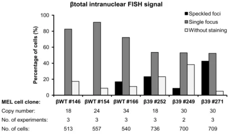
III.2.1.The subcellular localization of β-globin depends on the presence of a nonsense codon and transgene copy number ... 59
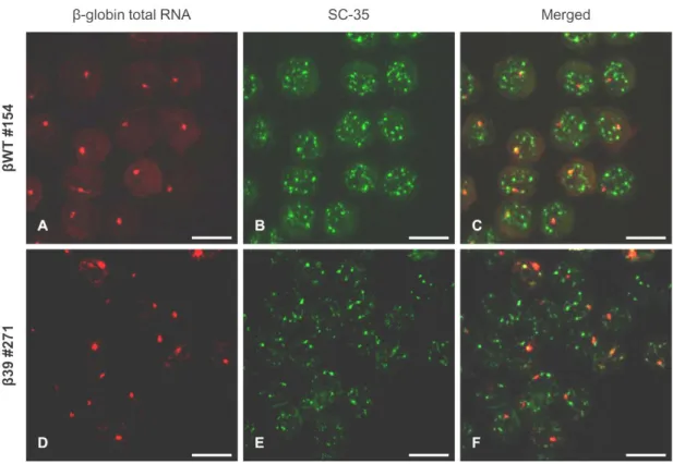
III.2.2.Beta-globin transcripts presenting a nuclear speckled-like FISH pattern do not colocalize with the SC-35 domains ... 62
III.2.3.Beta-globin speckled-like intranuclear RNA localization is due, at least in part, to unspliced transcripts ... 64
III.3.Analysis of the Nonsense-mutated β-globin Transcripts Processing Status in Erythroid Cells . ... 67
III.3.1.Human β-globin pre-mRNAs carrying a nonsense mutation accumulate at low levels ... 67
III.3.2.The low levels of the β39 pre-mRNAs are PTC-specific ... 69
III.3.3.The decreased β-globin pre-mRNA levels are specific for transcripts carrying NMD-sensitive nonsense codons ... 71
III.3.4.The presence of an NMD-sensitive nonsense codon does not affect the relative rates of removal of introns 1 and 2 in the human β-globin pre-mRNAs... 74
III.3.5.The reduced steady-state pre-mRNA level of NMD-sensitive transcripts does not reflect differential decay rates ... 76
CHAPTER IV. Discussion ... 81
CHAPTER V. Conclusions and Perspectives ... 91
Figure Index
Figure I.1 Overview of the gene expression steps. ... 5
Figure I.2 mRNA quality control systems in eukaryotes. ... 15
Figure I.3 Simplified model of the translation initiation mechanism. ... 21
Figure I.4 Splicing and the boundary rule for PTC definition in mammals. ... 24
Figure I.5 Mammalian NMD models. ... 29
Figure III.1 Human β-globin transgene integrity and copy number analysis in stably transfected MEL cell clones. ... 58
Figure III.2 Beta-globin transcripts subcellular localization depends on the presence of a PTC and probably on the transgene copy number. ... 60
Figure III.3 Summary of fluorescence in situ hybridization results for β-globin total RNA intranuclear localization in MEL cell lines... 62
Figure III.4 Beta-globin transcripts presenting a nuclear speckled-like pattern do not colocalize with the SC-35 domains. ... 63
Figure III.5 Beta-globin speckled-like intranuclear RNA localization is due, at least in part, to unspliced transcripts expressed in MEL cell lines. ... 65
Figure III.6 Summary of fluorescence in situ hybridization results for β-globin unspliced RNA intranuclear localization in MEL cell lines... 66
Figure III.7 Human β-globin pre-mRNAs carrying a nonsense mutation accumulate at low levels in MEL cells. ... 68
Figure III.8 The low levels of the β39 pre-mRNA are not due to the disruption of a regulatory element encompassing codon 39. ... 70
Figure III.9 The decreased β-globin pre-mRNA levels are specific for transcripts carrying NMD- -competent nonsense mutations. ... 72
Figure III.10 The presence of the nonsense codon equally decreases the abundance of intron 1 versusintron 2 containing human β-globin pre-mRNAs. ... 75
Figure III.11 The half-life of a pre-mRNA carrying an NMD-sensitive PTC is not significantly different from that of the wild-type control pre-mRNA. ... 76
Figure III.12 The nonsense codon effect on the β-globin pre-mRNA abundance exhibits cell line specificity. ... 78
Table Index
Table I.1 Nuclear mRNA quality control. ... 8
List of Abbreviations, Acronyms and Symbols
A adenine
Air1 Arginine methyltransferase-interacting RING finger protein 1
Air2 Arginine methyltransferase-interacting RING finger protein 2
A-site aminoacyl-site
ATCC American Type Culture Collection ATP adenosine triphosphate
ATPase adenosine triphosphatase bp base pairs
BRCA1 breast cancer 1 BTZ Barentz
C cytosine
CAF1 CCR4-associative factor 1 CBC cap-binding complex CBP cap-binding protein
CCR4 carbon catabolite repressor 4
cDNA mRNA-complementary deoxyribonucleic acid ChIP chromatin immunoprecipitation
CTD C-terminal domain
C-terminal carboxyl-terminal
CTP cytidine triphosphate Dbr1 debranching 1 DCP decapping protein
DCP1 decapping enzyme 1 DCP2 decapping enzyme 2 dCTP deoxycytosine triphosphate
Dis3 homolog of Schizosacharomyces pombe dis3 (chromosome disjunction)
DMEM Dulbecco’s modified Eagle’s medium DMSO dimethyl sulfoxide
DNA deoxyribonucleic acid DNase deoxyribonuclease
Dom34 duplication of multilocus region 34 EDTA ethylenediaminetetraacetic acid eEF eukaryotic translation elongation factor eIF eukaryotic translation initiation factor
EJC exon junction complex
ESE exonic splicing enhancer E-site exit-site
ESS exonic splicing silencer
F faraday
FISH fluorescence in situ hybridization
G guanine
GAPDH glyceraldehyde-3-phosphate dehydrogenase GPx1 glutathione peroxidase 1
GTP guanosine 5’-triphosphate GTPase guanosine triphosphatase Hba-a1 hemoglobin alpha, adult chain 1 HBB hemoglobin, beta
Hbs1 Hsp70 subfamily B suppressor
HeLa line of human epithelial cells derived from a cervical carcinoma HIV-1 human immunodeficiency virus type 1
hnRNP heterogeneous nuclear ribonucleoprotein
Hrp1 heterogeneous ribonucleoprotein 1 HS hypersensitive site
Ig immunoglobulin
IRES internal ribosome entry site
kb kilobase
LCR locus control region m7G 7-methylguanosine MAGOH mago-nashi homolog
MEL mouse erythroleukemia Met methionine
Met-tRNAi methionine-loaded initiator transfer RNA
Mex67 messenger RNA export factor of 67 kDa
Mlp myosin-like protein
mRNA messenger ribonucleic acid mRNP messenger ribonucleoprotein Mtr2 mRNA transport 2
Mtr4 mRNA transport 4 MVM minute virus of mouse
Nab2 nuclear polyadenylated RNA-binding 2 NaCl sodium chloride
NAS nonsense-mediated altered splicing NGD no-go decay
NLS nuclear localization signal
NMD nonsense-mediated mRNA decay
NMTGS nonsense-mediated transcriptional gene silencing NMUP nonsense-mediated upregulation of pre-mRNA
NPC nuclear pore complex
nt nucleotides N-terminal amino-terminus Nup60 nuclear pore 60
NXF1 nuclear RNA export factor 1 ORF open reading frame
P probability
Pab2 poly(A)-binding protein 2
PABP poly(A)-binding protein
PABPC1 poly(A)-binding protein, cytoplasmic 1 PABPN1 poly(A)-binding protein, nuclear 1 PAN poly(A) nuclease
PAN2 PABP-dependent poly(A) nuclease 2 PAN3 PABP-dependent poly(A) nuclease 3 PAP poly(A) polymerase
Pap2 poly(A) polymerase 2
P-bodies processing bodies
PBS phosphate-buffered saline PCR polymerase chain reaction
PIPES piperazine-1,4-bis(2-ethanesulfonic acid)
PM/Scl-100 polymyositis/scleroderma-100 Pml39 pre-mRNA leakage 39 Poly(A) polyadenylate
PP2A protein phosphatase 2A
pre-mRNA messenger ribonucleic acid precursor Prp43 pre-mRNA processing 43
P-site peptidyl-site
PTC premature translation-termination codon
puro puromycin
Rai1 Rat1p interacting protein Rat1 ribonucleic acid trafficking 1
REF RNA and export factor-binding protein
RNase ribonuclease
RNPS1 RNA-binding protein S1 RPA ribonuclease protection assay RPMI Roswell Park Memorial Institute
Rrp44 ribosomal RNA processing 44
Rrp6 ribosomal RNA processing 6 RT reverse transcription
RT-qPCR reverse transcription-coupled quantitative polymerase chain reaction
SC-35 splicing component of 35 kD SDS sodium dodecyl sulphate siRNA small interfering RNA Ski7 superkiller 7
SMG suppressor of morphological defects on genitalia snRNA small nuclear RNA
snRNP small nuclear ribonucleoprotein SR serine/arginine-rich domain
SSC saline-sodium citrate buffer Sub2 suppressor of Brr1-1 2
SURF SMG1-UPF1-eRF1-eRF3 complex Swt1 synthetically lethal with trex 1
T thymidine
THO suppressor of the transcriptional defects of Hpr1Δ by overexpression
Thy1 thymus cell antigen 1, theta TPI triosephosphate isomerase
TRAMP Trf4/Trf5-Air1/Air2-Mtr4 polyadenylation complex Trf4 Topoisomerase one-related function 4
Trf5 Topoisomerase one-related function 5
Tris tris(hydroxymethyl)aminomethane
Tris-HCl tris hydrochloride tRNA transfer ribonucleic acid U uracil
UAP56 56-kDa U2AF-associated protein
uORF upstream open reading frame UPF up-frameshift
UTP uridine 5’-triphosphate UTR untranslated region
V volt
WT wild-type
Xrn1 exoribonuclease 1 XRN2 exoribonuclease 2
Yra1 yeast RNA annealing protein 1
α alpha
α-32P alpha phosphorus-32
β beta
β127 human β-globin gene with nonsense mutation at codon 127
β26 human β-globin gene with nonsense mutation at codon 26
β39 human β-globin gene with nonsense mutation at codon 39
β39missense human β-globin gene with missense mutation at codon 39
β62 human β-globin gene with nonsense mutation at codon 62
βWT wild-type human β-globin gene
I.1. mRNA Biogenesis and Quality Control
I.1.1. Overview of the gene expression steps
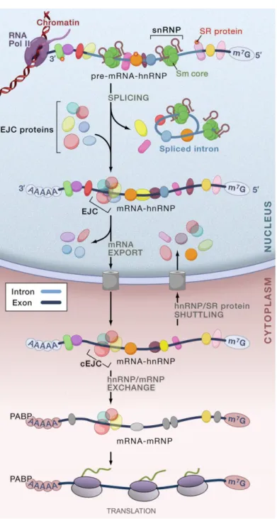
Gene expression comprises a series of interconnected events, in which genes are transcribed into messenger RNA (mRNA) and then translated into protein. In eukaryotic cells, a nuclear envelope separates DNA from the protein synthesis machinery, partitioning transcription from translation. During transcription, which occurs in the nucleus, the resulting mRNA precursors (pre-mRNAs) undergo several covalent chemical modifications, including 5’-end capping, splicing, and 3’-end cleavage and polyadenylation. These mRNA processing events take place with the aid of several specific factors (Orphanides and Reinberg, 2002; Moore, 2005; Moore and Proudfoot, 2009). Mature mRNAs are then exported to the cytoplasm, where they can be translated to protein, and ultimately, degraded (Reed, 2003; Dimaano and Ullman, 2004) (Figure I.1).
Eukaryotic mRNA precursors are synthesized by RNA polymerase II (RNA Pol II) in a process comprised of three stages: initiation, elongation and termination. Transcription starts with the assembly of the pre-initiation complex at the promoter of genes. This complex consists of RNA Pol II and several auxiliary proteins known as transcription factors, which recognize and bind to consensus sequences of the promoter located upstream of the start site for transcription (Proudfoot et al., 2002; Luna et al., 2008; Hocine et al., 2010). Also, the activity of the promoters may be greatly increased by enhancer sequences, which can act over distances of several kilobases, located either upstream or downstream of the gene. Transcription factors recruit and position RNA Pol II near the transcription start site and, subsequently, elongation occurs after transition to an RNA Pol II elongation complex. This switch is associated with alterations of chromatin structure and changes of the RNA Pol II C-terminal domain (CTD) phosphorylation state (Hocine et al., 2010). Also, productive transcriptional elongation is tightly coupled to mRNA
processing events such as 5’-capping and splicing (Luna et al., 2008; Hocine et al., 2010). RNA Pol II proceeds through the remainder of the gene until conserved polyadenylation signals direct
cleavage and polyadenylation at the 3’ end of the nascent transcript and transcription termination
occurs (Luna et al., 2008).
Capping takes place shortly after transcription initiation in which a 7-methylguanosine (m7G) cap
In the pre-mRNA processing step of splicing, noncoding intervening sequences, or introns, are removed and coding sequences, or exons, are spliced together by a two-step transesterification reaction catalysed by the spliceosome (Figure I.1). This complex consists of small nuclear ribonucleoproteins (snRNPs) comprising five small nuclear RNAs (snRNAs) - U1, U2, U4, U5 and U6 - in conjunction with a large number of additional proteins, like the Sm ribonucleoproteins, which assembles onto the pre-mRNA (Neugebauer, 2002; Soller, 2006). Formation of the
spliceosome complex at particular splice junctions relies on certain sequences, including the 5’
splice site, the branch point, a variable stretch of pyrimidines termed polypyrimidine tract, and the
3’ splice site (Neugebauer, 2002; Luna et al., 2008; Hocine et al., 2010). Additionally, in higher eukaryotes, flanking pre-mRNA regulatory elements, namely intronic and exonic splicing enhancers or silencers, bind trans-acting splicing factors that enhance or repress snRNP
recruitment to splice sites. Generally, exonic splicing enhancers (ESEs) are bound by serine/arginine-rich (SR) proteins, whereas exonic splicing silencers (ESSs) are bound by heterogeneous nuclear ribonucleoproteins (hnRNPs) (Hocine et al., 2010). Hence, splice site selection results from the cumulative effect of multiple factors.
Processing at the 3’ end of the pre-mRNA involves a cleavage step downstream a conserved polyadenylation signal (AAUAAA sequence). Endonucleolytic cleavage yields a free 3’-hydroxyl group to which a string of adenylic acid residues [poly(A) tail] is added by an enzyme called poly(A) polymerase (PAP). Generally, processive polyadenylation by PAP complexes is followed by rapid decoration of the poly(A) tail by poly(A)-binding proteins (PABPs), which protects the
transcripts from 3’ to 5’ exonucleolytic degradation (Neugebauer, 2002; Hosoda et al., 2006; Luna et al., 2008).
In addition to processing, nascent transcripts must be loaded with specific RNA-binding proteins to form messenger ribonucleoproteins (mRNPs). Several mRNP assembly factors, including the THO complex, mRNA export factors, namely the RNA helicase Sub2p (Saccharomyces cerevisiae)/UAP56 (human) and RNA-binding protein Yra1/ALY, the mRNA export receptor,
Figure I.1 Overview of the gene expression steps. The nascent transcript (pre-mRNA), capped with a 7-methylguanosine (m7G) cap, is bound by heterogeneous nuclear ribonucleoproteins (hnRNPs) and SR
(serine/arginine-rich domain-containing) proteins. Small nuclear ribonucleoprotein particles (snRNPs) with an extremely stable Sm core make up the spliceosome, which bind to the splice sites at the 5’ and 3’ ends of introns. The spliceosome catalyzes the removal of introns, which are excised as lariats and subsequently debranched and degraded, and the ligation of the flaking exons. The exon junction complex (EJC) assembles on spliced mRNA about 20 to 25 nucleotides upstream of joined exons, followed by the export of the mRNA to the cytoplasm. Many of the RNA-binding proteins shuttle between the nucleus and the cytoplasm. The poly(A)-binding protein (PABP) binds to the poly(A) tail of cytoplasmic mRNAs. cEJC indicates the remaining stable EJC on mRNA in the cytoplasm, which is removed by translating ribosomes.
Despite the fact that RNA processing and translation occurs in different compartments, several views portray gene expression as an organization of events physically and/or functionally connected, in which different steps are dependent or influenced by one another (Orphanides and Reinberg, 2002). For instance, the RNA polymerase II transcription machinery plays an active role in recruiting the cellular apparatus that caps and processes the nascent RNA transcript. On the other hand, the 5’-cap structure and the poly(A) tail are required to stabilize the mRNA and also play an essential function in translation initiation and termination (Proudfoot et al., 2002). Moreover, several evidences supports that pre-mRNA splicing and mRNP remodelling for export occur co-transcriptionally (Luna et al., 2008; Perales and Bentley, 2009). Likewise, pre-mRNA splicing promotes transcription elongation and is required for efficient export of the processed mRNA into the cytoplasm. Furthermore, the splicing event can also link the nuclear history of the mRNA to its cytoplasmic fate. During splicing, a set of proteins called exon junction complex (EJC) is deposited close to the splice sites (Le Hir et al., 2001b). Once bound, EJCs travel with the mRNA to the cytoplasm where most are removed as a consequence of the translation process (Figure I.1). Prior to their displacement, however, they can act as effectors of almost every aspect of mRNA metabolism, including subcellular mRNA localization, mRNA translational efficiency and mRNA decay (Kuersten and Goodwin, 2005; Moore and Proudfoot, 2009).
I.1.2. mRNA quality control
Considering the multitude of events involved in RNA biogenesis, as well as the series of transitions of RNAs between different complexes of proteins and subcellular compartments, the process of gene expression is susceptible to mistakes. For instance, errors in transcription, nuclear pre-mRNA processing or mRNP maturation can originate abnormal mRNAs which may be translated into deleterious proteins or impair mRNA metabolism and potentially lead to disease (Fasken and Corbett, 2005; Doma and Parker, 2007). In addition, mutations in genes can also give rise to aberrant mRNAs. To prevent these abnormal mRNAs from producing harmful proteins or effects, eukaryotic cells have developed multiple nuclear and cytoplasmic mRNA quality control mechanisms which recognize and degrade aberrant mRNAs (Doma and Parker, 2007; Isken and Maquat, 2007; Schmid and Jensen, 2008a; Fasken and Corbett, 2009; Houseley and Tollervey, 2009). These surveillance pathways generally intervene whether the mRNP production and transport is affected or if mRNP translation is disrupted (Mühlemann and Jensen, 2012). Hence, abnormal mRNAs are directly or indirectly recognized by means of specific factors, which in turn recruit ribonucleases that rapidly degrade the targeted transcripts (Houseley and Tollervey, 2009). Therefore, eukaryotic RNA processing steps and quality control mechanisms are deeply interconnected in order to ensure the fidelity of gene expression. Several events of mRNA processing have been described as checkpoints for mRNA quality control mechanisms, which
I.1.2.1. mRNA quality control in the nucleus
Multiple surveillance pathways appear to be active on eukaryotic mRNA within the nucleus, which have been described mostly in the yeast Saccharomyces cerevisiae and are likely to be
conserved in mammals and other higher eukaryotes (Doma and Parker, 2007; Schmid and Jensen, 2008a; Fasken and Corbett, 2009; Mühlemann and Jensen, 2012). Although the underlying molecular mechanisms are yet to be fully understood, generally, nuclear quality control systems either lead to rapid degradation of aberrant mRNAs or to retention of the targeted RNAs in a nuclear subdomain, in order to trigger subsequent processing or degradation. Specifically, different pathways have been described: (i) rapid RNA degradation in the nucleus, (ii) export to the cytoplasm for degradation, (iii) retention in transcription site-associated foci, (iv) retention at the nuclear pore, and (v) transcriptional downregulation of the genes from which abnormal RNAs are being produced (Doma and Parker, 2007; Schmid and Jensen, 2008a; Fasken and Corbett, 2009). Interestingly, these quality control mechanisms are not triggered exclusively by a single checkpoint or mRNA-processing event (Table I.1).
I.1.2.1.1. Nuclear degradation
This nuclear quality control mechanism targets aberrant transcripts for degradation via a few
conserved RNA-degrading enzymes or ribonucleases: endonucleases that cut RNA internally, 5’ to 3’exonucleases that hydrolyze RNA from the 5’ end, and 3’ to 5’ exonucleases that degrade RNA from the 3’ end (Houseley and Tollervey, 2009). In the nucleus, quality control is carried out by the exosome, which comprises an evolutionary conserved multiprotein complex containing two
active 3’ to 5’ exonucleases, Dis3p (also referred to as Rrp44p) and Rrp6p (PM/Scl-100 in humans) (Houseley and Tollervey, 2009; Lykke-Andersen et al., 2011). The nuclear exosome is
involved in the degradation of most nuclear RNAs, although Rat1p (XRN2 in humans), a 5’ to 3’
exonuclease, may also affect nuclear degradation (Doma and Parker, 2007; Schmid and Jensen, 2008a; Fasken and Corbett, 2009). For instance, yeast transcripts failing to receive a proper 5’- -cap structure are selectively degraded by Rat1p, which stimulates the activity of the decapping endonuclease Rai1, within a quality control process occurring during transcription elongation (Kim et al., 2004b; West et al., 2004; Jiao et al., 2010; Jimeno-González et al., 2010). Degradation by
the 5’ to 3’ exonuclease Rat1p was also reported to target abnormal transcripts originated from
Table I.1 Nuclear mRNA quality control.
Organism Defect Consequences of quality control
Yeast Capping Nuclear decapping and 5’ to 3’ degradation by Rat1p (Jiao
et al., 2010; Jimeno-González et al., 2010)
Yeast Splicing:
trapped lariat intermediate
Debranching, export, and cytoplasmic 5’ to 3’ decay by
Xrn1p(Hilleren and Parker, 2003; Mayas et al., 2010)
Nuclear decapping and 5’ to 3’ degradation by Rat1p
(Bousquet-Antonelli et al., 2000)
Rrp6p and/or core exosome-dependent nuclear degradation
(Bousquet-Antonelli et al., 2000)
Yeast
Splicing:
blocked spliceosome formation / first catalytic step or not recognized by spliceosome
Nuclear decapping and 5’ to 3’ degradation by Rat1p
(Bousquet-Antonelli et al., 2000)
Rrp6p and/or core exosome-dependent nuclear degradation
(Bousquet-Antonelli et al., 2000; Lemieux et al., 2011)
Retention in the nucleus by Mlp proteins near or at the nuclear pore complex (Galy et al., 2004; Palancade et al., 2005; Sommer and Nehrbass, 2005; Schmid and Jensen, 2008a; Fasken and Corbett, 2009; Dieppois and Stutz, 2010)
Yeast
mRNP assembly:
THO complex / Sub2p / Yra1p mutants
3’-end processing
Rrp6p and/or core exosome-dependent nuclear degradation and retention near the transcription site(Burkard and Butler, 2000; Hilleren et al., 2001; Jensen et al., 2001; Libri et al., 2002; Torchet et al., 2002; Thomsen et al., 2003;
Rougemaille et al., 2007; Assenholt et al., 2008; Saguez et al., 2008)
Retention in the nucleus by Mlp proteins near or at the nuclear pore complex (Palancade et al., 2005; Sommer and Nehrbass, 2005; Vinciguerra et al., 2005; Schmid and Jensen, 2008a; Fasken and Corbett, 2009)
Transcriptional downregulation (Jensen et al., 2004; Vinciguerra et al., 2005)
Fruit fly
Splicing:
not recognized by spliceosome
Rrp6p and/or core exosome-dependent nuclear retention near the transcription site (Eberle et al., 2010)
Transcriptional downregulation (Eberle et al., 2010)
Mammals
Splicing:
not recognized by spliceosome
absence of introns in a gene that normally contains introns
Accelerated nuclear degradation dependent on 3’ poly(A)
tail (Conrad et al., 2006)
Mammals
3’-end processing
Splicing:
not recognized by spliceossome
Rrp6 and/or core exosome-dependent nuclear retention of RNA near or at the transcription site (Custódio et al., 1999; de Almeida et al., 2010)
Transcriptional downregulation (Furger et al., 2002; Damgaard et al., 2008)
Several studies suggest that the nuclear exosome is associated with the elongating RNA polymerase II in active genes and functions together with a set of cofactors, which recognize aberrant RNAs by structure or sequence or are loaded onto abnormal mRNAs during defective mRNA processing, and subsequently stimulate the exosome to rapidly degrade such transcripts (Vanacova and Stefl, 2007; Schmid and Jensen, 2008b; Houseley and Tollervey, 2009). There are also evidence that polyadenylation can represent a path to nuclear RNA decay mediated by the exosome. For instance, defects in mRNP assembly may lead to improperly polyadenylated mRNAs with short poly(A) tails, which are recognized and degraded in an Rrp6p-dependent manner (Doma and Parker, 2007; Schmid and Jensen, 2008a; Fasken and Corbett, 2009). Degradative polyadenylation has been mainly associated with the activity of a nuclear exosome cofactor called TRAMP complex, comprised of a noncanonical poly(A) polymerase (Trf4 or Trf5), an RNA-binding protein (Air1 or Air2), and an RNA helicase (Mtr4), which adds short poly(A) tails to aberrant or unstable transcripts, forming a favourable substrate for rapid RNA degradation by the exosome (LaCava et al., 2005; Wyers et al., 2005; Vanacova and Stefl, 2007). Accordingly, inactivation of the TRAMP complex or exosome activities leads to the accumulation of abnormal RNAs originated from impaired splicing or 3’-end processing (Bousquet-Antonelli et al., 2000; Burkard and Butler, 2000; Torchet et al., 2002). In addition, yeast cells harbouring mutations in the THO complex and in the associated RNA helicase Sub2p, which are involved in mRNP assembly and transcription elongation, present rapidly degradation of several abnormal mRNAs
via a mechanism requiring Rrp6p and the TRAMP complex poly(A) polymerase Trf4 (also known
as Pap2) (Libri et al., 2002; Rougemaille et al., 2007; Assenholt et al., 2008; Saguez et al., 2008).
Interestingly, although polyadenylation-mediated exosome degradation via the TRAMP complex
has been widely associated with nuclear RNA quality control, recent evidences suggest that the nuclear exosome can also target transcripts polyadenylated by canonical poly(A) polymerases. A pre-mRNA nuclear decay pathway, targeting specific polyadenylated intron-containing transcripts in Schizosaccharomyces pombe yeast, was shown to involve the nuclear poly(A)-binding protein
Pab2 (PABPN1 in humans) and the nuclear exosome subunit Rrp6p (Lemieux et al., 2011).
I.1.2.1.2. Export to the cytoplasm for degradation
which would result in the production of non-functional or truncated proteins. Specifically, retention of intronic sequences likely result in the presence of a premature translation-termination codon (PTC) within the transcript. Therefore, unspliced pre-mRNAs are common substrates for the nonsense-mediated mRNA decay (NMD) in the cytoplasm, an surveillance mechanism that relies on translation and targets PTC-containing transcripts for rapid degradation in the cytoplasm (Isken and Maquat, 2007; Nicholson and Mühlemann, 2010). However, another RNA quality pathway targeting yeast aberrant unspliced precursors or splicing intermediates, which undergo export into
the cytoplasm for 5’ to 3’ digestion by Xrn1p, was described to occur independently from NMD (Hilleren and Parker, 2003). This pathway involving aberrantly processed nuclear transcripts is currently poorly understood. The splicing ATPase Prp43p is required for the release of lariat intermediates from the spliceosome and their export into the cytoplasm, where they are subjected to degradation with the aid of the debranching enzyme Dbr1p (Hilleren and Parker, 2003; Mayas et al., 2010).
I.1.2.1.3. Retention in transcription site-associated foci
Another quality control mechanism retains aberrant RNAs within the nucleus. For instance, mRNAs bearing defects in splicing or 3’-end processing are retained in the nucleus of both yeast
Saccharomyces cerevisiae and fruit fly Drosophila melanogaster cells (Hilleren et al., 2001;
Jensen et al., 2001; Eberle et al., 2010). Several data suggests that these aberrant RNAs are retained at the transcription site through a process involving the nuclear exosome and Rrp6 (Hilleren et al., 2001; Libri et al., 2002; Thomsen et al., 2003; Rougemaille et al., 2007; Eberle et al., 2010). In addition, other studies linked this pathway with the nuclear surveillance of mRNP assembly. In Saccharomyces cerevisiae THO/Sub2 mutants, a fraction of transcripts escapes
I.1.2.1.4. Retention at the nuclear pore complex
The perinuclear quality control mechanism plays a role in recognizing and concentrating correctly processed mRNAs at the nuclear pore for efficient export (Green et al., 2003; Sommer and Nehrbass, 2005). Retention of unspliced RNAs and malformed mRNPs at or near the nuclear pore complex, in order to prevent their escape to the cytoplasm, has been mostly described in
Saccharomyces cerevisiae (Green et al., 2003; Dziembowski et al., 2004; Galy et al., 2004;
Palancade et al., 2005; Vinciguerra et al., 2005; Lewis et al., 2007). This nuclear quality control mechanism comprises several nuclear proteins which retain abnormal mRNAs and mRNPs and potentially degrade them (Sommer and Nehrbass, 2005; Schmid and Jensen, 2008a; Fasken and Corbett, 2009). For instance, the nuclear pore-associated proteins Mlp1p, Mlp2p (Trp in vertebrates) and Pml39p proteins were described as major players in sorting aberrant mRNAs for retention (Galy et al., 2004; Fasken and Corbett, 2009). Additional NPC-associated proteins are involved, such as Esc1p and Nup60p proteins, probably indirectly because they are important for NPC assembly and Mpl1p anchoring, respectively (Fasken and Corbett, 2009). Furthermore, mRNPs may directly interact with Mlp1p via the poly(A)-binding protein Nab2, which is implicated
in mRNA export and poly(A) tail length control (Fasken and Corbett, 2009; Dieppois and Stutz, 2010). The underlying mechanism still requires further characterization, however it is though that Mlp1p, Mlp2p and Pml39p could recognize and retain abnormal RNAs or RNPs by interacting with mRNA splicing and mRNP assembly factors (Galy et al., 2004). Subsequently, a ribonuclease would be required to rapidly degrade these mRNAs before they can escape this perinuclear surveillance mechanism and reach the cytoplasm (Fasken and Corbett, 2009). Swt1, a
Saccharomyces cerevisiae endoribonuclease, was identified as a potential player in the
degradation of aberrant mRNAs at the nuclear pore. The mRNA products of Swt1 cleavage might then be subjected to further degradation by 5’ to 3’ exonucleases and the exosome (Fasken and Corbett, 2009; Dieppois and Stutz, 2010). As above mentioned, defects in mRNP assembly can also trigger exosome-dependent accumulation of the mRNA in association with the site of transcription. This linkage of the defective mRNP to the transcription site has been hypothesized to direct the entire locus towards the NPCs (Rougemaille et al., 2008a). Notably, Mlp proteins are present only in sections of the nuclear envelope adjacent to chromatin (Galy et al., 2004), suggesting that they may contact nascent transcripts, thereby linking mRNA synthesis to export and exerting surveillance very early on during mRNP formation (Rougemaille et al., 2008b; Schmid and Jensen, 2008a; Fasken and Corbett, 2009; Dieppois and Stutz, 2010).
I.1.2.1.5. Transcriptional downregulation
Several authors reported that the nuclear pore-associated proteins, Mlp1 and Mlp2, also play a critical role in a nuclear quality control mechanism that possibly feeds back on transcription in
Saccharomyces cerevisiae (Schmid and Jensen, 2008a). Indeed, the Mlp proteins were found to
transcription-coupled mRNA export protein, which triggered the transcriptional downregulation of a subset of genes (Vinciguerra et al., 2005). Conversely, the artificial decrease of transcription levels was shown to rescue the effects induced by quality control (i.e., nuclear retention) of mRNP
assembly mutants or 3’-end processing mutants (Jensen et al., 2004). Interestingly, a study in
Drosophila melanogaster cells reported that aberrant RNAs harbouring splice site mutations,
which undergo retention at the transcription site involving the nuclear exosome and Rrp6, also show transcriptional impairment of the corresponding gene due to chromatin modifications (Eberle et al., 2010). In overall, a nuclear quality control pathway acting on transcription may be part of a cellular response to provide a favourable environment for proper mRNP formation (Schmid and Jensen, 2008a; Mühlemann and Jensen, 2012).
I.1.2.1.6. Nuclear mRNA quality control in mammals
In higher eukaryotes, and specifically in mammals, research on nuclear mRNA quality control mechanisms is still scarce. Although the high conservation of complexes involved in RNA surveillance, namely the exosome and TRAMP complexes, strongly predicts the existence of similar pathways across species, a specific role of these complexes during mRNA quality control in mammals has yet to be established (Anderson and Wang, 2009; Lykke-Andersen et al., 2011). In addition, some differences could reflect distinct functional properties of the components of surveillance machineries. For instance, Rrp6p (PM/Scl-100) associates only with the nuclear exosome in yeast, whereas in human cells it is present both in the nucleus and in the cytoplasm (Lykke-Andersen et al., 2011).
processing factors, non-coding RNAs, snRNPs and many of their constituents work in concert to coordinate multiple steps of gene expression, including transcription, pre-mRNA processing and mRNA export (Misteli and Spector, 1998; Smith et al., 1999; Mao et al., 2011). It has been observed that within the interchromatin space, mRNPs move unrestricted in and out of interchromatin granule clusters, suggesting that, in general, mRNPs do not accumulate at specific regions for specific processing or modification steps (Molenaar et al., 2004; Politz et al., 2006). Nonetheless, specific spliced and unspliced RNAs have been described to colocalize at the periphery of these domains (Smith et al., 1999; Shopland et al., 2002; Handwerger and Gall, 2006).
In addition to the increased intricacy of the physical and/or functional organization of gene expression, widespread usage of alternative splice and polyadenylation sites can further complexify mRNP assembly in mammals (Kim et al., 2004a). Such increased complexity is probably met by more elaborate quality control mechanisms, which may also function in the regulation of mRNA metabolism (Schmid and Jensen, 2008a; Mühlemann and Jensen, 2012).
Nevertheless, several evidences support that some nuclear RNA quality control pathways might be conserved from yeast to higher eukaryotes. As in yeast, splicing defective transcripts can be subjected to rapid RNA decay in mammalian nuclei. Human β-globin transcripts harbouring 5’ or 3’ splice site mutations, or intronless polyadenylated β-globin cRNA, are more rapidly degraded in the nucleus of human cells and accumulate when degradation is inhibited (Conrad et al., 2006). In mammalian cells, defects in mRNA splicing and 3’-end formation can also cause accumulation of aberrant transcripts near the transcription site. For instance, fluorescence in situ hybridization
(FISH) experiments were performed to visualize the release of wild-type and mutated β-globin RNAs from their DNA template in stably transfected mouse erythroleukemia (MEL) cells (Custódio et al., 1999). In both conditions, β-globin RNAs visualized by FISH accumulate in a single nuclear
Transcriptional downregulation of genes bearing 5’ splice site mutations, from which abnormal
RNAs are being produced, is also described in mammalian cells. For example, human cells transfected with HIV-1 and β-globin genes containing altered splice donor sequences present impaired transcription of the corresponding nascent transcripts (Furger et al., 2002; Damgaard et al., 2008).
I.1.2.2. mRNA quality control in the cytoplasm
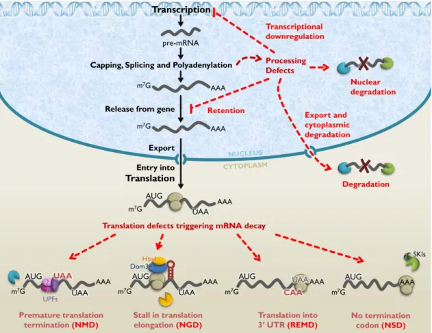
After leaving the nucleus, mammalian mRNPs may be further subjected to several RNA quality control mechanisms in the cytoplasm (Figure I.2). A general feature of these mechanisms comprises the discrimination of aberrant mRNAs from normal mRNAs by adaptor proteins, which interact with the translation apparatus and direct the aberrant mRNAs into a degradation pathway. The main event that triggers the rapid destruction of abnormal mRNAs in the cytoplasm is the failure of ribosomes to terminate translation correctly (Moore, 2005; Doma and Parker, 2007; Isken and Maquat, 2007). For instance, mRNAs with ribosomes stalled at stable secondary structures, or at a stretch of rare codons, are targeted for endonucleolytic cleavage in a surveillance pathway named no-go mRNA decay (NGD) (Doma and Parker, 2006). Aberrant mRNAs lacking an in-frame termination codon, such that translation continues to the end of the poly(A) tail, are targeted for degradation by the cytoplasmic exosome through the action of Ski7p
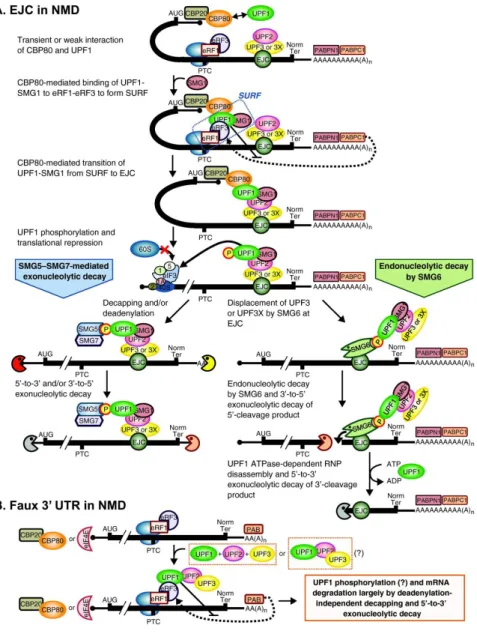
– a paralog of the eukaryotic translation elongation factor (eEF) 1A – and the eukaryotic translation release factor (eRF) 3, within a quality control mechanism referred to as nonstop mRNA decay (NSD) (Frischmeyer et al., 2002; Vasudevan et al., 2002; van Hoof et al., 2002). Recent evidences suggest that NSD and NGD processes are conserved from yeast to mammals and mechanistically related. For instance, translation of the poly(A) tail of a nonstop mRNA generates a polylysine chain that was reported to stall ribosomes by clogging their exit tunnels, which originates a no-go situation (Bengtson and Joazeiro, 2010). Moreover, both NSD and NGD are promoted by two eRF-mimicking proteins: the eRF1 paralog Pelota (Dom34p in yeast) and the eRF3 homologous GTPase Hbs1, which presumably interact with the stalled ribosome (Doma and Parker, 2007; Passos et al., 2009; Bengtson and Joazeiro, 2010; Shoemaker et al., 2010; Pisareva et al., 2011). Another cytoplasmic surveillance pathway, called ribosome extension- -mediated mRNA decay pathway (REMD), targets mRNAs when ribosomes inappropriately carry out translation throughout the 3’ untranslated region(3’ UTR) until another termination codon is encountered within this region. This quality control mechanism was reported to be specific to erythroid cells, triggering the destabilization of human α-globin mRNAs containing an antitermination mutation (Kong and Liebhaber, 2007). Finally, one of the best characterized surveillance mechanism is the nonsense-mediated mRNA decay (NMD), which targets aberrant transcripts containing a premature termination codon for decapping and 5’ to 3’ degradation by
between nuclear and cytoplasmic events in the eukaryotic gene expression process. Transcripts harbouring PTCs are distinguished from normal mRNAs in a process involving specific conserved factors, the UPF (up-frameshit) proteins, and their interactions with both the EJC, deposited during splicing, and the translation termination complex, triggering rapid decay of the aberrant mRNAs (Kashima et al., 2006).
I.2. Nonsense-mediated mRNA Decay
I.2.1. NMD targets and functions
Transcripts containing PTCs can arise from different biological processes in germ or somatic cells (Mühlemann et al., 2008). Namely, inherited genetic lesions or errors during replication can cause single base pair substitutions that change a sense codon to an in-frame PTC, commonly known as nonsense mutations. Also, insertion or deletion mutations can alter the ribosomal reading frame causing translating ribosomes to encounter a PTC, or introduce an in-frame PTC. Programmed somatic cell DNA rearrangements and hypermutations that occur in TCR (T-cell receptor) and Ig (immunoglobulin) genes in lymphocytes often generate frameshift mutations and downstream PTCs, which are also targeted to NMD (Li and Wilkinson, 1998). At the RNA level, PTCs can be generated by transcriptional errors and by abnormal pre-mRNA processing. For instance, mutations that alter splicing signals can produce PTCs, frequently due to the retention of intronic sequences containing in-frame nonsense codons (Mendell and Dietz, 2001; Holbrook et al., 2004; Egecioglu and Chanfreau, 2011). Moreover, it is estimated that approximately 60 to 70% of human pre-mRNAs undergo alternative splicing and, among these, 35% are predicted to have at least one splice variant that is expected to be targeted by NMD (Lewis et al., 2003; McGlincy and Smith, 2008).
PTCs can originate two outcomes on gene expression. Firstly, a PTC will terminate mRNA translation prior to completion of a full-length polypeptide, leading to the production of truncated proteins that are often non-functional and/or unstable. Secondly, mRNAs harbouring PTCs are also frequently unstable as they undergo rapid degradation via NMD, resulting in a drastic
reduction of steady-state mRNA abundance (Maquat, 1995). Therefore, by downregulating mRNAs bearing nonsense codons, NMD prevents the synthesis of C-terminally truncated proteins potentially toxic for the cell (Frischmeyer and Dietz, 1999; Khajavi et al., 2006). As about one third of all known disease-causing mutations originate a nonsense codon, NMD may function as a significant modulator of genetic disease phenotypes in humans (Frischmeyer and Dietz, 1999; Khajavi et al., 2006; Bhuvanagiri et al., 2010; Nicholson et al., 2010).
also found to play a role in regulating the expression of RNA transcripts involved in several biological processes, namely, stress responses, haemopoietic stem cell development, regulation of alternative splice forms, chromosome structure and function, cell-cycle progression, and embryonic development (Neu-Yilik et al., 2004; Rehwinkel et al., 2006; Bhuvanagiri et al., 2010; Gardner, 2010). NMD therefore could affect a large proportion of the transcriptome, which highlights the importance of this posttranscriptional mechanism in the quality control and regulation of eukaryotic gene expression.
I.2.1.1. NMD implications in disease
The biological and medical significance of the NMD pathway is pointed up by the fact that approximately 30% of all inherited genetic disorders are due to PTCs, as above mentioned, and in many of these cases, NMD influences the severity of the clinical phenotype (Holbrook et al., 2004; Stalder and Mühlemann, 2008). The majority of nonsense-associated diseases are caused either by insufficient levels of functional proteins that can result from degradation of the PTC-containing mRNAs by NMD or by the inability of the abnormally PTC-containing mRNAs, which are able to escape NMD, to generate full-length functional proteins (Lejeune and Maquat, 2005). Nevertheless, NMD can have beneficial effects by eliminating transcripts harbouring PTCs that would otherwise originate C-terminally truncated proteins, either with a complete loss of function or with a dominant-negative function leading to toxicity (Bhuvanagiri et al., 2010). An example of such beneficial effects on disease phenotype is β-thalassemia (Frischmeyer and Dietz, 1999; Holbrook et al., 2004). Beta-thalassemia is a hereditary form of anemia, characterized by the
absence or reduction in the synthesis of β-globin polypeptide chains, one of the hemoglobin subunits (Weatherall, 2000). Hemoglobin comprises a tetrameric complex required for oxygen
transport, composed of two α- and two β-globin subunits (Huisman, 1993). Defective β-globin production leads to subunit imbalance with an excess of the complementary α-globin chain and subsequent lack of functional hemoglobin in the cell (Weatherall, 2000). This condition can emerge due to the presence of PTCs in the β-globin transcripts (Huisman, 1993). If a PTC is located at a position that activates NMD, the production of truncated proteins is reduced and the possible deleterious effects due to their accumulation are minimized. Indeed, the excess of free α- -globin, as well as the limited amount of truncated β-globin protein that can be produced, are proteolytic degraded. As a result, individuals carrying only one affected allele present a clinically
asymptomatic phenotype of β-thalassemia trait, whose mode of inheritance is recessive, also known as “thalassemia minor” (Hall and Thein, 1994; Kugler et al., 1995; Holbrook et al., 2004). On the other hand, if a PTC is located at a position that does not induce NMD, such as nonsense
mutations in the third exon of the β-globin gene, substantial amounts of abnormal β-globin mRNAs are translated into truncated non-functional β-globin chains, which may overburden the cellular proteolytic system. Subsequent accumulation of these truncated products can often act in a dominant negative manner, leading to deleterious effects on the cell. This condition is correlated
intermedia”, presenting a dominant mode of inheritance (Hall and Thein, 1994; Holbrook et al., 2004; Neu-Yilik and Kulozik, 2008).
There are several other conditions where NMD exerts a protective impact and acts as a modulator of the disease phenotype. Examples of such conditions are the Marfan syndrome, retinal degeneration, von Willebrand disease, myotonia congenita and factor X deficiency (Frischmeyer and Dietz, 1999; Khajavi et al., 2006; Bhuvanagiri et al., 2010). A potential influence of NMD in cancer has also been suggested. In fact, transcripts of several mutant forms of the tumour suppressor proteins genes breast cancer 1 (BRCA1), TP53 and Wilms tumour (WT1) have been shown to be eliminated by NMD. The targeting for degradation of these PTC-containing transcripts, that would convert the tumour suppressors into dominant-negative oncoproteins, protects the heterozygous carriers from developing cancer (Holbrook et al., 2004).
However, it is important to note that NMD can also aggravate the disease phenotype by eliminating mRNAs that would otherwise support the synthesis of partially functional truncated proteins, leading to haploinsufficiency (Khajavi et al., 2006). Examples of such detrimental effect of NMD are Duchenne muscular dystrophy (DMD), cystic fibrosis, Hurler syndrome and X-linked nephrogenic diabetes insipidus (Holbrook et al., 2004). The aggravated clinical picture of protein deficiency induced by NMD is clearly illustrated in DMD, where NMD-insensitive PTCs located
near the 3’ end of the dystrophin gene result in variable mild phenotypes, whereas PTCs sensitive
to NMD are associated with a severe form of DMD (Khajavi et al., 2006). In addition, detrimental effects of the NMD activation were also reported in cancer conditions, namely in hereditary diffuse gastric cancer (HDGC) associated with PTC-causing mutations within the cadherin-1 (CHD1) gene (Bhuvanagiri et al., 2010). In these cases, where NMD has a detrimental effect on the disease phenotype, therapeutics that specifically modulates NMD would be clinically useful (Holbrook et al., 2004; Kuzmiak and Maquat, 2006; Bhuvanagiri et al., 2010). In the last decade, a therapeutic approach named suppression therapy has been developed that utilizes low molecular weight compounds to induce the translation machinery to recode a PTC into a sense codon. Suppression of translation termination at a nonsense codon enables translation elongation to proceed in the correct reading frame, which allows the production of a full-length protein and restore its function (Keeling and Bedwell, 2011).
I.2.2. Molecular basis of the NMD pathway in mammals
NMD has been extensively studied for decades in yeast, worms, fruit fly, plants and mammals, and several models have been proposed depicting different aspects of the NMD pathway, such as nonsense codon recognition or subcellular localization, amongst others (Isken and Maquat, 2007; Brogna and Wen, 2009; Rebbapragada and Lykke-Andersen, 2009; Nicholson and Mühlemann, 2010). In the overall, NMD starts with the recognition and discrimination of the PTC from the natural stop codon within a process dependent on mRNA translation and on highly conserved
surveillance complex assemble and interact to trigger NMD. UPF proteins form the core complex of the NMD machinery, linking premature translation termination to rapid mRNA degradation via
specific pathways of decay.
From early studies concerning the destabilization of mRNAs containing PTCs, several evidences suggested that NMD occurs during translation (Maquat, 2004). In relation to mammalian NMD, pharmacological inhibitors of translation, hairpin structures in the 5’ UTR (that prevent
translational initiation), and expression of suppressor tRNAs (which allow read-through of PTCs), were shown to inhibit NMD and stabilize mammalian transcripts harbouring PTCs (Urlaub et al., 1989; Nishimoto et al., 1991; Belgrader et al., 1993; Cheng et al., 1994). Furthermore, when translation initiation was prevented by the insertion of either a stem-loop structure or an iron- -responsive element into the 5’ UTR of mammalian transcripts, NMD was as well abrogated
(Belgrader et al., 1993; Thermann et al., 1998). Given the important role of translation on NMD pathway, a more detailed description of its mechanism is provided next.
I.2.2.1. Overview of the translation mechanism
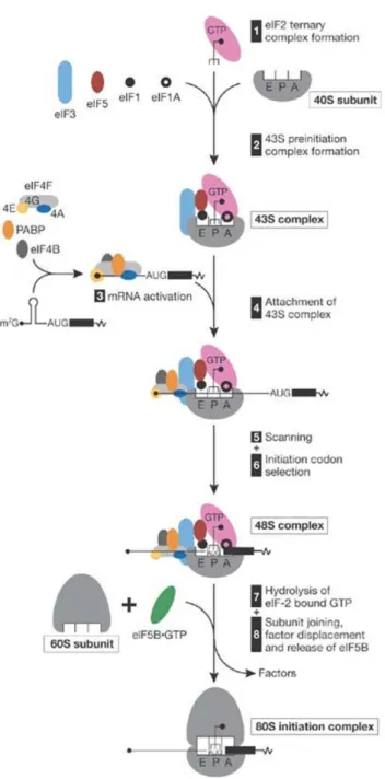
The translation process comprises three phases – initiation, elongation and termination. Translation initiation requires at least 11 initiation factors and occurs in two stages: formation of the 48S initiation complex at the initiation or start codon of mRNA, and its joining with a 60S ribosomal subunit, which results in the assembly of a competent 80S ribosome so it proceeds with elongation (Pestova et al., 2007; Sonenberg and Hinnebusch, 2009). On most mammalian mRNAs, the start codon is identified by a scanning mechanism, where the 43S pre-initiation
complex binds to the mRNA near the 5’ end and scans the 5’ UTR for an AUG codon. The 43S pre-initiation complex comprises the small (40S) ribosomal subunit, the initiation factors eIF1, eIF1A, eIF3 and eIF5, together with the so-called ternary complex. The ternary complex consists of the methionine-loaded initiator transfer RNA (Met-tRNAi), which will recognize the initiation
AUG codon, and eIF2 coupled to GTP (Gebauer and Hentze, 2004) (Figure I.3, steps 1 and 2). Binding of the 43S pre-initiation complex to the mRNA requires the cooperative action of eIF4F
and eIF4B or eIF4H, which unwind the 5’ UTR of the mRNA to allow ribosomal attachment. eIF4F
is composed by eIF4E (or CBP80:20), that physically binds the m7G cap structure, eIF4A and by
eIF4G, which functions as a scaffold protein promoting the assembly of the several factors involved in initiation (Gebauer and Hentze, 2004; Pestova et al., 2007). eIF4G interacts
simultaneously with eIF4E, eIF4A, eIF3, and with the 3’ end-associated cytoplasmic poly(A)- -binding protein (PABPC1) (Jackson et al., 2010) (Figure I.3, step 3). The eIF4G-mediated
interaction between eIF4E and PABP is thought to circularize the mRNA, bringing the 3’ UTR in close proximity to the 5’ end of the mRNA (Wells et al., 1998; Gebauer and Hentze, 2004). The
et al., 2010) (Figure I.3, step 4). The resulting 43S pre-initiation complex can now land next to the cap and scan the mRNA, in a 5’ to 3’ direction, until encountering the most 5’-proximal AUG start codon in a Kozak consensus sequence, where it forms a 48S initiation complex (Pestova et al., 2007) (Figure I.3, steps 5 and 6). Once the anticodon of the Met-tRNAi has engaged the start
codon, eIF5 triggers the eIF2-bound GTP hydrolysis, resulting in the release of eIF2-GDP and probably of other 40S-bound initiation factors (Gebauer and Hentze, 2004; Jackson et al., 2010). Finally, eIF5B catalyzes the joining of 60S ribosomal subunit (Figure I.3, steps 7 and 8). This event results in the assembly of an 80S ribosome at the initiation codon, and elongation can start to synthesize the polypeptide (Pestova et al., 2007).
In the elongation stage of translation, amino acids are added sequentially to the growing polypeptide chain (Abbott and Proud, 2004). The ribosome has three tRNA-binding sites through
which the tRNA substrates progress in a stepwise fashion: the Α-(aminoacyl) site, which accepts the incoming aminoacyl-tRNA, the P-(peptidyl) site, which holds the tRNA with the nascent peptide chain, and the E-(exit) site that holds the deacylated tRNA before it leaves the ribosome (Proud, 1994). The elongation process depends on the factor eEF1A, which mediates the delivery of the aminoacyl-tRNA to the Α-site of the ribosome where decoding takes place. Following a proofreading step to confirm the proper codon-anticodon interaction, the correct (cognate) aminoacyl-tRNA becomes accommodated into the Α-site. The ribosome then catalyses peptide bond formation between the aminoacyl-tRNA and the peptidyl-tRNA bound in the adjacent P-site,
resulting in the transfer of the peptide chain to the Α-site tRNA. Subsequently, eEF2 catalyses translocation of the peptidyl-tRNA and mRNA from the Α- to the P-site, and translocation of the deacylated tRNA from the P- to the E-site. The ribosome is moved along the mRNA such that the
next codon is positioned in the Α-site, and the elongation process is repeated (Abbott and Proud, 2004; Kapp and Lorsch, 2004).
Figure I.3 Simplified model of the translation initiation mechanism. eIF2-GTP/Met-tRNAiMet
ternary complex, eIF3, eIF1, eIF1A, eIF5 and a 40S subunit form a 43S pre-initiation complex, which initially