Resistance of mRNAs with AUG-proximal nonsense
mutations to nonsense-mediated decay reflects
variables of mRNA structure and translational activity
Francisco J.C. Pereira
1, Alexandre Teixeira
1,2, Jian Kong
3, Cristina Barbosa
1,4, Ana
Lu´ısa Silva
1, Ana Marques-Ramos
1, Stephen A. Liebhaber
3and Lu´ısa Rom ˜ao
1,4,*1Departamento de Gen ´etica Humana, Instituto Nacional de Sa ´ude Doutor Ricardo Jorge, 1649-016 Lisboa, Portugal,
2Centro de Investigac¸ ˜ao em Gen ´etica Molecular Humana, Faculdade de Ci ˆencias M ´edicas, Universidade Nova de
Lisboa, 1349-008 Lisboa, Portugal,3Departments of Genetics and Medicine, University of Pennsylvania, Philadelphia, PA 19104, USA and4BioISI - Biosystems & Integrative Sciences Institute, Faculdade de Ci ˆencias, Universidade de Lisboa, 1749-016 Lisboa, Portugal
Received March 26, 2015; Revised May 19, 2015; Accepted May 23, 2015
ABSTRACT
Nonsense-mediated mRNA decay (NMD) is a surveil-lance pathway that recognizes and selectively degrades mRNAs carrying premature termination codons (PTCs). The level of sensitivity of a PTC-containing mRNA to NMD is multifactorial. We have previously shown that human-globin mRNAs car-rying PTCs in close proximity to the translation ini-tiation AUG codon escape NMD. This was called the ‘AUG-proximity effect’. The present analysis of non-sense codons in the human ␣-globin mRNA illus-trates that the determinants of the AUG-proximity ef-fect are in fact quite complex, reflecting the ability of the ribosome to re-initiate translation 3′ to the PTC and the specific sequence and secondary structure of the translated ORF. These data support a model in which the time taken to translate the short ORF, impacted by distance, sequence, and structure, not only modulates translation re-initiation, but also im-pacts on the exact boundary of AUG-proximity pro-tection from NMD.
INTRODUCTION
The classical model of nonsense-mediated mRNA decay (NMD) in mammalian cells stipulates that the relationship of a nonsense codon to exon–exon junctions in a PolII tran-script dictates whether it will be recognized as ‘premature’ and trigger rapid decay. This decay, when it occurs, is trig-gered by an interaction of the translation termination plex at the stop codon with a retained exon junction com-plex (EJC) on the mRNA (1–6). These protein interactions appear to be critical to the discrimination of a premature
translation termination event from a normal one (1–3,5,6). The EJC is deposited 20–24 nucleotides (nts) upstream of the exon–exon junction(s) during splicing and remains as-sociated with the mRNA during its transport to the cyto-plasm (1–3). Translating ribosomes subsequently displace EJCs from the open reading frame (ORF) during the pio-neer round of translation. If a stop codon is located more than 50–54 nts upstream of at least one exon–exon junc-tion, the leading edge of the elongating ribosome will fail to displace it. In this case, when the ribosome reaches the termination codon, the translation eukaryotic release fac-tors eRF1 and eRF3 at the stop codon interactin ciswith the retained EJC(s)viabridging interactions between the re-lease complex associated proteins, UPF1 and SMG-1 and the EJC-associated factors, UPF2-UPF3 (7,8). This bridg-ing interaction triggers accelerated decay (i.e. NMD) of the nonsense-containing mRNA through the recruitment of ad-ditional factors (9–19).
In addition to the dependent NMD model, an EJC-independent NMD pathway postulates that identification of a stop codon as ‘premature’ depends on the physical dis-tance between the stop codon and the cytoplasmic poly(A)-binding protein 1 (PABPC1) bound to the poly(A) tail (20– 25). This ‘faux 3′UTR’ model proposes that PABPC1 and
UPF1 compete for interaction with eRF3 at the site of translational termination: if PABPC1 is in close proxim-ity to a stop codon, it interacts with the termination com-plex, stimulates translation termination (26), and represses NMD; alternatively, when the interaction of PABPC1 with the termination complex is reduced, for example due to a long 3′untranslated region (3′UTR), UPF1 interacts with
eRF3 and triggers NMD (20–25). Recent studies that map UPF1 binding throughout the mRNA (5′ UTRs, coding
regions and 3′ UTR) (27–29) irrespective of NMD (28)
seem to challenge this mechanistic model of NMD.
Nev-*To whom correspondence should be addressed. Tel: +351 21 750 8155; Fax: +351 21 752 6410; Email: [email protected]
C
The Author(s) 2015. Published by Oxford University Press on behalf of Nucleic Acids Research.
ertheless, elongating ribosomes displace UPF1 from cod-ing sequences causcod-ing its enrichment in 3′UTRs (28); thus,
transcripts with long 3′UTRs might increase the
probabil-ity that UPF1 will outcompete PABPC1 for release factor binding and trigger NMD.
Consistent with the faux 3′ UTR model of NMD is
the fact that endogenous NMD substrates are enriched in mRNAs containing long 3′UTRs (30–33). This model is
also supported by the observation that artificially tether-ing PABPC1 in close proximity to a premature termina-tion codon (PTC) can inhibit NMD through a mechanism that involves its eRF3-interacting C-terminal domain (21– 24,34). However, recent data have shown that interaction of PABPC1 with eRF3 is not strictly necessary for the tethered PABPC1 to suppress NMD (35), as NMD suppression may also be mediatedviaPABPC1 interaction with the eukary-otic initiation factor 4G (eIF4G) (36,37). Furthermore, it has been suggested that a key NMD determinant might be the efficiency of ribosome release at the PTC (38), which is an event where UPF1 seems to have a role (39). These and other observations (reviewed in reference38) reinforce the conclusion that the mechanisms that dictate NMD strength are complex and not well defined.
The pivotal role that PABPC1 plays in NMD suppression when in close proximity to a stop codon can also be high-lighted by the ‘AUG-proximity effect’. Studies from our lab-oratory have shown that human-globin (h-globin) mR-NAs containing nonsense mutations early in exon 1 accu-mulate to levels similar to those of wild-type (WT)-globin transcripts (40). This resistance to NMD is erythroid- and promoter-independent, and does not reflect translation re-initiation, abnormal RNA splicing, or impaired transla-tion (41). Instead, the observed NMD-resistance reflects the close proximity of the nonsense codon to the translation ini-tiation codon (41). This was called the ‘AUG-proximity ef-fect’ (21). Consistent with the repressive impact of PABPC1 on NMD (see above) (20–24) our mechanistic studies re-vealed that the AUG-proximity effect results the juxtaposi-tion of PABPC1 with the AUG-proximal PTC as a conse-quence of mRNA circularization and the inherent nature of the short ORF translation process (21,34).
In the present report, we carry out a detailed comparison of the AUG-proximity effect on NMD of the human␣- and
-globin mRNAs. While the data support a generality of the AUG-proximity effect on NMD, this detailed comparison also highlights variables of mRNA sequence and structure that factor into this pathway of NMD resistance. The im-pact of these variables appears to reflect the time taken for the 80S ribosome to translate the short ORF prior to en-countering the PTC.
MATERIALS AND METHODS
Construction of expression vectors
The 1677-bp EcoRI/RcaI fragment containing the whole wild-type ␣2-globin gene was obtained from plasmid pTet-␣WT (42) and sub-cloned into EcoRI/BspLU11I sites of the pTRE2pur vector (BD Biosciences), originating the pTRE-␣WT plasmid. Constructs ␣4 (CCU→UAG), ␣7 (AAU→UAG), ␣10 (GUC→UAG),
␣12 (GCC→UAG), ␣14 (UGG→UAG), ␣14/40
(UGG→UAG, AAG→UAG), ␣16 (AAG→UAA), ␣19 (GCG→UAG),␣21 (GCU→UAG), ␣23 (GAG→UAG),
␣25 (GGU→UAG), ␣27 (GAG→UAG), ␣30 (GAG→UAG), ␣32 (AUG→UAG), ␣36 (UUC→UAG),
␣40 (AAG→UAG), ␣45 (CAC→UAG), ␣50 (CAC→UAG), ␣55 (GUU→UAG), ␣60 (AAG→UAG),
␣65 (CGC→UAG), ␣70 (GUG→UAG), ␣73 (GUG→UAG), ␣76 (AUG→UAG), ␣78 (AAC→UAG),
␣80 (CUG→UAG), ␣82 (GCC→UAG), ␣84 (AGC→UAG), ␣86 (CUG→UAG), ␣93 (GUG→UAA),
␣101 (CUA→UAG) and␣116 (GAG→UAG), carrying the denoted nonsense mutations at codon positions indicated by the respective number, were created by site-directed mutagenesis, as recommended by the kit manufacturer (QuikChange Site-Directed Mutagenesis Kit; Stratagene), with mutagenic primers #1-#62 (Supplementary Table S1), using the plasmid pTRE-␣WT as DNA template. Potential re-initiation sites in ␣-globin codons 32 and 76 were sequentially mutated, from AUG (methionine: Met) to ACG (threonine: Thr), in cis by site-directed mutage-nesis with primers #63-#66, using pTRE-␣WT, ␣14,␣27 or ␣40 as templates, to create the constructs ␣WT.32– 76Met→Thr, ␣14.32–76Met→Thr, ␣27.32–76Met→Thr and␣40.32–76Met→Thr.
The␣WT-pseudoknot (pk),␣25-pk,␣27-pk and␣40-pk gene variants were prepared by replacing the first 19 codons of the native ␣-globin ORF by a 19-codon sequence re-sulting in a pseudoknot structure in the mRNA (43), by overlap-extension polymerase chain reaction (PCR) with overlapping primers #67 and #68 (Supplementary Table S1) and the␣WT,␣25,␣27 or␣40 genes as DNA templates. The ␣WT-(CAA)26 and ␣40-(CAA)26 gene variants were obtained by replacing the first 26 codons of the␣-globin ORF with 26 consecutive ‘CAA’ repeats, also by overlap-extension PCR, with overlapping primers #69 and #70 and the␣WT or␣40 genes as DNA templates. The␣27-(CAA)26 gene was created with overlapping primers #71 and #72 and the␣WT-(CAA)26 gene as DNA template. The␣ WT-(CAA)26–32–76Met→Thr, ␣27-(CAA)26–32–76Met→Thr and␣40-(CAA)26–32–76Met→Thr gene variants were pro-duced with overlapping primers #63 and #64 and the␣ WT-(CAA)26, ␣27-(CAA)26, ␣WT.32–76Met→Thr or ␣ 40-(CAA)26–32–76Met→Thr genes as DNA templates. For all the above mentioned ␣-globin gene variants prepared by overlapping PCR, flanking primers #73 and #74 were used and a 923-bp KpnI/ApaI fragment of each PCR product was cloned into the KpnI/ApaI sites of the pTRE-␣WT plasmid.
The wild-type -globin gene (WT), as well as the previously described human -globin variants 15 (UGG→UGA) and 39 (CAG→UAG) (41), were sub-cloned into the ClaI/BspLU11I sites of pTRE2pur vector (BD Biosciences) by PCR amplification of the 1806-bp ClaI/BspLU11I fragment, using primers with linkers for ClaI and BspLU11I (primers #75 and #76; Supplementary Table S1). The -globin variants 23 (GUU→UAG),
variants were constructed by replacing the first 19 codons of a native-globin ORF by a 19-codon sequence resulting in a pseudoknot structure in the mRNA (43), using the ExSite PCR-Based Mutagenesis Kit (Stratagene) as indi-cated by the manufacturer, with mutagenic primers #83 and #84, and theWT and39 genes as DNA templates, or primers #83 and #85 using the23 gene as DNA template. The WT-(CAA)25 and 39-(CAA)25 gene variants were obtained by replacing the first 25 codons of the native
-globin ORF with 25 consecutive ‘CAA’ repeats, by overlap-extension PCR with overlapping primers #86 and #87, and the WT or39 genes as DNA templates. The
26-(CAA)25 gene was prepared with overlapping primers #88 and #89, and the WT-(CAA)25 gene as template. Flanking primers #73 and #90 were used and a 478-bp ClaI/BbrPI fragment of each PCR product was cloned into the ClaI/BbrPI sites of theWT construct.
The hybrid genes 5′ UTR-/␣WT, 5′ UTR-/␣27, 5′
UTR-/␣40,WT26/␣,26/␣‘27’ andWT26/␣40 were obtained by replacing the 5′untranslated region (5′UTR)
or the first 26 codons plus the 5′ UTR of the
respec-tive␣-globin gene variants by the equivalent -globin se-quences. This was achieved by overlap-extension PCR with overlapping primers #91-#96 (Supplementary Table S1) and flanking primers #73 and #74, and the ␣WT, ␣27,
␣40,WT or26 genes as DNA templates. Additionally, 5′ UTR-␣/WT26/␣, 5′ UTR-␣/26/␣‘27’ and 5′
UTR-␣/WT26/␣40 genes were shaped by replacing the 5′UTR
of WT26/␣, 26/␣‘27’ and WT26/␣40, respectively, with the native 5′ UTR of␣-globin. Overlapping primers
#97 and #98 were used together with flanking primers #73 and #74 on␣WT,WT26/␣,26/␣‘27’ andWT26/␣40 template genes. A 1094-bp XbaI/BbrPI fragment of each PCR product was cloned into the XbaI/BbrPI sites of the␣WT construct. The hybrid genes 5′UTR-␣/WT, 5′
UTR-␣/26, 5′ UTR-␣/39, ␣WT27/, ␣27/‘26’ and
␣WT27/39 were produced reciprocally to/␣-globin hy-brid genes, with overlapping primers #97-#102 and flank-ing primers #73 and #90, and theWT,26,39,␣WT or ␣27 genes as DNA templates. Furthermore, the hy-brid genes 5′UTR-/␣WT27/, 5′UTR-/␣27/‘26’ and
5′ UTR-/␣WT27/39 were obtained using overlapping
primers #91 and #92, flanking primers #73 and #90, and theWT,␣WT27/,␣27/and␣WT27/39 genes as tem-plates. The 1110-bp XbaI/BbrPI fragment of each PCR product was inserted into the XbaI/BbrPI sites of theWT construct.
The 514-bp XhoI/HindIII fragment containing the
PhCMV*-1 tetracycline (tet)-responsive promoter was ob-tained from the pTRE2pur vector (BD Biosciences) and sub-cloned into XhoI/HindIII sites of the pGL2-Enhancer vector (Promega), which contains the firefly luciferase gene, and into XhoI/HindIII sites of the pGL4.70[hRluc] vector (Promega), which contains theRenillaluciferase gene, orig-inating the pGL2TRE and the pGL4TRE plasmids, respec-tively.
The ␣-globin/luciferase (Luc) hybrid genes, ␣14/Luc,
␣27/Luc, ␣27-(CAA)26/Luc and ␣27-pk/Luc, were ob-tained by replacing the ␣-globin sequence downstream to codon 32 with the firefly luciferase ORF, by overlap-extension PCR with overlapping primers #103 and #104
(Supplementary Table S1) and the␣14,␣27,␣27-(CAA)26 and ␣27-pk or pGL2TRE constructs as DNA templates. The respective positive and negative controls for luciferase activity were created with overlapping primers #105-#108 and the ␣14/Luc or ␣27/Luc genes as DNA templates. Flanking primers #73 and #109 were used and a 752-bp XhoI/XbaI fragment of each PCR product was cloned into the XhoI/XbaI sites of the pGL2TRE plasmid.
The-globin/Luc hybrid genes,15/Luc,23/Luc and
26/Luc, were prepared by replacing the-globin sequence downstream to codon 55 with the firefly luciferase ORF, by overlap-extension PCR with overlapping primers #110 and #111 (Supplementary Table S1) and the15,23,26 or pGL2TRE constructs as DNA templates. The respec-tive posirespec-tive and negarespec-tive controls for luciferase activity were produced with overlapping primers #112-#115 and the 15/Luc, 23/Luc or 26/Luc genes as DNA tem-plates. Flanking primers #73 and #109 were used and a 906-bp XhoI/XbaI fragment of each PCR product was cloned into the XhoI/XbaI sites of the pGL2TRE plasmid.
Cell culture and transfections
Mouse erythroleukemia (MEL) cells stably expressing the tet transactivator (MEL/tTA) (42) were used for condi-tional expression of human ␣-globin genes (previously cloned into the pTRE2pur vector). For transient transfec-tions, MEL/tTA cells were split 1 day before transfection and cultured in minimal essential medium (MEM) supple-mented with 10% (v/v) fetal bovine serum, and 100 ng/ml tetracycline (tet). Cells were transfected with 2g of
pTRE-␣WT, or each variant, as previously described. Then, cells were split into 60-mm-diameter dishes, and pulsed with␣ -globin mRNA for 4 h by growth in tet(-) media. Following this period, transcription from the plasmid was blocked by the addition of tet to the media. Cells from each culture dish were harvested in different time points for further analyses. HeLa cells, stably expressing the tet transactivator (HeLa/tTA) (42), were grown in Dulbecco’s modified Ea-gle’s medium (DMEM) supplemented with 10% (v/v) fe-tal bovine serum. Transient transfections were performed using Lipofectamine 2000 Transfection Reagent (Invitro-gen), following the manufacturer’s instructions, in 35-mm plates. For genes cloned into the pTRE2pur vector, 150 ng of the test construct DNA were cotransfected with 1850 ng of pEGFP plasmid DNA (BD Biosciences) as a con-trol for monitoring transfection efficiency, and cells were harvested after a 20 h transcription pulse. For gene con-structs cloned into the pGL2TRE plasmid, 2 g of the test construct DNA were cotransfected with 50 ng of the pGL4TRE plasmid as a control for luminescence, and cells were harvested after a 16 h transcription pulse. When ap-propriated, transcription was blocked by the addition of tet or 50 g/ml adenosine analogue 5,6-dichloro-1–3-D-ribofuranosylbenzimidazole (DRB) to the media and cells were harvested in different time points after treatment.
Transfection of siRNA
the manufacturer’s instructions in 35-mm plates using 100 pmol of siRNA oligonucleotides and 4 l of transfec-tion reagent. Twenty-four hours later, cells were transfected again with 50–75 pmol of siRNAs, 150 ng of the test con-struct DNA and 1000 ng of pEGFP vector. After additional 24 h, cells were harvested for analysis of RNA and protein expression. When appropriated, HeLa cells were treated with 50 g/ml DRB and RNA was thereafter extracted at different time points for further analysis. The siRNA oligonucleotides used for transfections [Luciferase (5′
-AA-CGUACGCGGAAUACUUCGA-3′) and hUPF1 (5′
-AA-GAUGCAGUUCCGCUCCAUU-3′)] were purchased as
annealed, ready-to-use duplexes from Dharmacon.
RNA isolation
Total RNA from transfected cells was prepared using the RNeasy mini kit (Qiagen) following the manufacturer’s indications. RNA samples were treated with RNase-free DNase I (Ambion) and purified by phenol:chloroform ex-traction. Before further analyses, mRNA samples were tested by reverse-transcription (RT) followed by PCR (RT-PCR) to reject the hypothesis of activation of cryptic splic-ing pathway(s), with consequent alteration in mRNA se-quence and possible circumvention of the premature ter-mination codon. From all transcript species a single full-length product was amplified (data not shown), demonstrat-ing that the studied nonsense transcripts present a normal splicing pattern.
Ribonuclease protection assay (RPA)
The␣-globin probe is a 174-bp fragment encompassing the 3′part of exon 3 inserted into the polylinker region of
pTRI-amp-18 (Ambion) and was generated byin vitro transcrip-tion, using a Maxiscript T7 or SP6 kit (Ambion), accord-ing to the manufacturer’s standard protocol. The-globin probe was produced using a Maxiscript SP6 kit (Ambion) and consists of a 170 bp fragment encompassing the last 20 bp of intron 2, the entire exon 3 coding region, and the first 21 bp of 3′ UTR, which was amplified by PCR and
inserted into the polylinker region of pGEM3 (Promega). The puroRprobe was generated using a Maxiscript T7 kit (Ambion), comprises a 280 bp puromycin-resistance gene fragment cloned into pGEM3 and protects 197 nt of the puromycin-resistance mRNA. Samples were processed as described elsewhere (41,44).
Western blot analysis
Protein lysates were resolved, according to standard pro-tocols, in 10% SDS-PAGE, and transferred to polyvinyli-dene difluoride (PVDF) membranes (Bio-Rad). Mem-branes were probed using mouse monoclonal anti-␣ -tubulin (Roche) at 1:10000 dilution, and goat polyclonal anti-hUPF1 (Bethyl Labs) at 1:500 dilution. Detection was carried out using secondary peroxidase-conjugated anti-mouse IgG (Bio-Rad) or anti-goat IgG (Sigma) antibodies followed by chemiluminescence.
Semi-quantitative RT-PCR
The reverse-transcription (RT) of 500 ng of RNA from HeLa cells cotransfected with pGL2TRE (or ␣- and  -globin/Luc hybrid variants; containing the firefly luciferase gene) and pGL4TRE (which contains theRenillaluciferase gene) was performed with Superscript II (Invitrogen), un-der conditions recommended by the manufacturer, using 2 pmol of reverse primers #109 and #116 (Supplementary Ta-ble S1) in a final volume of 10l. The PCR reactions for firefly and Renilla luciferases were executed in parallel at similar conditions: 1 l of the RT product was amplified in a 25l reaction volume using 0.2 mM dNTP mixture, 1.5 mM MgCl2, 10 pmol of each primer (primers #109 and #117 for the firefly luciferase and primers #116 and #118 forRenillaluciferase), 0.75 U of GoTaq DNA Polymerase (Promega) and 1x Reaction Buffer (Promega). Thermocy-cler conditions were 95◦C for 3 min, followed by 29 cycles
of 95◦C for 30 s, 58◦C for 45 s and 72◦C for 45 s, followed
by a final extension of 72◦C for 10 min. Samples were
re-solved by electrophoresis in 1% agarose gels stained with ethidium-bromide, which were then digitalized and densito-metric analysis was performed using ImageQuant software (Molecular Dynamics).
Reverse transcription-coupled quantitative PCR (RT-qPCR)
First-strand cDNA was synthesized from 2 g of total RNA using the SuperScript II Reverse Transcriptase (Invitrogen) according to the manufacturer’s instructions. Real-time PCR was performed with the ABI7000 Sequence Detection System (Applied Biosystems) using SYBR Green PCR Master Mix (Applied Biosystems). The relative expression levels of -globin and ␣-globin mRNAs were normalized to the internal control puromycin-resistance mRNA in HeLa cells and calculated using the com-parative CT method (2−CT) (45). The CT values of variant -globin and ␣-globin mRNAs amplicons were compared to the respective WT, ␣WT counterpart or to WT Luc siRNA, ␣WT Luc siRNA as indicated in figures and normalized with the reference amplicon CT value. The amplification efficiencies of the -globin,
␣-globin targets and puromycin-resistance reference am-plicons were determined for each assay by dilution series. The forward and reverse primers for human -globin mRNA were 5′
-GTGGATCCTGAGAACTTCAGGCT-3′ and 5′-CAGCACACAGACCAGCACGT-3′ and for
␣-globin were 5′
-CCCGGTCAACTTCAAGCTCC-3′ and 5′-CAGCACGGTGCTCACAGAAG-3′.
In addition, we have used the following primers: 5′-CGCAA CCTCCCCTTCTACG-3′ and 5′
-GGTGACGGTGAAGCCGAG-3′ for
Luminometry assays
HeLa cells were lysed with Passive Lysis Buffer (Promega) and luminescence was measured in a Lucy 2 luminometer (Anthos Labtec) with the Dual-Luciferase Reporter Assay System (Promega), according to the manufacturer’s stan-dard protocol, using automatic injectors.
RESULTS
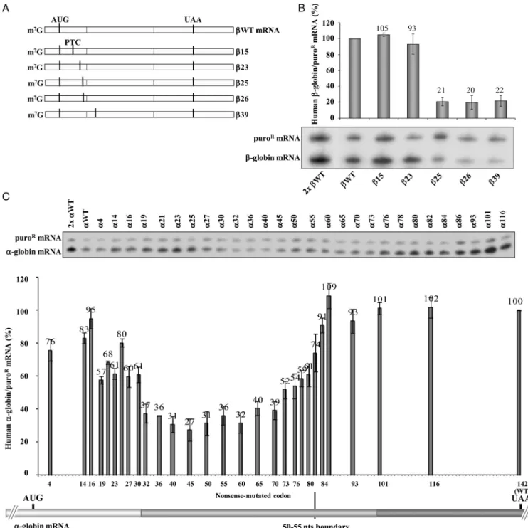
The AUG-proximity boundaries that dictate NMD resistance differ between human- and␣-globin mRNAs
We have previously reported that human-globin mRNAs carrying nonsense mutations in proximity to the initiation AUG are resistant to NMD and are expressed at levels ap-proaching those of the wild-type-globin (WT) mRNA (40,41). To further characterize this ‘AUG-proximity effect’ and to define its parameters, we compared the boundary of this effect in human-globin and␣-globin mRNAs.
The boundaries were mapped by assessing expression of corresponding series of ␣- and-globin mRNAs (Figure
1). The analysis of the-globin mRNAs (summarized in Figure 1B) revealed that 15 and 23 mRNAs accumu-late at normal levels, more specifically at 105% and 93% of the WT mRNA, respectively (Figure 1B), while lev-els of 25,26, and39 mRNAs were drastically lower, at 21%, 20% and 22% of WT mRNA, respectively (Fig-ure1B). The stabilization of25,26 and39 mRNAs in UPF1-depleted cells to WT levels confirmed that the low levels were due to NMD (Supplementary Figure S1A and B). Targeted comparison of15 and39 mRNAs further confirmed the distinction of NMD sensitive and NMD re-sistant mRNAs related to their AUG-proximity (Supple-mentary Figure S1C). Together, these data map the bound-ary of the AUG-proximity effect for NMD resistance of -globin mRNA between codons 23 and 25.
In a separate experiment, we mapped the boundary of the AUG-proximity effect for NMD resistance in the h␣-globin mRNA. Results revealed that␣-globin mRNAs carrying a PTC located at exon 1 (exon 1 encompasses 30 codons) ac-cumulate to steady-state levels of 57% to 95% of normal control that are significantly above typical NMD levels (35) (Figure1C). In contrast, mRNAs containing nonsense mu-tations in the first half of exon 2, between codons 32 and 70, appear to be fully sensitive to NMD with levels ranging from 27% to 40% of the␣WT levels (Figure1C). Nonsense mutations positioned further into exon 2 achieved higher levels, most likely reflecting an inhibition of NMD as the PTC comes within 50–55 nts of the last exon–exon junction (Figure1C). As predicted by the EJC model of NMD, the PTCs located 3′to codon 82 accumulated to normal levels
(Figure1C).
To confirm that the steady-state mRNA levels being mea-sured reflected the corresponding stabilities of the PTC-containing h␣-globin mRNAs, we measured the absolute half-lives of selected mRNAs. The half-life of the ␣32 mRNA (3.5 h) and ␣40 mRNA (2.0 h) (46) were consis-tent with a full sensitivity to NMD (Supplementary Figure S2A) and contrasted with the half-lives of␣27 (7.5 h) and
␣30 (7.0 h). The intermediate half-lives of␣73 (5.5 h) and
␣80 (6.2 h) mRNAs are likely to reflect stabilization as the PTC is moved closer to the terminal EJC (47,48).
To directly demonstrate that␣-globin transcripts carry-ing a PTC in exon 1 escape NMD, we analyzed the im-pact of UPF1-depletion in HeLa cells (Supplementary Fig-ure S2B). In cells treated with control Luc siRNAs, the␣7,
␣10,␣12,␣14, and␣16 mRNAs accumulated to levels 86% to 98% of the␣WT control, contrasting with 30% for the NMD-sensitive␣40 mRNA. UPF1 depletion had no appre-ciable impact on the levels of the␣7,␣10,␣12,␣14, and␣16 mRNAs (Supplementary Figure S2C). Levels of␣23,␣27 and␣30 mRNAs in the control siRNA-treated cells were intermediate (77%, 67% and 66%) (Supplementary Figure S2C), consistent with partial NMD sensitivity. Consistent with this conclusion was the observation that UPF1 deple-tion resulted in a significant increase in the accumuladeple-tion levels of␣23,␣27,␣30, and␣40 mRNAs (Supplementary Figure S2C), as well as in a significant increase in the half-lives of␣27, and␣40 mRNAs, while that of the␣16 mRNA did not significantly change (Supplementary Figure S2D).
Taken together, these data are consistent with NMD-resistance of PTCs located in close proximity to the AUG. In the case of the h␣-globin mRNA, PTCs extending to at least codon 16 escape the full impact of NMD and PTCs located more distal to the AUG in exon 1 demonstrate par-tial commitment to NMD. Of note, the boundary for the AUG-proximity effect appears to differ between the two hu-man globin mRNAs; the boundary of the AUG-proximity effect for NMD resistance in h-globin mRNA maps be-tween codons 23 and 25, contrasting with the boundary fur-ther 3′(between codons 30 and 32) in the h␣-globin mRNA.
Defining the basis for this difference has potential to the full understanding of the NMD pathway response.
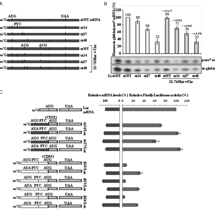
The human␣-globin mRNAs can activate efficient transla-tional re-initiation 3′to an AUG-proximal PTC
Translation re-initiation downstream of a PTC has the potential to inhibit NMD via disruption of downstream EJC(s) (49). We have previously shown that translation re-initiation does not occur 3′to AUG-proximal nonsense
mu-tations in h-globin mRNAs (for example, 15 mRNA) (41). In contrast, re-initiation can occur in the h␣-globin mRNA (46). Such re-initiation might account for the differ-ence in the NMD resistance boundary. To test this possibil-ity, we first focused our studies on the␣-globin mRNA with a PTC at the 3′part of exon 1, at codon 27. The PTC at this
position in h␣-globin mRNA is partially NMD-resistant and is 3′to the AUG-proximity boundary determined for
the h-globin mRNA. We mutated the putative initiating AUG codons located downstream of the PTC, which are lo-cated at positions 32 and 76. The 32Met and 76Met AUG-to-ACG conversions created the␣27.32–76Met→Thr gene (Figure 2A). Its expression was compared to that of the
␣WT.32–76Met→Thr, ␣14.32–76Met→Thr, and ␣40.32– 76Met→Thr control genes previously described (46). The
80% of␣14 mRNA, as previously described (46), showing that blocking potential translation re-initiation sites results in partial NMD triggering of the␣14 mRNA. Consistent with this conclusion is the observation that UPF1 deple-tion resulted in a significant increase in the accumuladeple-tion of
␣14.32–76Met→Thr mRNA (Supplementary Figure S3B). In addition, we observed that␣27.32–76Met→Thr mRNA accumulates at about 56% of␣WT, and at about 85% of␣27 mRNA (Figure 2B). Of note, expression of the ␣27 mR-NAs with the double missense mutation remains well above the levels of the␣40 mRNA, indicating that translation re-initiation does not fully explain their NMD resistance.
To better quantify the impact of translation re-initiation on␣14 and␣27 mRNA accumulation, we used a luciferase reporter assay. The first in site for potential re-initiation downstream of␣14 and␣27 is at codon 32Met. Thus, we created a set of␣14/Luc and␣27/Luc constructs in which the ␣-globin gene upstream of codon 32 was fused with the firefly luciferase ORF (Figure2C). After transient ex-pression of these constructs in HeLa cells, luciferase activ-ities were measured and normalized to the corresponding mRNA levels. Results show that the␣14/Luc and␣27/Luc constructs carrying the 0Met codon mutated to AUA, al-low for 104% and 108% of the uncoupled luciferase rela-tive activity, respecrela-tively. In addition, both ␣14/Luc and
␣27/Luc constructs lead to the production of consider-able high amounts of luciferase, being about 75% and 62% of the uncoupled luciferase activity, respectively (Figure
2C). Taken together, these results demonstrate that transla-tion can efficiently re-initiate at codon 32Met in h␣-globin mRNA. Our data further reveal that the efficiency of trans-lation re-initiation decreases when the ORF is lengthened and the intercistronic region is shortened, as has been pre-viously described (43,50).
An equivalent analysis was performed to show the in-effectiveness of -globin mRNA to re-initiate translation at codon 55Met as previously defined (41). Indeed, we ob-served that15/Luc and23/Luc constructs result in very low amounts of luciferase activity, at about 7% and 5%, re-spectively, when compared with the relative activity result-ing from the uncoupled luciferase gene (Figure2C). Also, these levels are similar to those obtained from the26/Luc construct, which shows a luciferase activity of 4% (Figure
2C). The very low luciferase activity given by26/Luc con-struct is not surprising, as26 transcripts are efficiently de-graded by NMD. Thus, NMD-resistant (15 and23 mR-NAs) and NMD-sensitive transcripts (26) show similar low levels of translation re-initiation.
This set of experiments lead us to conclude that the level of mRNA accumulation for h␣-globin mRNAs with AUG-proximal PTCs, and the corresponding resistance to NMD, is impacted to some extent by the potential to re-initiate translation 3′to the AUG-proximal PTC.
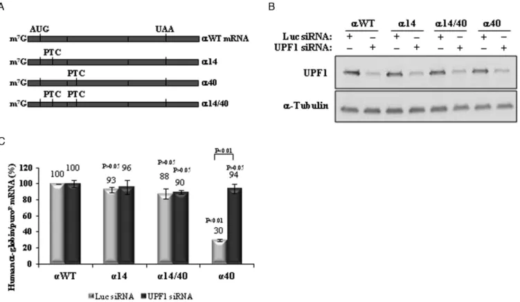
Human ␣-globin mRNAs carrying an AUG-proximal PTC do not allow for ribosomal read-through of the PTC
Ribosomal read-through of a nonsense codon can in theory inhibit NMD, by displacing the downstream located EJC(s) (49) and consequently preventing the interaction of UPF1 with the terminating complex and with the UPF2/UPF3
components of the EJC (7). We have previously shown that the h-globin mRNA carrying the15 mutation incisto the39 mutation does not allow ribosomal read-through of the15 PTC (41). These results have been subsequently confirmed by others (51). Here, we assessed the potential contribution of PTC read-through to NMD resistance of AUG-proximal PTCs in the h␣-globin mRNA. We con-structed an expression vector containing two PTCsin cis, one at codon 14 and another at codon 40 (construct␣14/40; Figure3A). The encoded mRNA was expressed in HeLa cells, in parallel with the␣WT, ␣14 and␣40 control con-structs (Figure3B). Read-through of the PTC at position 14 would be expected to destabilize the double mutant␣14/40 mRNA because the PTC at position 40 is NMD sensitive (Figures 1 and 3C). Contrary to this prediction, we ob-served that the␣14/40 mRNA is expressed at levels com-parable to those of the␣14 and␣WT mRNAs, while␣40 mRNA is expressed at 30% of the normal control (Figure
3C). UPF1 depletion of the transfected cells normalized the levels of␣40 mRNA as expected, but had no impact on the accumulation of the␣14/40 or␣14 mRNA. This set of data shows that PTC read-through does not occur and thus can-not account for the NMD resistance of the AUG-proximal nonsense-mutated␣-globin mRNAs.
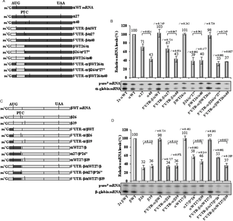
The boundary of the AUG-proximity effect for NMD resis-tance is impacted by the ORF sequence
Although human ␣- and -globin genes derive from the same ancestral gene and share a similar structure and func-tion, the respective mRNAs are distinct in sequence, with a strong divergence in the structures of the correspond-ing 5′ UTRs. This difference in sequence is likely to
con-tribute to the lower efficiency for initiating translation in
␣-globin mRNA (52). Knowing that NMD mechanisms are translation-dependent, we hypothesized that the differ-ence in the boundary of the AUG-proximity effect for␣ -and-globin mRNAs might reflect their respective trans-lation initiation efficiencies. This hypothesis was tested by replacing the 5′UTR of␣-globin with that of the-globin
mRNA in␣WT,␣27 and␣40 genes, generating three hy-brid mRNAs: 5′UTR-/␣WT, 5′UTR-/␣27 and 5′
UTR-/␣40 (Figure4A). mRNA quantification of these tran-scripts transiently expressed in HeLa cells showed that the partial NMD inhibition observed for␣27 mRNA––it accu-mulates at 71% of␣WT mRNA (Figure4B)––is maintained in␣27 mRNAs with the-globin 5′UTR (5′UTR-/␣27
mRNA is 67% of the␣WT).
Next, we tested whether the ORF sequence affects the boundary of the AUG-proximity effect. Thus, in ␣WT, the entire sequence upstream of codon 27 was replaced by the corresponding sequence of WT or 26, creating the WT26/␣ and 26/␣‘27’ constructs, respectively. In
␣40 gene, the entire sequence upstream of codon 27 was replaced by the corresponding sequence of WT, gener-ating theWT26/␣40 gene (Figure4A). Moreover, these latter hybrid genes were further altered by replacing the 5′ UTR of -globin for that of ␣-globin, originating the
5′ UTR-␣/WT26/␣, 5′ UTR-␣/26/␣‘27’ and 5′
Figure 3. The␣-globin mRNA does not allow PTC read-through. (A) Schematic representation of the studied human␣-globin mRNAs. Vertical lines represent translation initiation (AUG) or termination [native (UAA) or premature (PTC)] codons. The name of each transcript is specified on the right. (B) Western blotting analysis of protein samples obtained from HeLa cells transiently transfected with constructs carrying the wild-type␣-globin gene (␣WT), or a␣-globin gene nonsense-mutated at codons 14 (␣14), 40 (␣40) or 14 and 40 (␣14/40). Cells were subjected to single knockdown of UPF1 (UPF1 siRNA) or treated with control siRNA targeting firefly Luciferase (Luc siRNA). Anti-UPF1 and anti-␣-tubulin (control) antibodies were used as indicated. (C) Using these cells, mRNA levels were determined by RT-qPCR using primers specific for human-globin gene and for puromicin-resistance (puroR) gene. Quantification was performed by the relative standard curve method. Histogram represent fold-change of each sample relative to the control (WT Luc siRNA) arbitrarily set to 100%. All values are normalized internally to puroRmRNA levels [SD are shown (n=3)]. TheP-values from student’s
t-tests are also shown. Except otherwise indicated,P-values refer to the comparison with the wild-type control transcript levels treated with control siRNA (␣WT Luc siRNA).
carry the-globin ORF sequence, regardless of the 5′UTR
sequence (Figure4B). As a control for NMD, we observed that hybrid transcripts nonsense-mutated at position 40 (5′
UTR-/␣40,WT26/␣40 and 5′UTR-␣/WT26/␣40)
ac-cumulate at levels equivalent to those of␣40 mRNA (43%, 43%, 40% and 37% of the␣WT mRNA, respectively), all being fully committed to decay.
The reciprocal approach was also used. The 5′UTR of
-globin was replaced by the␣-globin 5′UTR in theWT,
26 and39 genes, creating the 5′UTR-␣/WT, 5′
UTR-␣/26 and the 5′UTR-␣/39 constructs, respectively
(Fig-ure4C). Analysis of these constructs revealed that the26 and39 low mRNA levels are preserved if the transcripts carry the␣-globin 5′UTR (Figure4D).
Moreover, in the WT gene, the entire sequence up-stream of codon 27 was replaced by the corresponding se-quence of ␣WT or␣27 genes, creating the␣WT27/and the␣27/‘26’ genes, respectively; in the39 gene, the entire sequence upstream of the codon 27 was replaced by the cor-responding sequence of␣WT, originating the␣WT27/39 gene (Figure4C). Additionally, the 5′UTR of␣-globin was
replaced by the -globin 5′ UTR in the latter three
hy-brid genes, which originated the 5′ UTR-/␣WT27/, 5′
UTR-/␣27/‘26’, and 5′UTR-/␣WT27/39 constructs
(Figure4C). Results show that if26 mRNA carries the
␣-globin ORF, irrespectively of the 5′ UTR, the mRNA
levels significantly increase from 32% to 58% (␣27/‘26’ mRNA) or 55% (5′ UTR-/␣27/‘26’ mRNA), relatively
to theWT mRNA level (Figure4D).
The analysis of these hybrid mRNAs leads us to make two conclusions. First, we find that the sequence of the 5′
UTR has no impact on determining the NMD sensitivity of the transcript. Second, these results reveal an important role of the ORF sequence in determining the full NMD commit-ment, and thus in determining the exact boundary of the AUG-proximity effect.
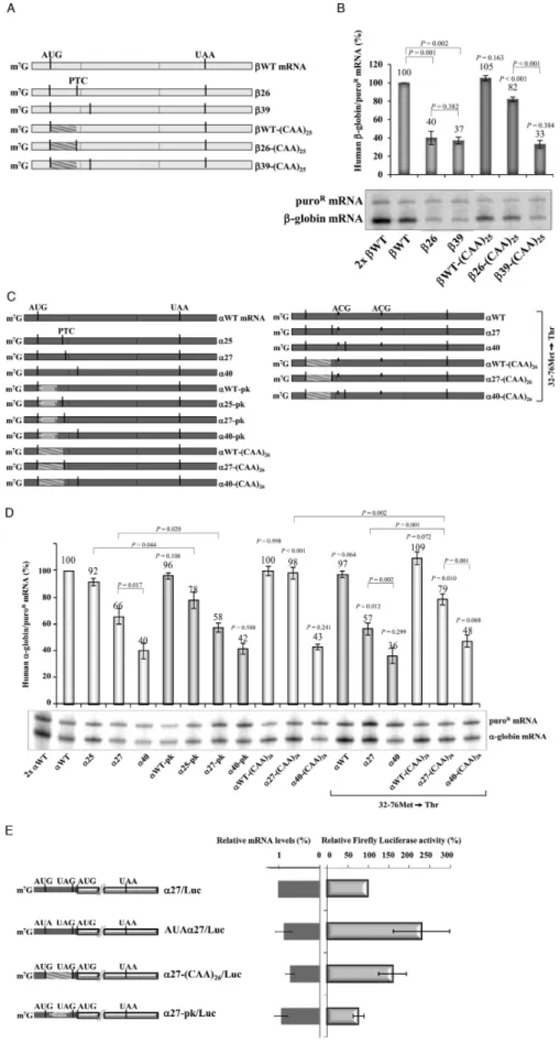
The mRNA secondary structure dictates the boundary of the AUG-proximity effect
sponse and data from Figure4, suggested a model in which the time taken by the ribosome to translate the ORF dic-tates the boundary of the AUG-proximity effect. To fur-ther explore this potential determinant of NMD and the determinants of the boundary of the AUG-proximity ef-fect, we next replaced the first 25 codons of the 26 gene by a tandem array of 25 ‘CAA’ repeats, which has been shown to facilitate rapid translation elongation (55), cre-ating the 26-(CAA)25 construct (Figure 5A). Knowing that the boundary of the AUG-proximity effect in the h -globin mRNA is located between codons 23 and 25, the unstructured sequence when inserted upstream of the26 PTC should inhibit NMD and shift the boundary of the AUG-proximity effect further downstream. The first 25 codons of the WT, and39 control genes were also re-placed, creating theWT-(CAA)25, and39-(CAA)25 con-structs, respectively (Figure5A). mRNA secondary struc-tures predicted by MFOLD (http://mfold.bioinfo.rpi.edu/ cgi-bin/rna-form1.cgi) show that while the26 mRNA has a minimal free energy of −232.50 kcal/mol, introduction of the (CAA)25unstructured segment makes this mRNA to increase its minimal free energy to−199.90 kcal/mol. Our analysis of mRNA accumulation shows that the altered26 mRNA [26-(CAA)25] increased from 40% to 82% of the wild-type control (Figure5B). In contrast, the unstructured insertion did not relieve NMD in the39 mRNA:39, and
39-(CAA)25 mRNAs accumulate at similar levels, respec-tively at 37%, and 33% of theWT transcript (Figure5B). As expected, expression of both39 mRNAs was brought to WT levels in UPF1-depleted cells (Supplementary Figure S4).
To further relate ORF structure to AUG-proximity boundary, using the␣-globin mRNAs, we introduced either a pseudoknot-inducing sequence or an unstructured CAA repeat in the ORF. The pseudoknot was introduced into
␣WT,␣25,␣27 and␣40 genes, replacing the first 19 codons of these constructs (Figure5C). MFOLD predicted that the pseudoknot induces a significant change in the minimal free energy from−231.60 kcal/mol (␣25) and−225.10 kcal/mol (␣27) to−241.20 kcal/mol and−228.40 kcal/mol, respec-tively. In parallel, the unstructured (CAA)26 repeats were introduced into the␣WT, ␣27 and␣40 mRNAs (see Fig-ure5C) changing the minimal free energy of the h␣-globin mRNAs from−225.10 kcal/mol to−185.70 kcal/mol. The analyses of mRNA accumulation revealed that the pseudo-knot structure introduced into the␣25 gene (␣25-pk gene) resulted in a significant reduction of the encoded mRNA accumulation levels (Figure 5D). Identical outcomes were obtained when the pseudoknot structure was inserted in cisto the PTC at position 27 (Figure5D). In contrast, the unstructured ORF consisting in 26 ‘CAA’ repeats inserted into the␣27 mRNA [␣27-(CAA)26mRNA] significantly in-creased the mRNA levels from 66% to 98%, relatively to the ␣WT (Figure 5D). As expected, in the context of a transcript with a PTC further downstream already NMD-sensitive (␣40), the pseudoknot structure, or the (CAA)26 unstructured ORF, does not affect levels of mRNA accu-mulation (Figure 5D). These results lead us to conclude that the sensitivity of mRNAs with AUG-proximal PTCs to NMD is impacted by the strength of secondary structure preceding the PTC; the presence of a pseudoknot increases
NMD sensitivity and the presence of an unstructured ORF decreases NMD sensitivity.
Knowing that h␣-globin transcripts carrying an AUG-proximal PTC, such as the ␣27 mRNA, allow for effi-cient downstream translation re-initiation, which is par-tially responsible for their NMD inhibition (Figure2), we next investigated the outcome of the (CAA)26 unstruc-tured ORF independently of the translation re-initiation ef-fect. For that, we introduced the 26 consecutive ‘CAA’ re-peats into the␣WT.32–76Met→Thr,␣27.32–76Met→Thr and ␣40.32–76Met→Thr genes. Under these conditions, the presence of the (CAA)26segment into the ORF of the
␣27 mRNA makes it to increase from 57% to 79% of the
␣WT mRNA level (Figure 5D). These results are parlel to those obtained when translation re-initiation is al-lowed (Figure5D). In fact, by comparing results obtained in both conditions, we observe that the presence of the un-structured ORF into the␣27 mRNA makes a 1.5-fold in-crease in mRNA levels. Thus, our data show that in the␣27 transcripts, the unstructured ORF enhances NMD inhi-bition independently of translation re-initiation. However, it is worth noticing that the accumulation levels for ␣27 and␣27-(CAA)26mRNAs are higher when translation re-initiation is allowed (Figure5D). These results show that under conditions where translation re-initiation can occur, NMD inhibition is stronger as it results from two different effects: translation re-initiation and the AUG-proximity ef-fect. In addition, the AUG-proximity effect for NMD in-hibition is more prominent when the ORF has an unstruc-tured sequence.
DISCUSSION
In general, nonsense codons located more than 50–55 nts upstream of the 3′-most exon–exon junction elicit NMD
in mammalian cells (47,48). However, our previously re-ported data have shown that human -globin transcripts carrying nonsense mutations in the 5′region of exon 1
accu-mulate to levels comparable to those of wild-type-globin mRNA (40). This ability of mutated-globin mRNA to es-cape NMD was demonstrated to depend on the distance of the nonsense codon to the initiator AUG (21,41,46) and, accordingly, it was named the ‘AUG-proximity effect’. Ad-ditional data have supported that this effect is due to the influence of PABPC1, which seems to be brought into the proximity of an early PTC during cap-dependent transla-tion and 43S scanning (21,34). Although this effect has not been observed in budding and fission yeast (56,57), it seems to be a general attribute in mammalian cells (21); these dif-ferences might reflect difdif-ferences in the NMD determinants that appeared during evolution. For example, contrary to what occurs in mammalian cells, inS. pombe, the important NMD determinant is the proximity of the PTC to an intron, being modest the contribution of the distance between the PTC and PABPC1 (57).
The aim of this study was to analyze if human␣- and
-globin mRNAs share similar NMD profiles. Being two highly related genes,␣- and-globin preserve a similar gen-eral organization, and the encoded peptides form a com-parable structure and accomplish an equivalent function. Therefore, it is not surprising that they share the overall NMD behavior, and the AUG-proximity effect for NMD inhibition observed in-globin (Figure1B) was also found in ␣-globin mRNA (Figure 1C). However, we have ob-served that this effect occurs for PTCs located further down-stream (until codon 30) in the␣-globin mRNA (Figure1). On the other hand, contrary to what is observed for  -globin mRNA, the human ␣-globin mRNA exhibits effi-cient translation re-initiation at a downstream AUG, for example at codon 32 (Figure 2 and reference 46). Trans-lation re-initiation contributes to NMD inhibition, as ob-served in Figure2B. However, the degree of translation re-initiation observed in short ORF-containing␣-globin tran-scripts is insufficient to explain the full outcome of NMD
inhibition (Figure2B). Indeed, our results show that NMD inhibition observed in␣-globin transcripts carrying a short ORF is due to both the AUG-proximity effect and transla-tion re-initiatransla-tion, while for-globin transcripts carrying an AUG-proximal PTC, NMD inhibition is fully explained by the AUG-proximity effect as translation re-initiation is very modest (Figure2C). The potential role of translation re-initiation in NMD resistance of AUG-proximal nonsense-mutated-globin mRNAs has been the subject of a pre-vious study (51). In that study, the NMD-resistance of -globin AUG-proximal nonsense-mutated mRNAs was at-tributed to efficient translation re-initiation downstream of the PTC. It may be of importance, however, to note that the interpretation of these studies is complicated by the use of a-globin gene in which exon 1 was transformed into a functional exon 2 by the introduction of an exogenous in-tron into the 5′UTR. The expression of this recombinant
-globin gene, even lacking a PTC, was repressed when com-pared to the normal-globin transcript (51). In contrast, by using globin genes with native structures, we have found that even in cases in which we do detect some level of trans-lation re-initiation downstream of the AUG-proximal PTC, site-specific elimination of the re-initiation codons fails to fully restore NMD-sensitivity (46). Thus, while a contribu-tion of translacontribu-tion re-initiacontribu-tion in NMD-evasion cannot be completely ruled out in any particular circumstance, our de-tailed analyses of the human-globin gene lead us to con-clude that the AUG-proximity is a major inhibitor of the NMD pathway.
Our results show that the boundary of the AUG-proximity effect for NMD inhibition in the human␣- and
-globin transcripts is determined by each of the ORF secondary structure (Figure 4) that, in turn, affects the time of translation elongation. In fact, an unstructured ORF speeds up progression of elongating ribosomes and inhibits decay of an otherwise NMD-sensitive nonsense-mutated transcript (Figure5). On the contrary, a complex secondary structure present in a short ORF retards pro-gression of elongating ribosomes and increases NMD effi-ciency in an otherwise NMD-insensitive nonsense-mutated transcript (Figure5). Knowing that the mRNA secondary structure is involved in determining the overall rate of
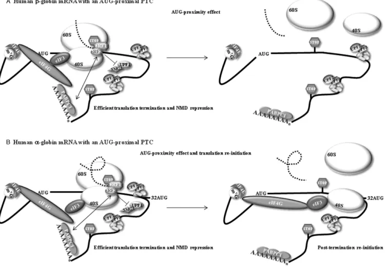
Figure 6. A model for the effect of an AUG-proximal premature termination codon (PTC or stop codon). (A) The effect in the human-globin transcript. During cap-mediated translation initiation, cytoplasmic poly(A) binding protein 1 (PABPC1) interacts with the eukaryotic initiation factor 4G (eIF4G). This interaction indirectly tethers PABPC1 to the 40S ribosomal subunitviathe interaction of eIF4G with eIF3, which interacts with the 40S. The resulting configuration brings PABPC1 into the vicinity of the AUG initiation codon as a consequence of 43S scanning. The maintenance of the PABPC1-eIF4G-eIF3 interactions with the 40S during the first steps of translation elongation brings PABPC1 into close proximity with the termination complex at an AUG-proximal PTC. This proximity allows PABPC1 to interact with the release factor eRF3, thus impairing the UPF1-eRF3 interaction, resulting in efficient translation termination and inhibition of NMD––this was called the ‘AUG-proximity effect’. (B) In the human␣-globin transcript, the AUG-proximity effect is observed for PTCs located further downstream because the open reading frame (ORF) is less structured. The more relaxed structure allows a faster elongation rate resulting in a longer window of the AUG proximity effect. In addition, the brief ORF translation also allows efficient translation re-initiation to occur at codon 32. This re-initiation diminishes the levels of residual exon junction complexes on the mRNA, thus contributing to the overall repression of NMD.
lation, since mRNA unwinding causes prolonged ribosome pausing (53,54), results herein suggest that the rate of trans-lation elongation might be higher in ␣- than in-globin transcripts, given that the boundary of the AUG-proximity effect is located further downstream in the␣-globin mRNA. In normal individuals, the average␣/-globin mRNA ra-tio is about 1.3 (58). Of interest, nearly 70% of ␣-globin mRNA is mainly found in pre-initiation complexes, whereas about 50% of total-globin mRNA is associated with ac-tively translating 80S ribosomes (52). These data suggest that ␣-globin manifests an inefficient or at least delayed translation initiation. Therefore,␣-globin mRNA may ac-tually have a higher translation elongation rate than  -globin mRNA to assure a correct balance between the syn-thesized␣- and-globin chains. On the other hand, the␣ -globin gene is known to possess a high G+C content and this feature leads to a strong union of the DNA double
a higher translation elongation rate than that observed for the-globin mRNA, and this supports our present data.
The relevance of the time taken to translate a short ORF is related to the fact that some initiation factors appear to remain ribosome-associated for the first moments of the ORF translation elongation (43,50,63,64). Thus, if the elon-gation phase is brief, the ribosome may reach the translation termination codon before it has had time to discharge initi-ation factors and other associated proteins. When the ribo-some arrives at the stop codon during translation of a short ORF, it might still hold the interaction with eIF4G, due to the brief translation, and PABPC1 might remain bound to eIF4G as well. The proximity of PABPC1 at the end of translation has been shown to promote correct termination and to inhibit NMD activation in human cells (21–24,65) as it competes with UPF1 to interact with eRF3 at the ter-mination complex (7,26,34). The inability of UPF1 to bind eRF3 prevents the interaction between the premature ter-mination complex and UPF2/UPF3 at a downstream EJC, which results in the repression of NMD. The hypothesis of the time taken by the ribosome to translate a short ORF being a major determinant of NMD inhibition can be sum-marized as follows: during a brief cap-mediated translation of a short ORF, PABPC1 might be in close proximity to the ribosome until it reaches the termination codon. In this sit-uation, PABPC1 may interact with eRF3 and preclude the binding of UPF1 to eRF3, thus preventing NMD activa-tion (Figure6A). In fact, this model is also in accordance with our previous data (34). This effect will be much more downstream extended if the short ORF is unstructured than if it is a well-structured one. In the unstructured ORF, the ribosome is faster and elongates too further downstream before the initiation factors, such as eIF4G-PABPC1, are disassembled; in addition, by the time the ribosome reaches the PTC, PABPC1 can interact with eRF3 at the terminat-ing complex, which inhibits NMD (Figure 6B). Alterna-tively, and in accordance with recent data (36,37), PABPC1-bound eIF4G might be the player that represses NMD. Ac-tually, a brief ORF translation, which allows the retention of some initiation factors by the time the ribosome reaches the stop codon, may explain both the AUG-proximity effect (reflecting the proximity of PABPC1 to the PTC) and trans-lation re-initiation, when the transcript is permissive to both mechanisms (Figure6B). In fact, the resumption of scan-ning and re-initiation also require a cap-mediated transla-tion initiatransla-tion with the participatransla-tion of eIF4F, and most likely depends on the preservation of eIF4F, at least the cen-tral one-third fragment of eIF4G, and eIF3 associated with the ribosome throughout translation of the ORF (50,64,66). Our observation that an unstructured ORF in the␣-globin mRNAs enhances NMD inhibition due to both the AUG-proximity effect and translation re-initiation further sup-ports this notion. However, for translation re-initiation to occur it is necessary that the intercistronic sequence and the re-initiation codon are favorable (50), and this does not seem to be the case in the -globin mRNA. In addition, UPF1 seems to play a role in the efficiency with which ribo-somes are released from the mRNA after translation termi-nation (39). This effect is likely to modulate the efficiency of translation re-initiation (39). Our results therefore sug-gest that␣- and-globin mRNAs differently interact with
UPF1. The identification of additional features that deter-mine efficient translation re-initiation in␣-globin but not in
-globin AUG-proximal nonsense-mutated transcripts will be further studied.
SUPPLEMENTARY DATA
Supplementary Dataare available at NAR Online.
FUNDING
Fundac¸˜ao para a Ciˆencia e a Tecnologia (FCT) [POCI/BIA-BCM/59140/2004 and UID/MULTI/04046/2013 to BioISI from FCT/MCTES/PIDDAC; SFRH/BD/14273/2003 to F.J.C.P. and SFRH/BD/63581/2009 to C.B.]. Funding for open access charge: Instituto Nacional de Sa ´ude Doutor Ricardo Jorge and Oxford Journals Publisher.
Conflict of interest statement.None declared.
REFERENCES
1. Le Hir,H., Izaurralde,E., Maquat,L.E. and Moore,M.J. (2000) The spliceosome deposits multiple proteins 20–24 nucleotides upstream of mRNA exon-exon junctions.EMBO J.,19, 6860–6869.
2. Le Hir,H., Gatfield,D., Izaurralde,E. and Moore,M.J. (2001) The exon-exon junction complex provides a binding platform for factors involved in mRNA export and nonsense-mediated mRNA decay.
EMBO J.,20, 4987–4997.
3. Alexandrov,A., Colognori,D., Shu,M.D. and Steitz,J.A. (2012) Human spliceosomal protein CWC22 plays a role in coupling splicing to exon junction complex deposition and nonsense-mediated decay.
Proc. Natl. Acad. Sci. U.S.A.,109, 21313–21318.
4. Metze,S., Herzog,V.A., Ruepp,M.D. and M ¨uhlemann,O. (2013) Comparison of EJC-enhanced and EJC-independent NMD in human cells reveals two partially redundant degradation pathways.
RNA,19, 1432–1448.
5. Kashima,I., Jonas,S, Jayachandran,U., Buchwald,G., Conti,E., Lupas,A.N. and Izaurralde,E. (2010) SMG6 interacts with the exon junction complex via two conserved EJC-binding motifs (EBMs) required for nonsense-mediated mRNA decay.Genes Dev.,24, 2440–2450.
6. Popp,M.W. and Maquat,L.E. (2014) The dharma of
nonsense-mediated mRNA decay in mammalian cells.Mol. Cells,37, 1–8.
7. Kashima,I., Yamashita,A., Izumim,N., Kataoka,N., Morishita,R., Hoshino,S., Ohno,M., Dreyfuss,G. and Ohno,S. (2006) Binding of a novel SMG-1-Upf1-eRF1-eRF3 complex (SURF) to the exon junction complex triggers Upf1 phosphorylation and nonsense-mediated mRNA decay.Genes Dev.,20, 355–367. 8. Hwang,J., Sato,H., Tang,Y., Matsuda,D. and Maquat,L.E. (2010)
UPF1 association with the cap-binding protein, CBP80, promotes nonsense-mediated mRNA decay at two distinct steps.Mol. Cell,39, 396–409.
9. Yamashita,A., Izumi,N., Kashima,I., Ohnishi,T., Saari,B., Katsuhata,Y., Muramatsu,R., Morita,T., Iwamatsu,A., Hachiya,T.
et al.(2009) SMG-8 and SMG-9, two novel subunits of the SMG-1 complex, regulate remodeling of the mRNA surveillance complex during nonsense-mediated mRNA decay.Genes Dev.,23, 1091–1105. 10. Okada-Katsuhata,Y., Yamashita,A., Kutsuzawa,K., Izumi,N.,
Hirahara,F. and Ohno,S. (2012) N- and C-terminal Upf1 phosphorylations create binding platforms for SMG-6 and SMG-5:SMG-7 during NMD.Nucleic Acids Res.,40, 1251–1266. 11. Cho,H., Kim,K.M. and Kim,Y.K. (2009) Human proline-rich nuclear
receptor coregulatory protein 2 mediates an interaction between mRNA surveillance machinery and decapping complex.Mol. Cell, 33, 75–86.
the helicase UPF1 with the NMD factors SMG5-SMG7 and SMG6.
Nucleic Acids Res.,42, 9447–9460.
13. Nicholson,P., Josi,C., Kurosawa,H., Yamashita,A. and M ¨uhlemann,O. (2014) A novel phosphorylation-independent interaction between SMG6 and UPF1 is essential for human NMD.
Nucleic Acids Res.,42, 9217–9235.
14. Eberle,A.B., Lykke-Andersen,S., M ¨uhlemann,O. and Jensen,T.H. (2009) SMG6 promotes endonucleolytic cleavage of nonsense mRNA in human cells.Nat. Struct. Mol. Biol.,16, 49–55.
15. Glavan,F., Behm-Ansmant,I., Izaurralde,E. and Conti,E. (2006) Structures of the PIN domains of SMG6 and SMG5 reveal a nuclease within the mRNA surveillance complex.EMBO J.,25, 5117–5125. 16. Schmidt,S.A., Foley,P.L., Jeong,D.H., Rymarquis,L.A., Doyle,F.,
Tenenbaum,S.A., Belasco,J.G. and Green,P.J. (2014) Identification of SMG6 cleavage sites and a preferred RNA cleavage motif by global analysis of endogenous NMD targets in human cells.Nucleic Acids
Res.,43, 309–323.
17. Fukuhara,N., Ebert,J., Unterholzner,L., Lindner,D., Izaurralde,E. and Conti,E. (2005) SMG7 is a 14–3–3-like adaptor in the nonsense-mediated mRNA decay pathway.Mol. Cell,17, 537–547. 18. Lejeune,F., Li,X. and Maquat,L.E. (2003) Nonsense-mediated
mRNA decay in mammalian cells involves decapping, deadenylating, and exonucleolytic activities.Mol. Cell,12, 675–687.
19. Unterholzner,L. and Izaurralde,E. (2004) SMG7 acts as a molecular link between mRNA surveillance and mRNA decay.Mol. Cell,16, 587–596.
20. Amrani,N., Ganesan,R., Kervestin,S., Mangus,D.A., Ghosh,S. and Jacobson,A. (2004) A faux 3′-UTR promotes aberrant termination and triggers nonsense-mediated mRNA decay.Nature,432, 112–118. 21. Silva,A.L., Ribeiro,P., In´acio,A., Liebhaber,S.A. and Rom˜ao,L.
(2008) Proximity of the poly(A)-binding protein to a premature termination codon inhibits mammalian nonsense-mediated mRNA decay.RNA,14, 563–576.
22. Ivanov,P.V., Gehring,N.H., Kunz,J.B., Hentze,M.W. and
Kulozik,A.E. (2008) Interactions between UPF1, eRFs, PABP, and the exon junction complex suggest an integrated model for mammalian NMD pathways.EMBO J.,27, 736–747.
23. Eberle,A.B., Mathys,H., Stalder,L., Orozco,R.Z. and M ¨uhlemann,O. (2008) Posttranscriptional gene regulation by spatial rearrangement of the 3′untranslated region.PLoS Biol.,6, e92.
24. Singh,G., Rebbapragada,I. and Lykke-Andersen,J. (2008) A competition between stimulators and antagonists of Upf complex recruitment governs human nonsense-mediated mRNA decay.PLoS Biol.,6, e111.
25. Silva,A.L and Rom˜ao,L. (2009) The mammalian nonsense-mediated mRNA decay pathway: to decay or not to decay! Which players make the decision?FEBS Lett.,583, 499–505.
26. Uchida,N., Hoshino,S., Imataka,H., Sonenberg,N. and Katada,T. (2002) A novel role of the mammalian GSPT/eRF3 associating with poly(A)-binding protein in Cap/Poly(A)-dependent translation.J. Biol. Chem.,277, 50286–50292.
27. Hogg,J.R. and Goff,S.P. (2010) Upf1 senses 3′UTR length to potentiate mRNA decay.Cell,143, 379–389.
28. Z ¨und,D., Gruber,A.R., Zavolan,M. and M ¨uhlemann,O. (2013) Translation-dependent displacement of UPF1 from coding sequences causes its enrichment in 3′UTRs.Nat. Struct. & Mol. Biol.,20, 936–943.
29. Kurosaki,T., Li,W., Hoque,M., Popp,M.W., Ermolenko,D.N., Tian,B. and Maquat,L.E. (2014) A post-translational regulatory switch on UPF1 controls targeted mRNA degradation.Genes Dev.,28, 1900–1916.
30. Mendell,J.T., Sharifi,N.A., Meyers,J.L., Martinez-Murillo,F and Dietz,HC. (2004) Nonsense surveillance regulates expression of diverse classes of mammalian transcripts and mutes genomic noise.
Nat. Genet.,36, 1073–1078.
31. Bruno,I.G., Karam,R., Huang,L., Bhardwaj,A., Lou,C.H., Shum,E.Y., Song,H.W., Corbett,M.A., Gifford,W.D., Gecz,J., Pfaff,S.L. and Wilkinson,M.F. (2011) Identification of a microRNA that activates gene expression by repressing nonsense-mediated RNA decay.Mol. Cell,42, 500–510.
32. Yepiskoposyan,H., Aeschimann,F., Nilsson,D., Okoniewski,M. and M ¨uhlemann,O. (2011) Autoregulation of the nonsense-mediated mRNA decay pathway in human cells.RNA,17, 2108–2118.
33. Hurt,J.A., Robertson,A.D. and Burge,C.B. (2013) Global analyses of UPF1 binding and function reveal expanded scope of
nonsense-mediated mRNA decay.Genome Res.,23, 1636–1650. 34. Peixeiro,I., In´acio,A., Barbosa,C., Silva,A.L., Liebhaber,S.A. and
Rom˜ao,L. (2012). Interaction of PABPC1 with the translation initiation complex is critical to the NMD resistance of AUG-proximal nonsense mutations.Nucleic Acids Res.,40, 1160–1173.
35. Roque,S., Cerciat,M., Gaugu´e,I., Mora,L., Floch,A.G., Zamaroczy,M., Heurgu´e-Hamard,V. and Kervestin,S. (2015) Interaction between the poly(A)-binding protein Pab1 and the eukaryotic release factor eRF3 regulates translation termination but not mRNA decay inSaccharomyces cerevisiae.RNA,21, 124–134. 36. Fatscher,T., Boehm,V., Weiche,B. and Gehring,N.H. (2014) The
interaction of cytoplasmic poly(A)-binding protein with eukaryotic initiation factor 4G suppresses nonsense-mediated mRNA decay.
RNA,20, 1579–1592.
37. Joncourt,R., Eberle,A.B., Rufener,S.C. and M ¨uhlemann,O. (2014) Eukaryotic initiation factor 4G suppresses nonsense-mediated mRNA decay by two genetically separable mechanisms.PLoS One,9, e104391.
38. Brogna,S. and Wen,J. (2009) Nonsense-mediated mRNA decay (NMD) mechanisms.Nat. Struct. & Mol. Biol.,16, 107–113. 39. Ghosh,S., Ganesan,R., Amrani,N. and Jacobson,A. (2010)
Translational competence of ribosomes released from a premature termination codon is modulated by NMD factors.RNA,16, 1832–1847.
40. Rom˜ao,L., In´acio,A., Santos,S., ´Avila,M., Faustino,P., Pacheco,P. and Lavinha,J. (2000) Nonsense mutations in the human beta-globin gene lead to unexpected levels of cytoplasmic mRNA accumulation.
Blood,96, 2895–2901.
41. In´acio,A., Silva,A.L., Pinto,J., Ji,X., Morgado,A., Almeida,F., Faustino,P., Lavinha,J., Liebhaber,S.A. and Rom˜ao,L. (2004) Nonsense mutations in close proximity to the initiation codon fail to trigger full nonsense-mediated mRNA decay.J. Biol. Chem.,279, 32170–32180.
42. Kong,J., Ji,X. and Liebhaber,S.A. (2003) The KH-domain protein alpha-CP has a direct role in mRNA stabilization independent of its cognate binding site.Mol. Cell. Biol.,23, 1125–1134.
43. Kozak,M. (2001) Constraints on re-initiation of translation in mammals.Nucleic Acids Res.,29, 5226–5232.
44. Pereira,F.J.C., Silva,M.C., Picanc¸o,I., Seixas,M.T., Ferr˜ao,A., Faustino,P. and Rom˜ao,L. (2006) Human alpha2-globin nonsense-mediated mRNA decay induced by a novel alpha-thalassaemia frameshift mutation at codon 22.Br. J. Haematol.,133, 98–102.
45. Pfaffl,M.W. (2001) A new mathematical model for relative quantification in realtime RT-PCR.Nucleic Acids Res.,29, e45. 46. Silva,A.L., Pereira,F.J., Morgado,A., Kong,J., Martins,R.,
Faustino,P., Liebhaber,S.A. and Rom˜ao,L. (2006) The canonical UPF1-dependent nonsense-mediated mRNA decay is inhibited in transcripts carrying a short open reading frame independent of sequence context.RNA,12, 2160–2170.
47. Thermann,R., Neu-Yilik,G., Deters,A., Frede,U., Wehr,K., Hagemeier,C., Hentze,M.W. and Kulozik,A.E. (1998) Binary specification of nonsense codons by splicing and cytoplasmic translation.EMBO J.,17, 3484–3494.
48. Zhang,J., Sun,X., Qian,Y. and Maquat,L.E. (1998) Intron function in the nonsense-mediated decay of-globin mRNA: Indications that pre-mRNA splicing in the nucleus can influence mRNA translation in the cytoplasm.RNA,4, 801–815.
49. Zhang,J. and Maquat,L.E. (1997) Evidence that translation re-initiation abrogates nonsense-mediated mRNA decay in mammalian cells.EMBO J.,16, 826–833.
50. P ¨oyry,T.A.A., Kaminski,A. and Jackson,R.J. (2004) What determines whether mammalian ribosomes resume scanning after translation of a short upstream open reading frame?Genes Dev.,18, 62–75. 51. Neu-Yilik,G., Amthor,B., Gehring,N.H., Bahri,S., Paidassi,H.,
Hentze,M.W. and Kulozik,A.E. (2011) Mechanism of escape from nonsense-mediated mRNA decay of human beta-globin transcripts with nonsense mutations in the first exon.RNA,17, 843–854. 52. Shakin,H. and Liebhaber,S.A. (1986) Translational profiles of alpha
53. Wen,J.D., Lancaster,L., Hodges,C., Zeri,A.C., Yoshimura,S.H., Noller,H.F., Bustamante,C. and Tinoco,I. (2008) Following translation by single ribosomes one codon at a time.Nature,452, 598–603.
54. Somogyi,P., Jenner,A.J., Brierley,I. and Inglis,S.C. (1993) Ribosomal pausing during translation of an RNA pseudoknot.Mol. Cell. Biol., 13, 6931–6940.
55. Pestova,T.V. and Kolupaeva,V.G. (2002) The roles of individual eukaryotic translation initiation factors in ribosomal scanning and initiation codon selection.Genes Dev.,16, 2906–2922.
56. Meaux,S., vanHoof,A. and Baker,K.E. (2008) Nonsense-mediated mRNA decay in yeast does not require PAB1 or a poly(A) tail.Mol.
Cell,29, 134–140.
57. Wen,J. and Brogna,S. (2010) Splicing-dependent NMD does not require the EJC in Schizosaccharomyces pombe.EMBO J.,29, 1537–1551.
58. Chaisue,C., Kitcharoen,S., Wilairat,P., Jetsrisuparb,A., Fucharoen,G. and Fucharoen,S. (2007) Alpha/beta-globin mRNA ratio
determination by multiplex quantitative real-time reverse transcription-polymerase chain reaction as an indicator of globin gene function.Clin. Biochem.,40, 1373–1377.
59. Fischel-Ghodsian,N., Nicholls,R.D. and Higgs,D.R. (1987) Long range genome structure around the human alpha-globin complex analysed by PFGE.Nucleic Acids Res.,15, 6197–6207.
60. Cooper,G.M., Brudno,M., Stone,E.A., Dubchak,I., Batzoglou,S. and Sidow,A. (2004) Characterization of evolutionary rates and
constraints in three mammalian genomes.Genome Res.,14, 539–548. 61. Jiang,C. and Zhao,Z. (2006) Mutational spectrum in the recent
human genome inferred by single nucleotide polymorphisms.
Genomics88, 527–534.
62. Tuan,D. and Forget,B.G. (1980) Physicochemical measurement of the base composition of mRNA-related sequences of the human alpha and beta globin genes.Mol. Biol. Rep.,6, 179–183.
63. Jackson,R.J. (2005) Alternative mechanisms of initiating translation of mammalian mRNAs.Biochem. Soc. Trans.,33, 1231–1241. 64. Szamecz,B., Rutkai,E., Cuchalov´a,L., Munzarov´a,V.,
Herrmannov´a,A., Nielsen,N.H., Burela,L., Hinnebusch,A.G. and Val´aˇsek,L. (2008) eIF3a cooperates with sequences 5′of uORF1 to promote resumption of scanning by post-termination ribosomes for re-initiation on GCN4 mRNA.Genes Dev.,22, 2414–2425. 65. Kahvejian,A., Roy,G. and Sonenberg,N. (2001) The mRNA
closed-loop model: the function of PABP and PABP-interacting proteins in mRNA translation.Cold Spring Harbor Symp. Quant. Biol.,66, 293–300.