Volume 2009, Article ID 157531,8pages doi:10.1155/2009/157531
Research Article
Isolation of Osteosarcoma-Associated Human Antibodies from
a Combinatorial Fab Phage Display Library
Carmela Dantas-Barbosa,
1Fabr´ıcia P. Faria,
2Marcelo M. Brigido,
3and Andrea Q. Maranh˜ao
3, 41Institut Gustave Roussy, UPRES EA 3535 PR2. 39, Rue Camille Desmoulins, 94805 Villejuif, France
2Departamento de Bioqu´ımica e Biologia Molecular, Instituto de Ciˆencias Biol´ogicas, Universidade Federal de Goi´as,
ICB II-Campus II Samambaia, 74001-970 Goiania, GO, Brazil
3Departamento de Biologia Celular, Instituto de Ciˆencias Biol´ogicas, Universidade de Bras´ılia, Campus Universit´ario Darcy Ribeiro,
Asa Norte, 70910900 Brasilia, DF, Brazil
4Laborat´orio de Imunologia Molecular, Universidade de Bras´ılia, Campus Universit´ario Darcy Ribeiro, Sala ASS 128, ICC Sul,
Asa Norte, 70910-900 Brasilia, DF, Brazil
Correspondence should be addressed to Carmela Dantas-Barbosa,carmeladantas@hotmail.com
Received 8 February 2009; Revised 29 June 2009; Accepted 9 September 2009
Recommended by Anthony L. DeVico
Osteosarcoma, a highly malignant disease, is the most common primary bone tumor and is frequently found in children and adolescents. In order to isolate antibodies against osteosarcoma antigens, a combinatorial osteosarcoma Fab library displayed on the surface of phages was used. After three rounds of selection on the surface of tumor cells, several osteosarcoma-reactive Fabs were detected. From these Fabs, five were better characterized, and despite having differences in their VH (heavy chain variable domain) and Vκ(kappa chain variable domain) regions, they all bound to a protein with the same molecular mass. Further analysis by cell ELISA and immunocytochemistry suggested that the Fabs recognize a membrane-associated tumor antigen expressed in higher amounts in neoplasic cells than in normal tissue. These results suggest that the human Fabs selected in this work are a valuable tool for the study of this neoplasia.
Copyright © 2009 Carmela Dantas-Barbosa et al. This is an open access article distributed under the Creative Commons Attribution License, which permits unrestricted use, distribution, and reproduction in any medium, provided the original work is properly cited.
1. Introduction
Osteosarcoma is the most common primary bone tumor that affects children and young adults [1]. It is highly malignant, characterized by a high propensity to metastasize [2]. Use of multiagent chemotherapy pre- and postoperatively has significantly improved the outcome in the last 30 years, leading to a 5-year disease-free survival for patients with localized tumors in the 20% to 60% range [3]. Despite the use of these drugs, approximately 30% of patients with localized disease and 60% of patients with pulmonary metas-tases succumb to the illness [4]. Osteosarcoma is a complex neoplasia. Cytogenetic analysis has revealed multiple chro-mosomal rearrangements without a typical translocation, as can be seen in Ewing’s sarcoma. In recent years, the immunocytochemical subclassification of osteosarcomas has
significantly enhanced the accuracy of pathological diagnoses [5], suggesting that new immunochemical markers could further improve diagnosis and prognosis. Recently, Salas et al. [6] introduced Ezrin expression as a prognostic factor for the survival rate of patients with conventional osteosarcoma, correlating its expression with tumor progression. It was also shown that Ezrin is useful as an immunohistochemical marker to differentiate between chondroblastic osteosarco-mas and conventional chondrosarcoosteosarco-mas [7]. However, other osteosarcoma markers are required to further characterize this complex, multifactorial neoplasia.
general it is expected that humanized or fully human anti-bodies are better tolerated than murine monoclonal ones. To isolate such potential biopharmaceuticals, many approaches can be used, including the phage display technique. The use of the phage display technique can lead to the isolation of either antibodies or peptides able to discriminate tumor cells [8]. In a traditional approach, the identification of the tumor marker is the first step, after that it is necessary to isolate an antibody against this marker. Using a Fab phage display library, these steps can be merged into one, and it is possible to identify antigens without previous knowledge. The use of complex antigens such as cell membranes broadens the possibility of finding differentially expressed markers. However, as a more complex antigen source is used, the techniques employed to separate bound phages from unbound phages become more difficult. This usually makes selection on cell surfaces much more complex than selection using purified antigens. Focusing on this challenge, different selection procedures have been developed. Among these procedures, a method called BRASIL (Biopanning and Rapid Analysis of Selective Interacting Ligands) was specifically designed to isolate phages able to bind to the cell surface [9]. In order to isolate anti-osteosarcoma fully human Fabs, we used a previously constructed combinatorial Fab phage display library produced with antibody repertoires from osteosarcoma patients, relying on the fact that these antibody repertoires could be enriched with antibodies against the patients’ own tumors [10]. Here, we report the utilization of this library for panning Fabs against the surface of osteosarcoma cells. The selection procedure was adapted from that of the BRASIL method for using a phage antibody library [9]. This is the first report of the use of the BRASIL method with a human Fab library. After three rounds of selection against the cell surface of an osteosarcoma cell line, we were able to identify many reactive Fabs, most of them sharing similar V genes. Five Fabs were chosen for further analysis, including western blot, cell ELISA, and immunocytochemistry. Our data revealed that these Fabs recognize a membrane antigen that is more widely expressed in the osteosarcoma cells compared to normal osteoblast and fibroblast cells. These results suggest that these human Fabs could be further used to improve the diagnosis and prognosis of osteosarcoma.
2. Materials and Methods
2.1. Cell Lines. The human osteosarcoma cell lines U2
OS (HTB-96) and Saos-2 (HTB-85), and the fetal normal osteoblast cell line hFOB1.19 (CRL-1172) were purchased from ATCC and cultured as recommended. A short-term culture of normal human fibroblasts was cultured at 37◦C
in 5% CO2 with Ham’s F10 medium supplemented with 100 U/mL of penicillin, 100µg/mL of streptomycin, and 10% FBS.
2.2. Panning of the Fab Phage Library with Osteosarcoma
Cells. A combinatorial Fab phage display library generated
from the antibody V regions of eleven osteosarcoma patients, containing 2.7×10⁸different forms, was constructed
previ-ously [10] and used for selection according to the BRASIL method [9]. Briefly, the cultured cells were collected with PBS and 5 mM EDTA and then washed with the medium defined for each lineage, resuspended in culture medium containing 1% BSA at a cell density of approximately 1×10⁶
cells per mL, and incubated with 1×10¹¹ plaque forming
units (pfu) of phage for approximately 1.5 hours on ice with sporadic gentle mixing. After that, the cell-phage suspension was transferred to the top of a nonmiscible organic lower phase (a 9 : 1 mixture of dibutyl phthalate : cyclohexane) and centrifuged at 1000 g for 10 minutes at room temperature. The tube was snap frozen in liquid nitrogen, and the cell-phage pellet was used to infect a log-phase culture of
Escherichia coli(strain XL1-Blue). In the selection procedure
using the surface of U2-OS cells, we also performed a preclearing step before the third selection cycle. To do this, the phages were incubated with normal hFOB 1.19 osteoblasts, and, after separation by centrifugation through the organic phase, the supernatant containing the unbound phages (in the aqueous upper phase) was incubated with U2-OS osteosarcoma cells as described above. The selected phages were amplified byE. coliinfection according to the procedure described elsewhere [11] and were then used in the next selection cycle.
2.3. Expression of Soluble Fab Fragments. DNA was extracted
from the bacterial pellet obtained after each round of selection using a QIAprep spin miniprep Kit (Qiagen),
and then used to transform HB2151 E. coli cells for
production of soluble Fab without the filamentous phage Gene III fusion protein partner. Individual carbenicillin-resistant HB2151 colonies were picked and grown overnight (ON) at 37◦C in a 96 deep well plate in 1 mL SB medium
containing carbenicillin (100µg/mL) and glucose (1%). After this incubation, the cultures were used to produce two replica plates: one for a glycerol stock and the second for the production of the soluble Fabs themselves. The original ON culture plate was centrifuged, and plasmid DNA was extracted from the E. coli using the QIAprep Kit. Fab production was performed by inoculation of 1.5 mL of SB medium containing carbenicillin (100µg/mL) and glucose (1%) with 50µL of the ON culture. Cultures were incubated at 37◦C, shaking at 250 rpm, until they reached an OD of
0.9 at 600 nm. The cultures were then centrifuged, and fresh SB medium containing carbenicillin (100µg/mL) and IPTG (2 mM) was added, followed by incubation at 30◦C for an
additional 18 hours. Supernatants containing the soluble Fabs were used in the dot blot and ELISA assays.
For large-scale Fab production, the procedure was the same as described above, except that phages were produced in 100 mL of SB mediumE. coliculture. Supernatants were concentrated using Centriprep 10 filter units (Millipore) and buffer exchanged in PBS. Purification of Fabs was performed using Ni-NTA resin (Qiagen) under nondenaturing condi-tions according to the manufacturer’s instruccondi-tions. Proteins were analyzed by SDS-PAGE [12].
2.4. Sequence Analysis. Sequencing reactions were prepared
(CH: 5′CGCCTGAGTTCCACGACACC3′, MMB4: 5′
GCT-TCCGGCTCGTATGTTGTGT3′, Cκ: 5′
AGAGGAGTC-CAGATTTCA3′, and MMB5: 5′
CGTTTGCCATCTTTT-CATAATC3′) [10] and were performed using ET Terminator
(GE-Healthcare) in a MegaBace 500 plus sequencer (Molec-ular Dynamics). Alignments were done using IgG Blast at the NCBI blast server (http://www.ncbi.nlm.nih.gov/blast) and with the program BioEdit [13]. The Kabat numbering and CDR definitions were adopted from Andrew Martin’s web site (http://www.bioinf.org.uk/abs/).
2.5. Analysis of Binding of Selected Fabs to Total Protein
Extracts and to the Cell Surface by ELISA. Ninety-six well
plates were coated with 100µL of a 10µg/mL total protein solution prepared from osteosarcoma and osteoblast cells as described in the next section. Human serum albumin was used as a mock protein. The coating was carried out overnight at 4◦C. The plates were then blocked with 3%
BSA and incubated with 100µL of the induced supernatant from each selected clone. The supernatant from the empty parental plasmid (pComb3X) E. coli culture was assayed as a negative control. Fab binding was detected with a rat anti-HA (hemagglutinin decapeptide tag) HRP-conjugated antibody from Roche, followed by the ABTS peroxidase substrate. For selecting individual clones, a threshold value was arbitrarily set at ten times greater than the value obtained for the empty vector and at least three times greater than the value obtained for the incubation with proteins from normal osteoblast cells.
Cell ELISA was performed according to the protocol adapted from Williams and Sharon (2002) [14]. Briefly, polystyrene 96-well tissue culture plates were seeded with 2 × 10⁶ U2-OS, Saos-2, or fibroblast cells in 200µL of
culture medium defined for each cell lineage supplemented with 10% (v/v) FBS and incubated for 30 hours. The cells were fixed with 4% formaldehyde in PBS for 20 minutes at room temperature followed by two washes with PBS. The washed cells were incubated with 100µL of the induced supernatant from each clone or with the purified Fab. The empty parental pComb3X plasmid supernatant was used as a negative control. Detection of Fab binding was performed as described above.
2.6. Immunodetection of Osteosarcoma Fab Ligands. Protein
extracts from U2-OS and fibroblast cells were prepared in cold lysis buffer (0.5% NP-40, 0.3% Triton X 100, 50 mM
Tris HCl pH 8.0, 5 mM MgCl2, 0.5 mM EDTA, 1 mM ATP,
1 mM DTT, 10% glycerol) and complete mini-EDTA-free protease inhibitor cocktail tablets from Roche. Total protein extracts were separated by SDS-PAGE under reducing con-ditions and transferred to nitrocellulose membranes (GE-Healthcare). After blocking with 5% skim milk—TBST at room temperature for 1 hour, the membranes were probed with one of the five purified Fabs (0.02µg/mL) for 2 hours at RT and washed 3 times with TBST (50 mM Tris-HCl, pH 7.5, 150 mM NaCl, 0.05% tween 20). The membranes were then incubated with a rabbit anti-HA (hemagglutinin decapeptide tag) antibody (Santa Cruz) diluted 1 : 1000 for 1
hour, washed again and incubated with anti-rabbit HRP at a 1 : 5000 dilution. Bands were visualized using an ECL Super-Signal West Pico Chemiluminescent Substrate detection kit (Thermoscientific). To control loading differences, a rabbit anti-alpha-tubulin antibody (AbCAM) was used instead of the selected Fabs.
2.7. Immunocytochemistry. Cultures of U2-OS and human
fibroblast cells grew until they reached 70–90% confluency on a slide chamber. The cells were then washed twice with PBS and fixed and permeabilized by immersion in precooled methanol for 6 minutes at −20◦C. After two washes with
PBS, the slides were incubated with 2% BSA (w/v) and 5% normal goat serum (v/v) for 30 minutes. After an overnight incubation at 4◦C with 15µL of purified Fabs, the slides were
washed and incubated with 1 : 200 anti-HA antibody (Santa Cruz) for 45 minutes, followed by an incubation with a goat anti-rabbit IgG-FITC at 1 : 2000 (Sigma) in a moist chamber in the dark. After addition of DAPI I, the slides were observed with an Axiolab fluorescence microscope (Zeiss) using the Applied Imaging Cytovision software.
2.8. Immunohistochemistry. Archived, formalin-fixed,
para-ffin-embedded tissue sections (4µm) were deparaffinized in xylene and rehydrated using a series of alcohol and distilled water solutions. Endogenous peroxidases were quenched by incubation with 10 V H2O2 for 10 minutes, followed by
microwave antigen retrieval in 10 mM sodium citrate buffer, pH 6.0. Purified anti-osteosarcoma Fabs at 200µg/mL, diluted to 1 : 200 (N-H7) and 1 : 20 000 (O-G11), were incubated with tissue sections for 1 hour at room temperature. After rinsing with phosphate-buffered saline, the slides were incubated for 30 minutes with rabbit anti-HA antibody (Santa Cruz), diluted 1 : 200 in 1% BSA-PBS, and then incubated with a biotinylated goat anti-rabbit IgG (Rockland). After the antibody incubations, the slides were washed with Tris-based buffer and then incu-bated with streptavidin-conjugated horseradish peroxidase
(LSAB+ kitTM, Dako) and DAB as the chromogen. The
sections were counterstained with Harry’s hematoxylin and covered with Entellan (Merck).
3. Results
3.1. Selection of Fabs that Bind to Osteosarcoma Cells. To
Table1: Reactivity of randomly selected Fabs toward tumor antigens in ELISA assays.
Cycle of selection(System) Clones assayed Positive clones in Cell ELISAa Positive clones on Total Protein Extractsb
2nd (N) 40 10 28
3rd (N) 39 NTc 13
2nd (O) 48 0 1
3rd (O) 40 1 4
aA clone was considered positive if the absorbance at 405 nm was three times greater than the absorbance found for hFOB 1.19 cell surface binding. bA clone was considered positive if the absorbance at 405 nm was ten times greater than the absorbance observed for empty vector supernatant.
cNT
=Not tested.
We found that 67% of the N clones and 46% of the O clones gave positive signals, demonstrating that these clones express reactive Fabs (data not shown). Individual Fab-expressing clones were tested for the ability to bind to osteosarcoma cell proteins by ELISA using two different strategies. The first approach evaluated Fab binding to fixed osteosarcoma cell monolayers, and the second approach evaluated Fab binding to total protein extracts from osteosarcoma cells. Seventy-nine clones from the N system and eighty-eight from the O system were tested.Table 1summarizes these results. Clones showing an A450 of less than 0.1 (similar to supernatants
without Fab) were considered nonbinders.
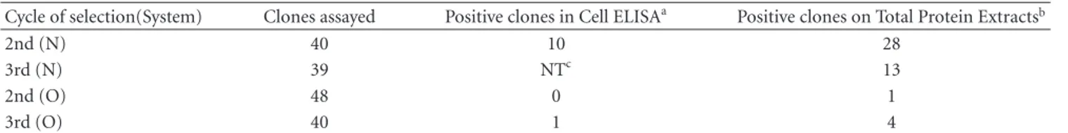
The most reactive clones were tested further in another ELISA assay using protein extract from osteosarcoma cells and normal osteoblast cells. An unrelated protein was included as a negative control. Triplicates of 27 clones were tested in this second trial, including 16 from the second and 6 from the third rounds of the N system and 2 clones from the second and 3 from the third round of the O system. The results of this assay confirmed the reactivity of the Fabs with osteosarcoma cells (Figure 1).
3.2. VH and VL Repertoire Usage of Selected Fabs. The
VH and VL coding regions of the 20 most reactive clones were determined by sequencing (Figure 2). Analysis of the deduced amino acid sequences of the corresponding V genes indicated that all VH and Vκchains were distinct from the sequences (51 VH and 74 Vκ) previously described from the original library of randomly chosen clones [10]. The VH sequences revealed that all clones belonged to the VH3 family and that their CDR3 varied in length from 6 to 14 amino acid residues. The majority of the clones (12/20) had a VH3-7 gene segment and a CDR3H of eight amino acids in length. An unusual histidine residue was predicted at position 52a of clone O-G11. This was first reported in the original library. The presence of the amino acid cysteine (C) at position 79 in framework 3 is a striking characteristic, and it was found in 12 clones harboring the VH3-7 gene segment, as can be seen in the N-H7 clone.
The amino acid sequences encoded by the Vκgenes also differed from those reported for the unselected repertoire. The O2 Vκfamily clones N-E8 and O-C10 had a conservative valine substitution at position 48 (I48V), and clones
O-F4 and O-G11 (B3 Vκ family) had a nonconservative
phenylalanine substitution at this same position (I48F). This rare mutation is reported to occur in less than 1% of the Vκ
sequences [15], and was present in different clones of the
original library [10]. The B3 family clones also presented a previously observed conservative change at the key position 64 (G64A). The predominance of clones harboring the VH3.7 (12/20) and A17 (11/20) gene segments supports the specific ligand selection.
Based on the results of the ELISA and the DNA sequen-cing, we chose five different Fabs for further analysis empha-sizing their reactivity. The clones chosen were: N-H7 (VH3-7—A17, frequency 11/20), N-E8 (VH3-23—O2, frequency 1/20), N-H10 20—A30, frequency 1/20), O-F4 (VH3-23—B3, frequency 1/20) and OG-11 (VH3-74—B3, fre-quency 2/20). The Fabs were produced in 100 mL cultures and purified using metal affinity columns before use in further analyses.
3.3. Clones N-H7, N-E8, N-H10, O-F4, and O-G11 Recognize
the Same Overexpressed Osteosarcoma Protein. Immunoblot
experiments were carried out for an initial characterization of the molecules in whole cell lysates derived from the osteosarcoma cell line U2-OS or from normal fibroblasts that were detected by the chosen Fabs. From this analysis, an intense 80 kDa band and a weaker 50 kDa band were observed for all analyzed Fabs, suggesting that all clones recognized the same proteins and that these proteins are present in both osteosarcoma cell lines (Saos-2 and U2-OS). The same pattern was observed at a lower intensity when normal fibroblasts were probed, suggesting that these proteins are overexpressed in malignant bone cells (Figure 3).
3.4. Pattern of Fab Staining of Osteosarcoma Cells and
Tissue. Further analysis of Fab reactivity was addressed by
comparing the ability of the antibody fragments to bind to monolayers of osteosarcoma cells and normal fibroblasts by cell surface ELISA.Figure 4shows the confirmation that the purified Fabs recognized a protein present in both kinds of cells (tumor and normal human fibroblast). However, the signal obtained was at least twice as strong for the tumor cells as for the normal fibroblasts, suggesting overexpression of the antigen in the osteosarcoma cells. The clones obtained from selection in Saos-2 cells (the N system) were tested against Saos-2 and U2-OS osteosarcoma cell lines. The clones from the O system were assessed using only U2-OS cells in addition to the normal human fibroblasts.
0 0.005.1 0.015.2 0.025.3 0.035.4 0.045
.5
N-B7 N-A8 N-H7 N-G9 N-C8 N-H9 N-A11 N-F11 N-C10 N-F10 N-D7 N-D8 N-E8
N-H10 N-G11 N-C11 N-B1 N-E4 N-G5 N-B3 N-B4 N-E3 pComb
Saos-2 hFOB HSA
(a)
0 0.05 0.1 0.15 0.2 0.25
O-F4 O-D6 O-G11 O-H11 O-C10 pComb
U2-OS hFOB HSA
(b)
Figure1: Selection of Fab clones able to recognize osteosarcoma cell protein extracts. Protein extracts from Saos-2 cells (horizontal line
bars, panel (a)), U2-OS cells (horizontal lines, panel (b)), and h FOB 1.19 (normal human osteoblast, black bars, both panels) were used to coat a 96-well plate.E. colisupernatants from the N system (panel (a)) or the O system (panel (b)) Fab clones were added to the plate. After washing, the bound Fabs were detected using rabbit anti-HA antibody followed by an HRP-conjugated anti-rabbit antibody. The experiments were performed in triplicate. The Fabs were also added to HSA- (human serum albumin) coated wells (diagonal line bars, both panels) as a negative control. Supernatant from an empty vectorE. coliculture was also used as negative control (pComb). The absorbances were measured at 405 nm.
. . . . . . . | . . . | . . . | . . . | . . . | . . a b . . . | . . . | . . . | . . a b c . . . | . . . | a b c d e f . . | . . . | . . E VQL V E S GGG L VQP GG S L R L S CGA S GV T F S THWMA WV RQA P GKG L EWVA S I KGHG S I KNY VD S VKG R F A I S RDNAKN S L C L QMN S L R A E DT AV Y Y CA T HTDWR F DY WGQGT L V T V S Q VQL VQ S GGGVV R P GG S L R L S CAA S G F T F D DY GMS WV RQA P GKG L EWV S G INWNGG S TGY AD S VKG R F T I S RDNAKN S L Y L QMN S L R A E DT A L YHCA R DP S RGG S GWYWF DP WGQGT L V T V S S L GGK F GG S V L VQP GG S L R L S CAA S G F T F S S Y AMS WV RQA P GKG L EWV S A I S G S GG S T Y Y AD S VKG R F T I S RDN S KNT L Y L QMN S L R A E DT AV Y Y CAK P F HQ S D S S S W E VQL V E S GGG L VQP GG S L R L S CAA S G F T F S S Y AMT WV RQA P GKG L EWV S A I S G S GG S T Y Y AD S VKG R F T I S RDN S KNT L Y L QMN S L RG E DT AV Y Y CGK P F HQ S D S S S W Q VQL QE S GGG L VQP GG S L R L S CVA S G F D F S R YWMH WV RQA P GKG L EWV S H I H S DG I P T A Y AD S V RG R F T I S RD I AKNT L Y L QMNN L R P E DT AV Y Y CV T F I V E S K WGQGT L V T V S
. . S P
S
. . . . . . . | . . . | . . .
. . .
. . . . a b c d e f . . | . . . | . . . | . . . | . . . | . . . | . . . | . . . | . . . . E L VMTQ S P L S L P V T L GQP A S I S C T S S Q S L V Y S DG NT Y L N WF HQR P GQ S P R R L I Y KV S NRD S GV P DR F S G S G S GTD F T L K I S RV E A E DVG I Y Y C MQGTHWP T T F GGGT KV E I K R E L VMTQ S P A T L S A S VGDT VA I T C R A S G S I G THL N WYQHK P GKA P KV L V S S A S T L QT GV P S R F S G S G S GTD F T L T I S S L QP E D F A T Y Y C QQ S Y S I Y A L T F GGGT KV E L K R E L VMTQ S P A T L S A S VGDT VA I T C R A S G S I G THL N WYQHK P GKA P KV L V S S A S T L QT GV P S R F S G S G S GTD F T L T I S S L QP E D F A T Y Y C QQ S Y S I Y A L T F GGGT KG S S N E E L QMTQ S P S S L S A S VGDRV T I A C R A S QD I R NDL N WYQQK P R KA P K R L I Y VA S N L Q S GV P S R F S G S G S GT E F T L T I S S L QA E DVAV Y Y C QQY S QY S S H F GQGT R L E I K R E L QMTQ S P S S L S A S VGDRV T I A C R A S QD I R NDL N WYQQK P R KA P K R L I Y VA S N L Q S GV P S R F S G S G S GT E F T L T I S S L QA E DVAV Y Y C QQY Y S I P L T F GQGT R L E I K R E L VMTQ S P D S L AV S L G E R A T I NC K S S R S V L Y S VN S KNY L G WF QQK P GQP P K L L F Q WA S I R E S GV P DR F S A CG S GTD F T L T I S S L QA E DVAV Y Y C HQY AAV P F T F G P GT KGT S N E E L VMTQ S P D S L AV S L G E R A T I NC K S S R S V L Y S VN S KNY L G WF QQK P GQP P K L L F Q WA S I R E S GV P DR F S A CG S GTD F T L T I S S L QA E DVAV Y Y C HQY AAV P F T F G P GT KVD I K R
Clone Family Frequency
VH3-7 VH3-20 VH3-23 VH3-23 VH3-74
12/20 3/20 N-H7
N-H7 N-E8 N-H10
N-H10 N-B3 N-E8 O-F4
O-F4 O-G11
O-G11 O-C10
1/20 2/20 2/20
A17 11/20 O2 1/20 O2 1/20
A30 1/20
1/20 A30
3/20 B3 B3 2/20
VH
FR1 CDR1 FR2 CDR2 FR3 CDR3 FR4
Clone Family Frequency FR1 CDR1 FR2 CDR2 FR3 CDR3 FR4
10 20 30 40 50 60 70 80 90 100 103 110
30 40 50 60 70 80 90 100
10 20
Vκ
Figure2: Variable gene repertoires of selected Fabs. Twenty different reactive Fab genes were sequenced, and the amino acid sequences of
25 32.5 47.5 62 83 (kDa)
175
32.5 47.5 62 83 175 (kDa) O-F4 N-B7 N-H10 N-E8 O-G11
1 2 1 2 1 2 1 2 1 2
α-tubulin
Figure 3: Detection of osteosarcoma antigens by selected Fabs.
A western blot using five selected purified Fabs was performed. Protein extracts from U2-OS cells (lane 1) and normal human fibroblasts (lane 2) were transferred to nitrocellulose membranes. The membranes were probed with the O-F4, N-H7, N-H10, N-E8, and O-G11 Fabs. For loading comparison, an anti-alpha tubulin antibody was also used.
0 0.05 0.1 0.15 0.2 0.25
N-H7 N-H10 N-E8 O-G11 O-F4
Figure 4: Cell surface ELISA. The selected purified Fabs were
added to monolayers of Saos-2 cells (light gray bars), U2-OS cells (dark gray bars), and human fibroblasts (black bars). The bound Fabs were detected using rabbit HA and HRP-conjugated anti-rabbit antibodies. Human serum albumin (white bars) was used as negative control. The absorbances were measured at 405 nm.
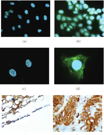
investigations were done mainly with the N-H7 (Figure 5, panels (a) and (b)) and O-G11 (Figure 5, panel (c), (d), (e) and (f)) Fabs. These Fabs were chosen based on the frequency of N-H7, whose VH and VL sequences were shared with 10 other selected clones, and on singularity of O-G11, as it came from the third round of the O system, after depletion on normal osteoblast cells. The immunofluorescence staining pattern suggested that these two Fabs interact with a cell membrane component. The staining was dot like for the osteosarcoma cell line U2-OS (Figure 5, panels (b) and (d)), but only a vague staining was observed for the normal fibroblasts (Figure 5, panels (a) and (c)).
Immunohistochemistry analyses performed using
archived normal bone and osteosarcoma sections confirmed the membrane staining pattern. Panels (e) and (f) of
Figure 5show the reactivity of the O-G11 Fab in a normal
bone section and in an osteosarcoma section, respectively. There was visible staining in the bone marrow adipocytes and in the osteoblasts in the normal tissue (Figure 5,
(a) (b)
(c) (d)
(e) (f)
Figure5: Indirect immunofluorescence staining and
immunohis-tochemistry were performed with the selected Fabs. Methanol-fixed fibroblasts ((a) and (c)) or U2-OS osteosarcoma cells ((b) and (d)) were stained with Fab N-H7 ((a) and (b)) or Fab O-G11 ((c) and (d)). Fab O-G11 was also used to stain to normal bone (e) (200x) and osteosarcoma tissue (f) (400x) sections. The osteoblasts in normal bone are arranged in lines; the empty region corresponds to bone trabecules dislocated during washing. These results are representative from four different osteosarcoma cases analyzed.
panel (e)), but staining was less intense than the staining observed in osteosarcoma tissues (Figure 5, panel (f)). This demonstrated the preference of O-G11 for tumor cell membranes.
4. Discussion
Among the successful approaches using whole cells is the BRASIL method, described as a very efficient way to rescue ligand phages [9]. Until now, this technique has been used only with peptide libraries [18, 19]. We successfully adapted the BRASIL protocol for use with a Fab library. We performed three rounds of selection, and the reactivity analysis of the second and third rounds revealed that after two rounds we were able to identify Fabs that bind to osteosarcoma cells. The ability to select specific tumor binders is usually attributed to the depletion step [20], but the omission of this step does not impair the isolation of specific binders [21, 22]. Our data suggest that the introduction of a negative selection step (only before the third selection cycle for the O system) did not significantly improve the isolation of specific Fabs, since the N and O Fabs apparently recognized the same antigen (Figure 3). The lack of more effective selection in the O system could also be a consequence of the small number of positive clones in round 2, just prior to the depletion step (Table 1).
Selected Fab clones were chosen based on their Fab coding sequences (Figure 2) and on their reactivity to tumor antigens assayed as a monolayer of osteosarcoma cells
(Table 1) and total protein extracted from cells (Table 1, and
Figure 1). These assays were carried out to favor the binding
of antibodies that recognize the physiological conforma-tions of the antigen(s). The five clones that were further characterized all recognized the same antigen (Figure 3), despite the fact that some of them reacted differently in the cell ELISA or protein ELISA assays, suggesting that these Fabs may recognize different epitopes [23]. Comparing the ELISA results (Figures1and4), we observed that the results were similar if we used fibroblasts or normal osteoblasts, as negative controls.
Another important finding was that the same clones were recovered after selection. Also, it is remarkable that the majority of these clones (11 out of 20) harbored the same sequence (A17, for Vκand VH3.7 for the heavy chain,
Figure 2). This cannot be attributed to a bias in the Fab
library, since all of the sequenced clones from rounds two or three had different VH and Vκ sequences compared to the original library [10]. The emergence of clones with a restricted diversity of V gene fragments is a feature that accounts for the selection of specific ligands, as was reported when other antibody phage display libraries were used to isolate high affinity immunoglobulins [22, 24]. Clones sharing VH3-7 and A17 were the majority of the clones identified in our selection process. There was one exception among the 20 sequenced clones: clone N-B1, which has the VH3-7 gene associated with an A30 Vκgene. The effect of this rare association on affinity and selectivity can be further addressed.
Apparently, all of the five chosen clones, each repre-senting a distinct gene family, recognized the same protein profile. This was also true for Fabs selected against different osteosarcoma cell lines, Saos2 and U2-OS (clones from the N and O systems, resp.). Similar results have been reported by Aryee and coworkers regarding six different scFvs selected against FLI1 [25]. Our data point to the existence of an immunodominant antigen. The immunodominance
phe-nomenon has been frequently observed in phage display analysis. In two separate studies with melanoma, one using a scFv phage display library constructed from blood from melanoma patients and the other using an Fab library, the authors were able to isolate only one positive scFv and Fab, respectively [26, 27]. This is in agreement with our results using two different cell lines, Saos-2 and U2-OS, as the antigen source. This does not exclude the possibility of binding to different epitopes on the same protein, and further epitope mapping must be carried out. Another interesting characteristic was the ability of the selected antibody to bind to the antigen in both native and denaturing conditions (Figures 1(a), 1(b) and 3), suggesting a linear rather than a conformational epitope.
The immunofluorescence staining pattern (Figure 5) was typical for Fabs recognizing a membrane-associated ligand, as expected for a cell surface selection procedure. The background in immunohistochemestry was high, and although it was possible to see staining of normal cells, the staining was stronger with the osteosarcoma tissues. The O-G11 Fab gave a very strong background even when used as much as 1 : 20 000 dilution (estimated concentration of 0.1 ng/mL). Usually the concentration of commercial antibodies is about 200µg/mL, and they are used at dilutions of 1 : 50–1 : 200 in immunohistochemistry assays. O-G11 recognized the osteoblasts in a very strong way and also reacted with adipocytes in the bone medulla but did not stain the blood vessel (data not shown).
It is difficult to measure the strength of the sig-nal in osteosarcoma cells and compare it to the sigsig-nal strength from normal controls in cell imaging approaches, such as immunofluorescence and immunohistochemistry. This became more evident when a quantitative assay was employed (Figure 3). The higher level of the antigen in the osteosarcoma cells was confirmed by cell surface ELISA
(Figure 4). In these assays our results showed that the antigen
recognized by these Fabs was expressed at a level two times higher in the osteosarcoma cells than in normal tissue. This antigen is a potential marker for osteosarcoma, and investi-gation into the identity of this antigen should be completed. In conclusion, this work describes the isolation of five dif-ferent human Fabs that recognize osteosarcoma-associated antigens. These monoclonal antibody fragments harbored different VH and VL domains but apparently recognized the same protein. Our data suggest that this protein epitope is found in lower amounts in normal cell lines than in osteosarcoma cell lines and is an immunodominant antigen. As these are fully human antibodies, they can be further characterized, aiming their use as biopharmaceuticals.
Acknowledgments
References
[1] H. D. Dorfman and B. Czerniak, “Bone cancers,”Cancer, vol. 75, no. 1, pp. 203–210, 1995.
[2] D. C. Dahlin and K. K. Unni, “Osteosarcoma of bone and its important recognizable varieties,”The American Journal of
Surgical Pathology, vol. 1, no. 1, pp. 61–72, 1977.
[3] G. Saeter, “Osteosarcoma: ESMO clinical recommendations for diagnosis, treatment and follow-up,”Annals of Oncology, vol. 18, supplement 2, pp. ii77–ii78, 2007.
[4] P. A. Meyers and R. Gorlick, “Osteosarcoma,”Pediatric Clinics
of North America, vol. 44, no. 4, pp. 973–989, 1997.
[5] ˇZ. Pohar-Marinˇsek, “Difficulties in diagnosing small round cell tumours of childhood from fine needle aspiration cytology samples,”Cytopathology, vol. 19, no. 2, pp. 67–79, 2008. [6] S. Salas, C. Bartoli, J.-L. Deville, et al., “Ezrin and
alpha-smooth muscle actin are immunohistochemical prognostic markers in conventional osteosarcomas,”Virchows Archiv, vol. 451, no. 6, pp. 999–1007, 2007.
[7] S. Salas, G. De Pinieux, A. Gomez-Brouchet, et al., “Ezrin immunohistochemical expression in cartilaginous tumours: a useful tool for differential diagnosis between chondroblastic osteosarcoma and chondrosarcoma,” Virchows Archiv, vol. 454, no. 1, pp. 81–87, 2009.
[8] R. Brissette, J. K. A. Prendergast, and N. I. Goldstein, “Identification of cancer targets and therapeutics using phage display,”Current Opinion in Drug Discovery and Development, vol. 9, no. 3, pp. 363–369, 2006.
[9] R. J. Giordano, M. Card ´o-Vila, J. Lahdenranta, R. Pasqualini, and W. Arap, “Biopanning and rapid analysis of selective interactive ligands,”Nature Medicine, vol. 7, no. 11, pp. 1249– 1253, 2001.
[10] C. Dantas-Barbosa, M. M. Br´ıgido, and A. Q. Maranh˜ao, “Construction of a human Fab phage display library from antibody repertoires of osteosarcoma patients,”Genetics and
Molecular Research, vol. 4, no. 2, pp. 126–140, 2005.
[11] C. Rader, R. P. Steinberger, and C. F. Barbas III, “Selection from antibody libraries,” in Phage Display : A Laboratory
Manual, C. F. Barbas III, D. R. Burton, J. K. Scott, and
G. J. Silverman, Eds., pp. 10.2–10.20, Cold Spring Harbor Laboratory Press, Cold Spring Harbor, NY, USA, 1st edition, 2000.
[12] J. Sambrook and D. W. Russel,Molecular Cloning: A Laboratory
Manual, Cold Spring Harbor Laboratory Press, New York, NY,
USA, 3rd edition, 2001.
[13] T. A. Hall, “BioEdit: a user-friendly biological sequence alignment editor and analysis,” Nucleic Acids Symposium
Series, vol. 41, pp. 95–98, 1999.
[14] B. R. Williams and J. Sharon, “Polyclonal anti-colorectal cancer Fab phage display library selected in one round using density gradient centrifugation to separate antigen-bound and free phage,”Immunology Letters, vol. 81, no. 2, pp. 141–148, 2002.
[15] I. M. Tomlinson, J. P. L. Cox, E. Gherardi, A. M. Lesk, and C. Chothia, “The structural repertoire of the human V(κ)
domain,”The EMBO Journal, vol. 14, no. 18, pp. 4628–4638, 1995.
[16] S. Mao, C. Gao, C.-H. L. Lo, P. Wirsching, C.-H. Wong, and K. D. Janda, “Phage-display library selection of high-affinity human single-chain antibodies to tumor-associated carbohydrate antigens sialyl Lewisx and Lewisx,”Proceedings of the National Academy of Sciences of the United States of
America, vol. 96, no. 12, pp. 6953–6958, 1999.
[17] B. M. Edwards, S. H. Main, K. L. Cantone, S. D. Smith, A. Warford, and T. J. Vaughan, “Isolation and tissue profiles of a large panel of phage antibodies binding to the human adipocyte cell surface,”Journal of Immunological Methods, vol. 245, no. 1-2, pp. 67–78, 2000.
[18] P. U. Ardelt, C. G. Wood, L. Chen, et al., “Targeting urotheliumml: ex vivo assay standardization and selection of internalizing ligands,”Journal of Urology, vol. 169, no. 4, pp. 1535–1540, 2003.
[19] W.-D. Jia, H.-C. Sun, J.-B. Zhang, et al., “A novel peptide that selectively binds highly metastatic hepatocellular carcinoma cell surface is related to invasion and metastasis,” Cancer
Letters, vol. 247, no. 2, pp. 234–242, 2007.
[20] U. B. Rasmussen, V. Schreiber, H. Schultz, F. Mischler, and K. Schughart, “Tumor cell-targeting by phage-displayed peptides,”Cancer Gene Therapy, vol. 9, no. 7, pp. 606–612, 2002.
[21] T. Oyama, K. F. Sykes, K. N. Samli, J. D. Minna, S. A. Johnston, and K. C. Brown, “Isolation of lung tumor specific peptides from a random peptide library: generation of diagnostic and cell-targeting reagents,”Cancer Letters, vol. 202, no. 2, pp. 219– 230, 2003.
[22] R. Mutuberria, S. Satijn, A. Huijbers, et al., “Isolation of human antibodies to tumor-associated endothelial cell mark-ers by in vitro human endothelial cell selection with phage display libraries,”Journal of Immunological Methods, vol. 287, no. 1-2, pp. 31–47, 2004.
[23] H. Wu, J. D. Pancook, G. Beuerlein, et al., “Cloning, isolation and characterization of human tumor in situ monoclonal antibodies,”Cancer Immunology, Immunotherapy, vol. 51, no. 2, pp. 79–90, 2002.
[24] Y. Shen, X. Yang, N. Dong, X. Xie, X. Bai, and Y. Shi, “Generation and selection of immunized Fab phage display library against human B cell lymphoma,”Cell Research, vol. 17, no. 7, pp. 650–660, 2007.
[25] D. N. T. Aryee, M. Kreppel, R. Bachmaier, et al., “Single-chain antibodies to the EWS NH2terminus structurally discriminate
between intact and chimeric EWS in Ewing’s sarcoma and interfere with the transcriptional activity of EWS in vivo,”
Cancer Research, vol. 66, no. 20, pp. 9862–9869, 2006.
[26] X. Cai and A. Garen, “Anti-melanoma antibodies from melanoma patients immunized with genetically modified autologous tumor cells: selection of specific antibodies from single-chain Fv fusion phage libraries,” Proceedings of the
National Academy of Sciences of the United States of America,
vol. 92, no. 14, pp. 6537–6541, 1995.
Submit your manuscripts at
http://www.hindawi.com
Hindawi Publishing Corporation
http://www.hindawi.com Volume 2014
Anatomy
Research International
Peptides
Hindawi Publishing Corporation
http://www.hindawi.com Volume 2014
Hindawi Publishing Corporation http://www.hindawi.com
International Journal of
Volume 2014
Zoology
Hindawi Publishing Corporation
http://www.hindawi.com Volume 2014 Molecular Biology International
Genomics
International Journal of
Hindawi Publishing Corporation
http://www.hindawi.com Volume 2014
The Scientific
World Journal
Hindawi Publishing Corporation
http://www.hindawi.com Volume 2014
Hindawi Publishing Corporation
http://www.hindawi.com Volume 2014
Bioinformatics
Advances inMarine Biology
Journal of Hindawi Publishing Corporationhttp://www.hindawi.com Volume 2014 Hindawi Publishing Corporation
http://www.hindawi.com Volume 2014
Signal Transduction
Journal ofHindawi Publishing Corporation
http://www.hindawi.com Volume 2014 BioMed
Research International
Evolutionary Biology
International Journal of
Hindawi Publishing Corporation
http://www.hindawi.com Volume 2014
Hindawi Publishing Corporation
http://www.hindawi.com Volume 2014
Biochemistry Research International
Archaea
Hindawi Publishing Corporation
http://www.hindawi.com Volume 2014
Hindawi Publishing Corporation
http://www.hindawi.com Volume 2014
Genetics
Research International
Hindawi Publishing Corporation
http://www.hindawi.com Volume 2014
Advances in
Virology
Hindawi Publishing Corporation http://www.hindawi.com
Nucleic Acids
Journal ofVolume 2014
Stem Cells
International
Hindawi Publishing Corporation
http://www.hindawi.com Volume 2014
Hindawi Publishing Corporation
http://www.hindawi.com Volume 2014
Enzyme
Research
Hindawi Publishing Corporation
http://www.hindawi.com Volume 2014
International Journal of