FACULDADE DE FARMÁCIA
DEPARTAMENTO DE MICROBIOLOGIA E IMUNOLOGIA
Analysis of antibody neutralization specificities in
Human Immunodeficiency Virus Type 2 infection
Cheila Cristina Pereira Alves Rocha
Tese orientada pelo Prof. Doutor Nuno Taveira e co-orientada pelos Prof. Doutora Helena Barroso e Prof. Doutor José Moniz Pereira, especialmente
elaborada para a obtenção do grau de doutor em Farmácia, ramo da Microbiologia
Todas as afirmações efectuadas no presente documento são da exclusiva responsabilidade da sua autora, não cabendo qualquer responsabilidade à Faculdade de Farmácia de Lisboa pelos conteúdos nele apresentados.
Cheila Rocha teve o apoio financeiro da Fundação para a Ciência e Tecnologia através de uma bolsa de doutoramento (SFRH/BD/41328/2007).
“It is not the strongest of the species that survives, nor the
most intelligent, but the one most responsive to change”
ACKNOWLEDGEMENTS
I would like to thank everyone that accompanied me in this journey and directly or indirectly made this thesis possible.
I begin by thanking Prof. Doutor Nuno Taveira, my mentor for nine years, for his supervision, guidance and for continuously pushing me forward. Thank you for all the knowledge and expertise you shared with me, for the never ending scientific discussions, but mostly for believing in me even at times when I did not.
I would like to thank Prof. Doutora Helena Barroso, my co-supervisor, for teaching me most of the lab protocols, for all the scientific guidance and for keeping me on track. And, of course, thank you for your friendship.
To Professor Doutor José Moniz Pereira, Coordinator of the Departament of Microbiology and Imunology at Faculty of Pharmacy – Universty of Lisbon, and my co-supervisor, thank you for receiving me and for allowing me to develop my scientific work since 2004. I would also like to thank all the teachers that work in the department, particularly Prof. Doutor João Gonçalves, Prof. Doutor José Miguel Pereira, Prof. Doutora Isabel Portugal, Prof. Doutora Madalena Pimentel, Prof. Doutora Aida Duarte and Prof. Doutora Elsa Anes, and also Prof. Doutor Jorge Vitor from the Department of Biochemistry and Human Biology.
To my supervisor at University California – Irvine, Prof. Don Forthal and to Gary Landucci, thank you for welcoming me and teaching me what you know best. And to Sandeep Gupta a special thanks for being my weekend adventure buddy.
To Fundação para a Ciência e Tecnologia for the financial support that allowed the development of this work.
Thank you to all the patients and doctors that provided the samples and information much needed to develop this project.
To all my co-workers and friends in the lab. Thank you for all the help in the lab and out of it. Special thanks to Marcelino for the endless discussions about everything, Andreia for her laud laughter, Rita for her “British humor”, Joana for introducing me to the cell world, Pedro for all the singing and mood lifting interventions, and last but certainly not least, Inês for her long date friendship. Thank you all for the dinners, lunches, picnics and carnival celebrations! It is a joy and continuous pleasure to work in such an environment!
Thank you to all the other colleagues with whom I have shared the lab, the P3 and the corridors of the department: João, Carla, Marta Calado, Quirina, Marta Gíria, Joana Vital and Cláudia. I would like to thank everyone from Prof. Doutor João Gonçalves’ Lab. The ones that stayed and the ones that flew away: Sylvie, Andreia, Paula, Acilino, Inês, Catarina, Luís, Pedro, André, Carina, Patrícia, Ana Catarina and a special thank you to Mariana for all her advices and help.
Thank you to all my friends outside the lab world, especially those who accompanied closely my tears and joys during these years.
To my family, always in my hart, a special thank you for all the support and distractions at the right time and in the proper amounts! Thank you little brother for all the snails and airplanes you made with play dough to entertain your nephews!
Obrigado papi e mami por tudo o que sempre fizeram e continuam a fazer por mim! Sempre foram os melhores pais do mundo e são os melhores avós que alguém pode desejar! Mil vezes obrigado!
To my children, Tiago and Clara, for brightening my days and bringing healthy craziness into my life! Bruno, thank you for existing and being who you are! A!
The research described in this thesis was performed from January of 2008 to December of 2012 under the supervision of Prof. Doutor Nuno Taveira and the co-supervision of Prof. Doutora Helena Barroso and Prof. Doutor José Moniz Pereira.
The studies described in this thesis were performed at the Unidade de Retrovírus e Infecções Associadas – Centro de Patogénese Molecular in Faculdade de Farmácia de Lisboa. The results obtained were described in manuscripts submitted for publication or in preparation:
Rocha C, Calado R, Borrego P, Marcelino JM, Bártolo I, Rosado L, Cavaco-Silva P, Gomes P,
Família C, Quintas A, Skar H, Leitner T, Barroso H and Taveira N. 2013. Neutralizing antibody
response and virus evolution in early HIV-2 infection. (Manuscript accepted for publication
in Retrovirology)
Rocha C, Duarte J, Calado R, Marcelino JM, Borrego P, Palladino C, Tendeiro R, Foxall RB,
Valadas E, Sousa AE and Taveira N. 2013. Potent and broadly neutralizing antibodies are
produced in chronic HIV-2 patients with marked memory B cell depletion. (Manuscript
submitted to AIDS)
Rocha C, Borrego P, Calado R, Maltez F, Barroso H and Taveira N. 2013. X4 primary isolates of HIV-2 are less susceptible to antibody neutralization than R5 isolates. (Manuscript in
Other Publications
Published manuscripts:
Borrego P, Calado R, Marcelino JM, Bártolo I, Família C, Rocha C, Cavaco-Silva P, Doroana M, Antunes F, Maltez F, Caixas U, Barroso H, Taveira N. 2012. Baseline susceptibility of primary
Human Immunodeficiency Virus Type 2 to entry inhibitors. Antiviral Therapy.
17(3):565-570;
Oliveira V, Bártolo I, Borrego P, Rocha C, Valadas E, Barreto J, Almeida E, Antunes F, Taveira N. 2011. Genetic diversity and drug resistance profiles in HIV-1 and HIV-2 infected patients
from Cape Verde Islands. AIDS Research and Human Retroviruses. 28(5):510-522;
Marcelino JM, Borrego P, Rocha C, Barroso H, Quintas A, Novo C, Taveira N. 2010. Potent
and broadly reactive HIV-2 neutralizing antibodies elicited by a vaccinia virus vector prime-C2V3C3 polypeptide boost immunization strategy. J Virol 84(23):12429-12436;
Bártolo I, Casanovas J, Bastos R, Rocha C, Abecasis A B, Folgosa E, Mondlane J, Manuel R and Taveira N. 2009. HIV-1 genetic diversity and transmitted drug resistance in 2002-2004 in Maputo, Mozambique. J Acquir Immune Defic Syndr 51: 323-331;
Bártolo I, Rocha C, Bartolomeu J, Gama A, Fonseca M, Mendes A, Cristina F, Thamm S, Epalanga M, Cavaco-Silva P and Taveira N. 2009. Antiretroviral drug resistance surveillance among treatment-naive HIV-1-infected individuals in Angola: evidence for low level of transmitted drug resistance. Antimicrob Agents Chemother 53: 3156-3158;
Bártolo I, Rocha C, Bartolomeu J, Gama A, Marcelino R, Fonseca M, Mendes A, Epalanga M, Cavaco-Silva P, Taveira N. 2009. Highly divergent subtypes and new recombinant forms
prevail in the HIV/AIDS epidemic in Angola: New insights into the origins of the AIDS pandemic. Infection, Genetics and Evolution 9:672–682;
Borrego P, Marcelino JM, Rocha C, Doroana M, Antunes F, Maltez F, Gomes P, Novo C, Barroso H, Taveira N. 2008. The role of the humoral immune response in the molecular
evolution of the envelope C2, V3 and C3 regions in chronically HIV-2 infected patients.
Retrovirology 5:78
Book chapters:
Taveira N, Rocha C, Pádua E, Jani I. 2012. Retrovirus. In Microbiologia Médica. Helena Barroso, Nuno Taveira, António Meliço-Silvestre (ed). Capítulo 57 (editing).
Rocha C, Bártolo I, Barroso H e Taveira N. 2008. New insights into the relationship between HIV-2 evolution and disease progression. VIII HIV/AIDS Virtual Congress – Current research
insights into HIV/AIDS and related diseases. SIDAnet, Associação Lusófona.
Abstracts in conference proceedings:
Bártolo I, Rocha C, Casanovas J, Bastos R, Abecasis AB, Folgosa E, Mondlane J, Manuel R, Taveira N. 2009. HIV genetic diversity and transmitted drug resistance in 2002-2004 in
O Vírus da Imunodeficiência Humana do tipo 2 (HIV-2) foi identificado pela primeira vez como agente causal da Síndrome de Imunodeficiência Adquirida (SIDA) em 1986 numa colaboração entre cientistas e clínicos portugueses (Professora Maria Odette Santos Ferreira da Faculdade de Farmácia de Lisboa e Professor José Luís Champalimaud do Hospital Egas Moniz) e franceses (equipe de investigadores liderada pelo Professor Luc Montagnier do Instituto Pasteur em Paris). Embora este vírus seja semelhante ao HIV-1 em termos de organização estrutural e genómica, muitos aspectos distinguem as infecções provocadas por estes dois vírus. O HIV-2 é apenas responsável por pequenas epidemias nos países onde teve origem e em países vizinhos ou com relações históricas com estas regiões, em oposição ao HIV-1 que é responsável pela pandemia mundial. A maioria dos indivíduos infectados com HIV-2 apresenta cargas virais indetectáveis, contagem normais de células T CD4+ e ausência de progressão clínica. A resposta imunitária do hospedeiro ao HIV-2 também parece ser melhor comparativamente ao HIV-1, uma vez que a maioria dos indivíduos mantém fortes respostas celulares e humorais contra o vírus durante a fase crónica da infecção por HIV-2. Conhecer e compreender as interacções vírus-hospedeiro na infecção por HIV-2 poderá ser importante no desenho de vacinas, uma vez que as respostas imunitárias geradas contra o HIV-2 poderão ser mimetizadas na vacinação não só contra este vírus mas também contra o HIV-1.
Uma das maiores diferenças entre as duas infecções é a produção de anticorpos neutralizantes de elevado espectro e potência nos indivíduos infectados por HIV-2. Estes anticorpos e outras respostas imunitárias poderão ser fundamentais no controlo da infecção e a base para a progressão mais lenta para SIDA observada nestes doentes. Contudo, muitos aspectos da infecção por HIV-2 permanecem por esclarecer, nomeadamente como é que os anticorpos neutralizantes controlam o vírus e como o vírus reage a estes anticorpos, em particular na infecção aguda. Perante este cenário, o objectivo geral desta tese foi conhecer melhor a resposta dos anticorpos neutralizantes na infecção por HIV-2 e explorar a forma como eles influenciam a evolução genética e fenotípica do vírus.
A forma como o hospedeiro reage ao primeiro contacto com um agente infeccioso poderá ser determinante para o controlo e evolução da infecção. Desta forma, estudar a infecção primária por HIV-2 poderá elucidar sobre quais os mecanismos envolvidos no melhor controlo deste vírus comparativamente ao HIV-1. Uma vez que a maioria dos doentes infectados com HIV-2 são apenas diagnosticados na fase crónica da infecção, muito depois da seroconversão, torna-se extremamente difícil estudar a infecção aguda. Devido a este facto, ao contrário do que se passa na infecção por HIV-1, nada se sabe sobre a resposta imunitária e a evolução viral na infecção aguda por HIV-2. Esta lacuna no conhecimento sobre a infecção por HIV-2 levou ao primeiro objectivo desta dissertação: caracterizar a resposta em anticorpos neutralizantes e a evolução molecular e fenotípica do HIV-2 desde as fases iniciais e ao longo da infecção (capítulo 3). Estudar crianças infectadas por transmissão vertical constitui uma oportunidade única para conhecer a infecção aguda por HIV-2. Apesar de ser um evento raro, existem casos documentados de transmissão mãe-filho de HIV-2. Foram colhidas à nascença e ao longo de vários anos amostras de sangue de duas crianças nascidas de mães infectadas com HIV-2. A criança 1, nascida em 1998, foi diagnosticada com HIV-2, por PCR, aos 39 dias de vida e a criança 2, nascida em 1992, aos 27 dias de vida. A criança 1 nasceu com contagens de células T CD4+ normais e ausência de viremia, mas aos cinco anos de idade ocorreu um aumento drástico na carga viral em paralelo com o declínio das células T CD4+. Iniciou-se o tratamento, o que levou à recuperação virológica e imunológica. A criança 2 nasceu com encefalopatia apesar ter contagens de células T CD4+ normais e ausência de viremia. O tratamento foi iniciado de imediato, no entanto as opções terapêuticas existentes nessa altura (1992) eram muito limitadas. A carga viral aumentou e as células T CD4+ diminuíram, acabando a criança por falecer aos 9 anos. Foi extraído DNA de células de final de cultura de ambas as crianças de vários anos de infecção. O gene env foi amplificado, clonado e sequenciado. As sequências geradas foram analisadas em termos de diversidade genética, pressão selectiva, taxa de evolução nucleotídica e locais de glicosilação. Igualmente a partir das sequências geraram-se modelos estruturais da glicoproteina Env por homology modelling. Determinou-se o tropismo viral dos vírus de ambas as crianças à nascença e ao longo da infecção. Efectuaram-se ensaios de
neutralização com os plasmas das crianças dos vários anos de infecção contra os seus vírus autólogos e, no caso da criança 1, também contra 5 vírus heterólogos, três R5 e dois X4. A diversidade nucleotídica e aminoacídica e os locais sobre selecção positiva foram significativamente maiores na criança 1 comparativamente à criança 2. Da mesma forma, a taxa de evolução viral na criança 1 foi quase o dobro da criança 2 e semelhante à taxa de evolução viral em indivíduos com infecção crónica por HIV-2 a fazerem tratamento antirretroviral, de onde se conclui que a rápida evolução molecular do HIV-2 começa logo no início da infecção. Ambas as crianças foram infectadas com vírus que utilizavam o co-receptor CCR5 (vírus R5) mas aos 5 anos de idade já possuíam vírus com tropismo para células exprimindo o co-receptor CXCR4 (vírus X4). Este foi o primeiro estudo em que se observou efectivamente uma alteração de tropismo R5 para X4 na infecção por HIV-2. De salientar que a transição de tropismo observada nestas crianças foi extremamente rápida, uma vez que vírus HIV-2 que utilizam o co-receptor X4 são geralmente encontrados em indivíduos infectados há vários anos e em fases avançadas da infecção. Desde o início de vida, a criança 1 apresentou uma forte resposta em anticorpos neutralizantes, tanto autóloga como heteróloga, particularmente contra vírus R5. A resposta contra vírus X4 foi significativamente mais fraca e diminuiu ao longo da infecção concomitantemente com o aparecimento das estirpes utilizadoras do co-receptor X4. A criança 2 desenvolveu uma resposta muito fraca contra vírus X4 autólogos, que decresceu rapidamente com a progressão da doença. Em ambas as crianças, a alteração no tropismo de R5 para X4, a diversificação das regiões V1 e V3 e a conversão da estrutura secundária da região V3 para conformação em β-hairpin foram associadas ao escape aos anticorpos neutralizantes. Os resultados indicam que na presença de uma forte resposta dos anticorpos neutralizantes (criança 1) a diversidade e a taxa de evolução viral são muito elevadas, ao passo que quando a pressão imposta pelos anticorpos neutralizantes é menor (criança 2) estes marcadores de evolução são semelhantes à infecção aguda por HIV-1 (tanto adultos como crianças). Concluindo, estes dados apoiam a hipótese de os anticorpos neutralizantes serem responsáveis pela rápida evolução molecular e fenotípica do HIV-2 logo no início da infecção.
A produção de anticorpos neutralizantes está a cargo das células B. Após encontro com um antigénio, as células B naïve sofrem maturação e diferenciação em plasmócitos e células B de memória, capazes de produzir anticorpos específicos contra esse antigénio. Na infecção por HIV-1 e HIV-2 há diminuição acentuada do número de células B de memória. Esta depleção é irreversível mesmo com terapia antirretroviral. No entanto, e contrariamente à infecção por HIV-1, a maioria dos indivíduos infectados com HIV-2 produzem anticorpos neutralizantes durante a fase crónica da infecção. Qual será a relação entre as células B de memória e a produção de anticorpos neutralizantes na infecção por HIV-2? Conhecer, nesta infecção, qual a população de células B responsável pela produção dos elevados níveis de anticorpos neutralizantes poderá ser importante para a produção de uma vacina contra o HIV-1 e o HIV-2. Neste contexto, o segundo objectivo desta tese foi investigar a associação entre a resposta em anticorpos neutralizantes e as células B de memória na infecção por HIV-2 (capítulo 4). Foram estudados 37 indivíduos infectados com HIV-2, 73% dos quais nunca tinham recebido tratamento antiretroviral, 76% tinham carga viral indetectável e 59% tinham contagem de células T CD4+ igual ou superior a 350 células/µl. Os doentes apresentavam níveis variados de depleção das células B de memória sem alteração de classe (CD19+CD27+IgD+) e com alteração de classe (CD19+CD27+IgD-) directamente associada à diminuição do número de células T CD4+. Conhecia-se igualmente a resposta em anticorpos de ligação contra as regiões C2V3C3 e gp36 do invólucro. Os ensaios de neutralização foram efectuados com os plasmas dos indivíduos em estudo contra quatro vírus R5 heterólogos. Verificou-se que todos os doentes produziam anticorpos neutralizantes de elevada potência. Estes anticorpos eram também de largo espectro uma vez que, com duas excepções, todos os plasmas neutralizavam pelo menos dois vírus (55% neutralizavam três ou quatro vírus). O título de anticorpos neutralizantes não estava associado à contagem de células T CD4+, à carga viral ou ao tratamento antirretroviral. Contudo, o título de anticorpos neutralizantes estava associado ao nível de anticorpos de ligação contra a região C2V3C3 nos doentes com contagem de células T CD4+ ≥ a 350 células/µl, e aos anticorpos de ligação contra a região gp36 em doentes em estados mais avançados da doença (células T CD4+ < 350 células/ µl). Além disso, o título de anticorpos neutralizantes estava inversamente associado à depleção das células B de memória (sem e com alteração de classe) no grupo de doentes não tratados.
Os resultados obtidos sugerem que, apesar da diminuição das células B de memória com a progressão da doença, continuam a ser produzidos anticorpos neutralizantes de elevado espectro e potência ao longo da infecção por HIV-2. Estudos recentes com o HIV-1 mostraram que, apesar de serem raros os doentes que produzem anticorpos neutralizantes, a produção de anticorpos não específicos é mantida pelos plasmócitos, que não necessitam de constante exposição ao antigénio para produzirem anticorpos. Os resultados apresentados nesta tese apontam para que outra população de células B (provavelmente os plasmócitos) seja responsável pela produção e perpetuação dos anticorpos na infecção por HIV-2, com a principal diferença, face ao HIV-1, destes serem neutralizantes. Estes anticorpos são maioritariamente dirigidos contra a região C2V3C3 e, em fases mais avançadas da infecção, contra a gp36.
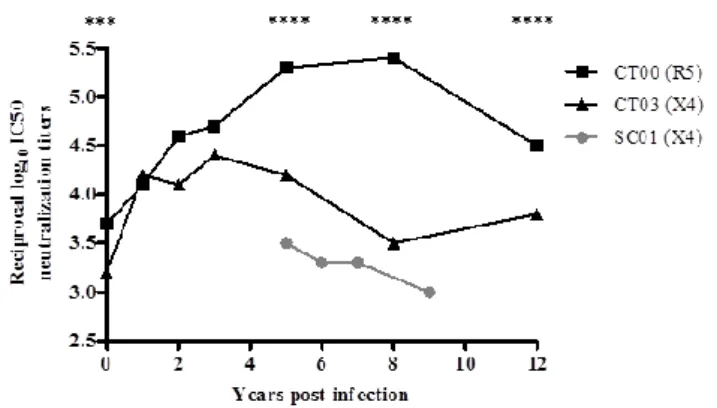
Estudos recentes verificaram que vírus HIV-2 com tropismo para células que expressam o coreceptor CXCR4, isolados de doentes a fases avançadas da infecção, são resistentes aos anticorpos neutralizantes. Foi também descrito no Capítulo 3 que vírus X4 isolados de crianças recentemente infectados são mais resistentes à neutralização. Estes resultados sugerem uma associação entre escape aos anticorpos neutralizantes e alteração de tropismo de R5 para X4. No entanto, permanece a dúvida se os vírus com tropismo para X4 são intrinsecamente resistentes à resposta em anticorpos neutralizantes. O terceiro objective deste trabalho foi caracterizar o fenótipo de neutralização de isolados primários utilizadores do coreceptor X4 isolados de doentes infectados com HIV-2 em várias fases da infecção de forma a determinar se todas as estirpes X4 são resistentes aos anticorpos neutralizantes independentemente do estadio da doença. Foram estudados cinco vírus X4 isolados de doentes em estados avançados de doença (mediana de células T CD4+=78 células/μl) e dois vírus X4 isolados de crianças infectadas por transmissão vertical (ver capítulo 3) em fase inicial da infecção (5 anos) (mediana de células T CD4+=320 células/μl). A sensibilidade dos vírus X4 à neutralização foi comparada com três vírus R5 usados como controlo (mediana de células T CD4+=275 células/μl). Os ensaios de neutralização foram efectuados contra 16 isolados clínicos heterólogos. Observou-se que os vírus X4 eram significativamente mais resistentes à neutralização que os vírus R5, independentemente da fase da infecção em que
foram isolados. O facto de os vírus X4 das crianças serem também mais resistentes à neutralização que os vírus R5, demonstra que a resistência aos anticorpos neutralizantes pode surgir muito cedo após a transmissão. Além disso, os vírus X4 de indivíduos em estados avançados de doença eram significativamente mais resistentes aos anticorpos neutralizantes que os vírus X4 isolados das crianças (infecção recente), o que sugere que a resistência aos anticorpos neutralizantes é um processo gradual que vai ocorrendo ao longo da infecção. Ao analisar-se a região V3 dos vírus em estudo, detectou-se que todos os vírus X4 apresentavam as mutações nesta região anteriormente associadas a alteração de tropismo de R5 para X4 (uma mutação na posição 18, a mutação V19K/R, uma inserção na posição 24 e uma carga global igual ou superior a 7). A avaliação das estruturas secundárias desta região revelou igualmente diferenças importantes entre vírus R5 e X4. Os vírus R5 na infecção aguda e crónica são caracterizados por ausência de estrutura secundária regular, ao passo que os vírus X4 de infecção recente apresentam uma estrutura secundária em β-hairpin. Os vírus X4 de indivíduos em fase avançada caracterizam-se pelas conformações β-α-β ou helix-loop-helix. Estes dados sugerem um modelo de evolução da estrutura secundária da região V3 no qual, ao longo da infecção, a pressão exercida pelos anticorpos neutralizantes sobre a V3 força o vírus a escapar e a alterar esta região de forma que deixe de ser reconhecida pelos anticorpos neutralizantes. Estas alterações favorecem igualmente a mudança de tropismo para X4 o que por sua vez está associado ao decréscimo das células T CD4+. Estes dados mostram que a resistência aos anticorpos neutralizantes é uma característica intrínseca dos vírus X4, provavelmente determinada por alterações na sequência e conformação da região V3, que dificultam o reconhecimento desta região pelos anticorpos.
Em conclusão, este trabalho permitiu demonstrar que a resposta dos anticorpos neutralizantes surge muito cedo na infecção aguda e persiste na fase crónica da infecção mesmo após significativa depleção das células B de memória, e ainda que os Nabs são responsáveis pela evolução viral, por alterações estruturais na região V3 e pela alteração do tropismo que leva à resistência à neutralização. Os resultados constituem também um novo e potencialmente relevante contributo na área das vacinas. Em primeira análise, o escape à acção dos anticorpos neutralizantes existe na infecção por HIV-2 e por conseguinte uma
vacina necessita de geral respostas contra estirpes R5 e X4. Os anticorpos são principalmente dirigidos contra a região V3 nas fases inicial e crónica da infecção. Em estádios mais avançados, a resposta contra esta região diminui mas surgem anticorpos cujo alvo é a gp36. Estes resultados confirmam que a V3 é um bom imunogénio a ser usado no desenho de vacinas mas salientam o facto de a gp36 poder conter epitopos importantes a serem incluídos numa vacina. Outra importante contribuição destes estudos foi a descoberta de que é possível manter uma forte resposta em anticorpos neutralizantes através de populações de células B que não as de memória, nomeadamente plasmócitos. Uma vacina contra o HIV-2 e o HIV-1 idealmente estimularia respostas deste tipo de células B. A dificuldade persiste em escolher o imunogénio ou grupo de imunogénios capazes de suscitar estas respostas. Destes estudos conclui-se que imunogénios baseados na região V3 e na gp36 do HIV-2 poderão ser bons candidatos para o desenho de vacinas.
ABSTRACT
Dynamics of the neutralizing antibody response and resulting HIV-2 escape during acute and chronic infections and their impact on viral evolution and disease progression remain unknown. The aims of this thesis were: characterize Nab response and molecular and phenotypic evolution of HIV-2 in early infection, investigate Nab responses in HIV-2 chronically infected patients with memory B cell imbalances and characterize the neutralization phenotype of HIV-2 X4-tropic isolates from diverse disease stages.
Broad and potent Nabs are elicited very early in HIV-2 infection, the potency of this response being associated with high evolutionary rates. Nab escape was associated with R5-to-X4 switch, increased diversity in V1 and V3 regions and changes in V3 conformation. These findings show that Nabs are the main driver of the rapid molecular and phenotypic evolution of HIV-2 in early infection.
Despite the loss of memory B cells observed with disease progression, broad and potent Nabs were elicited throughout HIV-2 infection. Nabs were found to target the C2V3C3 envelope region and, in advanced disease stage, the gp36 ectodomain. These data suggest a role for other B cell subsets in the production and perpetuation of Nabs.
HIV-2 X4-tropic viruses were found to be significantly more Nab resistant than R5 viruses (independently of disease stage) and late infection X4 isolates were significantly more Nab resistant than early infection X4 viruses. X4-tropism was associated with sequence changes and significant gain in the V3 loop secondary structure. The results prove that Nab resistance is an intrinsic feature of X4-tropic HIV-2 isolates, acquired through infection period, and is associated with amino acid and conformational changes in the V3 loop that favour R5-to-X4 switch.
In conclusion, Nab responses emerge very early in infection, persist despite memory B cells imbalances and drive tropism switch, supporting a major role for Nabs in HIV-2 evolution.
ABBREVIATIONS
µl Microliters
0C Celsius degree
ADCC Antibody-dependent cell-mediated cytotoxicity
ADCMI Antibody-dependent complement-mediated inactivation
ADCVI Antibody-dependent cell-mediated viral inhibition
AIDS Acquired Immunodeficiency Syndrome
ART Anti-retroviral therapy
BSA Bovin serum albumin
CDC Centers for Disease Control
CO2 Carbon dioxide
CRF Circulating recombinant form
CTL Cytotoxic T lymphocyte
DC Dendritic cells
DMEM Dulbecco's minimal essential medium
dN Rate of nonsynonymous substitutions
DNA Desoxiribonucleic acid
dS Rate of synonymous substitutions
ELISA Enzyme-linked immunosorbent assay
Fab Fragment antigen-binding
FBS Fetal bovine serum
Fc Fragment crystallizable region
FcγRs Fc receptor
GALT Gut associated lymphoid tissue
GM Growth medium
GTR General time reversible model
h hour
HAART Highly active anti-retroviral therapy
HIV-1 Human Immunodeficiency Virus type 1
HIV-2 Human Immunodeficiency Virus type 2
IC50 50% inhibitory concentration
IDU Injection drug users
IFN Interferon
Ig Imunoglobulin
IN Integrase
KDa Kilodalton
LTRs Long terminal repeats
mRNA messenger RNA
MSM Men who have sex with men
MTCT Mother to child transmission
Nabs Neutralizing antibodies
NK Natural killer
PBMC Peripheral blood mononuclear cell
PBS Phosphate buffered saline
PCR Polymerase chain reaction
PR Protease
PT Portuguese patients
RNA Ribonucleic acid
RT Retrotranscriptase
SIDA Síndrome de Imunodeficiência Adquirida
SIV Simian Immunodeficiency Virus
SIVcpz SIV from Pan troglodytes troglodytes chimpazees
SIVgor SIV from Western lowland gorillas
SIVsmm SIV from Cercocebus torgnatus atys sooty mangabeys
SU Surface glycoprotein
TCID50 50% tissue culture infectious dose
TLR Toll-like receptor
TNF Tumor necrosis factor
USA United States of America
TABLE OF CONTENTS
ACKNOWLEDGEMENTS
iPREFACE
iiiRESUMO
viiABSTRACT
xvABBREVIATIONS
xviiCHAPTER 1
General introduction 1The origin and discovery of the Human Immunodeficiency Virus type 2 (HIV-2) 3
The global spread of HIV-2 4
Biology of HIV-2 6
Genome and structure 6
Life cycle 8
HIV-2 envelope 10
Molecular and structural organization 10
Interaction between the Env protein and the cell 11
HIV-2 coreceptor usage and pathogenesis 13
Transmission 14
Pathogenesis of HIV-2 infection 15
HIV-2 molecular evolution 18
Mechanisms of viral evolution 18
Evolution of the env gene and disease progression 20
Human responses against HIV-2 22
Innate responses 22
Celular responses 24
Humoral responses 25
CHAPTER 2
Aim of the studies and work plan 37
CHAPTER 3
Neutralizing antibodies drive the molecular and phenotypic evolution of the
human immunodeficiency virus type 2 envelope 43
Title page 45
Abstract 46
Background 47
Materials and Methods 48
Results 54 Discussion 62 Conclusions 67 Competing interests 68 Authors' contributions 68 Acknowledgements 68 Additional material 69
CHAPTER 4
Potent and broadly neutralizing antibodies despite marked memory B cell
depletion in chronic HIV-2 infection 71
Title page 73
Abstract 74
Introduction 75
Materials and Methods 76
Results 78
Discussion 84
Acknowledgements 86
CHAPTER 5
Primary isolates of HIV-2 that use the CXCR4 co-receptor are intrinsically
resistant to antibody neutralization 93
Title page 95 Abstract 96 Introduction 97 Methods 98 Results 99 Discussion 102 Acknowledgements 105
CHAPTER 6
General discussion and conclusions 107
CHAPTER 7
General Introduction
The origin and discovery of the Human Immunodeficiency Virus
type 2 (HIV-2)
Immunodeficiency viruses, belonging to the Lentivirus genus, can be found in several species including non-human primates [1, 2]. These simians are the natural reservoir of many different specific variants of Simian Immunodeficiency Virus (SIV). Several independent zoonotic transmission events lead to the introduction of these viruses to humans. Human Immunodeficiency Virus type 1 (HIV-1) was introduced to humans from SIVcpz that infects West Central African chimpanzees (Pan troglodytes troglodytes) and from SIVgor that infects Western lowland gorillas (Gorilla gorilla gorilla) [1-6]. HIV-2 is closely related to SIVsm which is found in sooty mangabey monkeys (Cercocebus atys atys) [7, 8]. Phylogenetic analysis shows that there have been many cross-species transmissions to humans, with each successful event resulting in a specific form (group) of HIV [1, 9, 10]. In HIV-2 transmission events are thought to be at least eight, giving rise to groups A to H [8, 11], whereas in HIV-1 tree cross-species events occurred from chimpanzees to humans (groups M, N and O) [12, 13], and one from gorillas (group P) [5, 6]. Transmission from simians to man is estimated to have occurred in the late nineteenth through early twentieth century but its consequences have only be detected in 1981 for HIV-1 and 1985 for HIV-2 [14-16]. Indeed, the Acquired Immunodeficiency Syndrome (AIDS) was first described in 1981 in the United States of America (USA), after the observation of opportunistic infections along with immune suppression in young men who have sex with men (MSM) in New York City and California [17, 18]. Soon after similar observations were made in patients from Haiti, Africa and Europe [19-21] and it became clear that the new disease with unknown cause struck also haemophiliacs, injection drug users (IDU), women and infants (mother to child transmission - MTCT) [22-25]. In May 1983, at the Pasteur Institute (Paris), Luc Montagnier and Françoise Barré-Sinoussi isolated a new retrovirus from an AIDS patient [26] and in the following year a similar retrovirus was isolated by American investigators [27]. These findings proved that this retrovirus, later classified as HIV-1, was the causative agent of AIDS [28].
In the early 80’s, patients from Guinea-Bissau and Cape Verde Islands were admitted in a Portuguese hospital in Lisbon. These patients presented clinical symptoms similar to AIDS
but with constant negative serologic tests. At the time Maria Odette Santos Ferreira from the Faculty of Pharmacy of Lisbon took the blood samples from these patients to Luc Montagnier at the Pasteur Institute (Paris). The collaboration between Portuguese and French researchers lead to the characterization of a second retrovirus, distinct from HIV-1, classified as HIV-2 [29-31].
The global spread of HIV-2
According to UNAIDS, globally there has been a decline the number of newly infected adults and children in the last 10 years [32]. With the scale up of antiretroviral therapy (ART) over the past few years, the number of AIDS-related deaths has also decreased, but with the significant reductions in mortality the number of people living with HIV worldwide has increased. By the end of 2011, there were an estimated 34 million people living with HIV, 2.5 million newly infected adults and children and 1.7 million AIDS-related deaths [32]. Sub-Saharan Africa still accounts for the highest HIV burden, with 69% of the total number of HIV infected individuals and an average prevalence among adults of 4.9%. However, in 23 Sub-Saharan countries, the incidence of HIV has decreased more than 25% [32].
Despite the existence of eight HIV-2 groups, only groups A (HIV-2A) and B (HIV-2B) are considered endemic, with group A frequently found in the western part of West Africa (Guinea-Bissau, Senegal, The Gambia, Ivory Coast and Cape Verde) and group B more restricted to Ivory Coast and Ghana [11, 29, 33-38]. The remaining groups have only been identified in a few individuals from Sierra Leone, Liberia and Ivory Coast [7, 39-45]. The first HIV-2 A/B recombinant was isolated from a patient from Ivory Coast and recently three other A/B recombinants have been described in Japan, which lead to the classification of the first circulating recombination form (CRF) of HIV-2, the HIV-2 CRF01_AB [43, 46, 47].
Besides its confined geography, recent reports indicate that HIV-2 prevalence is now decreasing even in countries where the number of cases used to be high [48-52]. For instance, in Guinea-Bissau the prevalence of HIV-2 decreased from 8.3% in 1990 to 4.7% in 2007, and in The Gambia it decreased from 7.0% in 1988-91 to 4.0% in 2001-03. The prevalence of HIV-1 increased in these regions in the same period of time [49, 51, 52].
The origins of HIV-2 groups A and B are estimated to be around 1938 and 1945, respectively [15, 16, 53]. The epicentre of the HIV-2B epidemic is most likely Ivory Coast, whereas some doubts remain about the region where the cross-species event took place for HIV-2A. There is strong phylogenetic evidence that trace both Ivory Coast and Guinea-Bissau as the epidemic centres: serologic data tends to favour Guinea-Bissau, but the recent discovery of SIVsmm strains closely related to HIV-2A in faecal samples from sooty mangabeys in Ivory Coast suggests that this region is also the epicentre of HIV-2A [8, 49, 53, 54]. Whether the cross-species event took place in Ivory Coast and Guinea-Bissau was where the epidemic was established very early after transmission or Guinea-Bissau is indeed the epicentre of HIV-2A remains to be clarified [8, 53, 55]. From these two countries, or because of commercial relations, sex trade and migration between them, HIV-2A spread to other countries in West Africa, like Senegal, The Gambia, Cape Verde, Nigeria and Burkina Faso [53]. The viral migration outside West Africa most likely happened through immigration and socio economic connections with high-prevalence countries. For instance, past relations between France and Ivory Coast and Senegal led to multiple viral introductions in France. Furthermore, HIV-2A is thought to have spread from Guinea-Bissau and Cape Verde to Portugal, mainly during the independence war [15, 53, 56]. Within Europe, strong evidences point to transmission from Portugal to the main immigration destinations, like Luxemburg and the United Kingdom (UK), but most likely to Switzerland, Belgium and Germany as well [53, 56]. Portugal is also thought to be responsible for transmitting HIV-2A outside of Europe to other countries with socio economically linkage such as India, Mozambique and Brazil [11, 50, 57, 58].
Portugal is one of the few countries outside West Africa with a significant number of HIV-2 cases. In December 2011 the total number of AIDS cases associated with HIV-2 was 527, which represents 3.1% of the total number of AIDS cases [59]. Most of the cases (73.1%) were associated with heterosexual transmission and were in individuals with ages between 35 and 54 years (60.4%). Parenteral transmission through blood transfusions or surgical procedures during the independence ward against Guinea-Bissau (between 1960 and 1974) might have been an important transmission route since sexual transmission of HIV-2 is less efficient than for HIV-1 [60], and multiple exposures to HIV-2 might be necessary to facilitate
infection [61, 62]. The number of new diagnosed infections has been decreasing in the last ten years (from 23 new infections in 2001 to 8 in 2011) [59]. Several studies addressed HIV-2 epidemic in Portugal over the past few years. Gomes et al found that in Lisbon and the Southern part of the country, between 1997 and 2002, 57% of infected patients were from either Guinea-Bissau or Cape Verde and the incidence was similar between genders [61]. A few months later, Mota-Miranda and collaborators performed a similar study in the North region of Portugal, with data from 1985 to 2003, and concluded that 95% of infections were among Portuguese individuals but in 51% of the cases a connection with West Africa was established [63]. Another study performed in a Lisbon hospital between 1987 and 2006 showed a majority of infections in patients from West Africa (67.5%) and predominantly in women (66.9%) [64]. More recently, a large study involving several hospitals across the country, with data from 1985 to 2007, detected a mobility pattern of the epidemic before and after the year 2000. In the beginning most infected patients were Portuguese men, probably due to the return of Portuguese soldiers after the independence war in the late 70’s. After 2000 a change was observed towards women of West African origin, most likely because of the increase in migration from West Africa to Portugal in the late 90’s [62].
Biology of HIV-2
Genome and structure
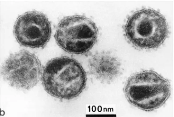
HIV-2 is a spherical enveloped virus with a diameter of approximately 110 nm (Figure 1.1) [65-67]. The envelope comprises a lipid bilayer derived from the host cell plasma membrane at budding, and therefore may also contain some host cell proteins from the human leukocyte antigen (HLA) system class I and II. Embedded in the lipid bilayer is the transmembrane glycoprotein (TM) bound non covalently to the outer surface glycoprotein (SU), this complex is arranged in trimers. Internally the viral particle is coated by the matrix proteins, essential to stabilize the spherical structure. Within the matrix resides the cone shaped capsid which contains two copies of a positive sense single stranded RNA associated with the nucleocapsid proteins. Inside the capsid there are also all the necessary enzymes to
viral maturation and early phases of replication, such as protease (PR), reverse transcriptase (RT) and integrase (IN) and the accessory proteins Nef, Vif, Vpr and Vpx [65-67].
Figure 1.1 - Electron Micrograph of HIV. The virus is more about 110 nm wide. (Adapted from
http://www.histology.leeds.ac.uk)
Each RNA molecule is about 9800 nucleotides long and is flanked by long terminal repeats (LTRs) at both ends (5’ and 3’). Nine genes are encoded in the compact genome, tree of them encode for structural or enzymatic proteins (gag, pol and env), two for regulatory proteins (tat and rev) and four for the mentioned accessory proteins (nef, vif, vpr and vpx) (Figure 1.2). The three open reading frames are used to translate all the proteins [65, 66, 68]. The gag encodes the polyprotein precursor Pr55Gag that is then cleaved into the proteins p26 (capsid), p16 (matrix), p6 (nucleocapsid) and p6 (C-terminal protein) by the viral PR. The Gag proteins are essential for virion assembly and release. The gag and pol genes produce a Pr160GagPol precursor polyprotein, which is then processed by the viral PR. The pol encodes for tree enzymes necessary for replication: the RT (p53), the PR (p11) and the IN (p34). The
env gene encodes for the polyprotein precursor Pr140Env, cleaved by PR in the glycoproteins
SU (gp125) and TM (gp36). These glycoproteins are essential for viral attachment and fusion to the host cell membrane [65, 66, 68].
Figure 1.2 – Genomic organization of HIV-2. (Adapted from Taveira et al, Manual sobre SIDA 2011 [65])
Life cycle
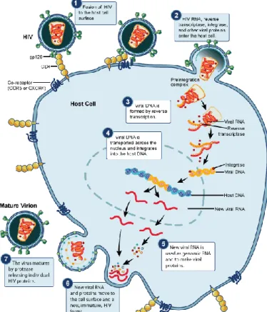
HIV-2’s life cycle usually begins with the binding of the virus to a specific receptor, the CD4, present in the cellular surface of the host cell [T-lymphocytes, monocytes, macrophages, dendritic cells (DC) and brain microglia] (Figure 1.3) [65, 68]. Besides CD4, other molecules expressed in the cell surface are essential to the interaction between virus and host cell. These are the chemokine receptors and work as coreceptors for HIV entry in the host cell [65, 68].
After entry and uncoating of the virus in the cytoplasm, RT starts reverse transcription of the viral RNA into double stranded DNA, that together with the proteins IN, RT, matrix and Vpr form the pre-integration complex. The translocation of this complex to the nucleus is mediated by IN and Vpx [69]. Once in the nucleus the IN integrates the viral DNA into an open region of the host chromosomal genome. The proviral DNA can remain latent (transcriptionally silent) in the host cell or be immediately transcribed by the cellular machinery continuing the virus life cycle [65, 69, 70].
The transcription of the proviral DNA is mediated by the promoter region within the 5’ LTR and originates tree classes of messenger RNA (mRNA): completely spliced mRNA or early transcripts (Rev, Tat, Nef), incompletely spliced mRNA or late transcripts (Env, Vif, Vpr and Vpu/Vpx) and unspliced mRNA or late transcripts (precursor polyproteins Gag and Gag-Pol). All these mRNA are later incorporated in the viral particles as genomic RNA. Early transcripts are transported outside the nucleus like any cellular RNAm and are needed to complete the expression of late transcripts. Rev is responsible for the transport of late transcripts. This
protein binds to the Rev responsive Element (RRE) in the RNA env region and carries the late transcripts to the cytoplasm to be translated [65, 70].
The Env precursor polyproteins are translated and glycosylated before they oligomerize in trimers. Then the polyproteins are cleaved in SU and TM glycoproteins and transported to the cytoplasmic membrane. The viral RNA and proteins are directed to the cellular surface, where new immature viral particles are formed and released by gemmulation of the cytoplasmic membrane, thus acquiring the lipid envelope containing the SU/TM trimers. Final maturation occurs outside the cell with the cleavage of the precursor protein Gag-Pol by PR and posterior structural rearrangement and repositioning of the viral proteins, giving rise to mature and infectious viral particles [65, 70].
HIV-2 envelope
Molecular and structural organization
As mentioned above, the envelope glycoproteins SU and TM are encoded by the env gene, and are responsible for the fusion between viral and host cell membranes, allowing the release of the viral capsid into the host cell cytoplasm. The SU and TM glycoproteins are associated by non covalent bounds forming trimers or spikes [71, 72]. HIV-2 spikes have been reported to be more prominent and stable after budding [73-75], whereas in HIV-1 they drop immediately after budding and during maturation [71, 76].
Despite the high variability of SU, some structural and functional elements are conserved, which allowed the classification of five hypervariable regions (V1 to V5), separated by five more conserved regions (C1 to C5). This glycoprotein has a complex secondary structure, with variable regions V1 to V4 forming loops stabilized by disulphide bridges. In its native conformation, SU has two domains, one internal and one external [77]. The external domain is highly glycosylated, has most of the antigenic determinants, including neutralizing epitopes, and is involved in the interaction between the SU and the cellular receptor and coreceptors. The internal domain is hydrophobic and is essential for the SU and TM association. Connecting the external and internal domains there is a smaller domain designated bridging sheet [65, 72, 77, 78].
The TM glycoprotein is divided in one extracellular ectodomain, one transmembrane region (insertion in the host cell membrane) and one intracytoplasmatic domain. The ectodomain has several domains common to other fusion proteins: a hydrophobic region rich in glycines at the N-terminal end called fusion peptide, followed by two α-helices containing leucine-zipper motifs designated heptad repeat 1 (HR1) and 2 (HR2). The fusion peptide is essential for attachment to the host cell membrane. The HR motifs present repeated patterns of seven amino acids, being the first and fourth residues hydrophobic, mainly leucines. These motifs are arranged in a thermostable structure in sextuple helix, formed by trimers of HR1 and HR2. This structure is directly involved in the fusion to the host cell. The
intracytoplasmatic domain mediates the binding of the envelope to the matrix protein, necessary for the maturation of new viral particles [65, 72, 77, 79].
Interaction between the Env protein and the cell
The entry of HIV into the host cell generally involves three steps mediated by the envelope glycoproteins: binding of the SU to the CD4 receptor, binding of the SU to the coreceptor and fusion of the viral envelope with the host cell membrane (Figure 1.4).
Figure 1.4 – Model of the multi-step process of HIV entry. (Adapted from http://www.virology.uzh.ch/)
The process begins with the interaction between the external domain of the SU and the CD4 receptor. After binding to the CD4, SU suffers major conformational changes, with the formation of a bridging sheet and increased exposure of V1, V2, V3 and C4 regions (Figure 1.5) [65, 72]. The folding of the bridging sheet is necessary to form the conserved part of the coreceptor binding site and the repositioning of the V1/V2 loop is thought to uncover this site [80]. All these rearrangements allow the stabilization of the SU-CD4 bond and leads to an approximation between the viral envelope and the host cell membrane and consequent interaction with the coreceptor [65, 72, 79, 81, 82]. Some HIV-2 primary isolates are known to infected cells independently of CD4, which implies that the coreceptor binding site in these isolates is already formed or exposed prior to the CD4 binding [82, 83]. Therefore it is
thought that the V3 region in some HIV-2 has a more open and exposed conformation, which allows it to induce conformational changes in the V1/V2 loop during fusion without the need to previously bind to the CD4 [82, 84, 85]. The SU-CD4 binding also induces changes in the TM glycoprotein: the fusion peptide becomes exposed and is inserted in the host cell membrane, and HR1 and HR2 fold in an antiparallel form, originating a six-helix bundle. The viral envelope and the host cell membrane are brought together during this process, leading to the formation of the fusion pore, allowing the entrance of the viral capsid into the cytoplasm of the target cell [65, 86, 87]. After CD4 binding, the exposure of the coreceptor binding site seems to be faster in HIV-2, dues leading to a more rapid fusion between the viral envelope and the host cell membrane, despite the lower affinity of gp125 (HIV-2) to the CD4 receptor compared to gp120 (HIV-1) [85, 88]. This faster exposure of the coreceptor binding site might be due to differences in orientation of the V1/V2 loop and folding of the bridging sheet, as mentioned above [80].
HIV-2 coreceptor usage and pathogenesis
In vivo, the main coreceptors of HIV are the CCR5 and CXCR4. However, HIV has been shown to use other coreceptors [89, 90]. HIV’s tropism to cells expressing one of the coreceptors is related with the replication capacity of the virus in different cell lines. Viruses that infect preferentially macrophage cell lines, without the capacity to induce syncytia and with low/slow replication rates typically use the CCR5 coreceptor and are called R5 viruses. On the other hand, viruses that infect mainly lymphocytic cell lines, with the ability to induce syncytia and with high/fast replication rates, are named X4 viruses and use the CXCR4 coreceptor. Finally, R5X4 viruses are variants with identical capacity of replicating in macrophage and lymphocytic cell lines and use indifferently the CCR5 and the CXCR4 coreceptors [91-94].
HIV-2, like HIV-1, uses mainly CCR5 and/or CXCR4 as coreceptors to enter CD4+ T lymphocytes. However, many HIV-2 isolates can use a wider variety of alternative coreceptors (like CCR1, CCR2, CCR3 and CCR8), though less efficiently than CCR5 and CXCR4 [95]. The use of a broader range of coreceptors does not seem to be related to HIV-2’s pathogenicity and the relevance of using alternative coreceptors in vivo is not clarified [83, 89, 91, 95, 96].
In HIV-2 infection, R5 viruses are isolated from asymptomatic patients or in early stages of disease, and X4 variants are found in advanced AIDS [97, 98]. The emergence of X4 variants in HIV-2 infection seems to be related with escape from neutralizing antibodies (Nabs) directed against the V3 region since X4 viruses are more resistant to neutralization [97]. In HIV-1 infection, R5 viruses are also found in acute and asymptomatic phases. In 50% of infected patients, the viruses evolve to an X4 phenotype with a rapid decline of CD4+ T cells and progression to AIDS. However, R5 viruses can also be responsible for CD4+ T cell depletion and persist in advanced disease. Contrarily to HIV-2, R5X4 and X4 HIV-1 variants seem to be more sensitive to Nabs, which might explain why the phenotype change does not always occur, and when it does it’s in a stage of immune system failure [93, 99-101].
The major determinants for CCR5 or CXCR4 coreceptor usage by HIV-2 are located in the C-terminal region of the V3 loop. An increase in charge on this region, by the presence of
positively charged amino acids is correlated with CXCR4 phenotype [84, 102-104]. The most relevant residues are in positions 18, 19 and 24 [104]. In HIV-1 the glycosylation pattern of V3 and V1/V2 regions also seem to influence the coreceptor phenotype [105-108]. The degree of glycosylation is lower in HIV-2 but the impact on coreceptor usage is still elusive since no clear association has been made between glycosylation of V1/V2 or V3 and coreceptor phenotype of HIV-2 [84, 109].
Transmission
HIV spreads through sexual contacts, contaminated blood or blood products (medical injections, blood transfusions and injection drug usage) and from mother to child (pregnancy, delivery and breast feeding) [57]. The most common route of transmission of HIV-2 is through heterosexual contact, as is for HIV-1, but transmission rates are 3 to 6 folder lower [60]. Similarly, MTCT is rare in HIV-2 with a rate below 5% compared to almost 25% in HIV-1 in untreated pregnant women [110-113]. A Portuguese study found a MTCT rate of 1.5% for HIV-2 and 3.4% for HIV-1 between 1999 and 2005 and transmission was associated with absence of ART [114]. The reasons for the reduced transmission rates of HIV-2 are still not fully understood, but are probably linked to the lower plasma viremia [49, 110] and reduced viral shedding in the genital tract seen during HIV-2 chronic infection [115, 116]. A study from The Gambia found a 37-fold difference in plasma RNA levels between HIV-2 and HIV-1 infected untreated pregnant woman (410 versus 15,100 RNA copies/ml) [110]. On the other hand, a study in Ivory Coast observed that 24% of HIV-1 infected women had detectable RNA in vaginal secretions compared to only 5% of HIV-2 infected pears [117]. Detectable RNA in the semen is also lower in HIV-2 compared to HIV-1 infected men (2.6log10 versus 4.4log10 RNA copies/ml) [115].
Despite being a rare event, HIV-2 MTCT has been document in both epidemiologic [111, 112, 118-122] and molecular studies [123-125]. There is evidence that the survival rate of HIV-2 infected is higher than their HIV-1 infected pears [126]. In fact, contrarily to HIV-1 infected children [127-129], disease progression and the first clinical manifestations might take many years to arise in HIV-2 infected infants [122, 130]. Furthermore, and also in opposition to
HIV-1 [127, 128], maternal plasma viral load does not seem to determine disease progression [126]. In HIV-1 infection, immaturity of the children’s immune system is thought to account for the fast disease progression, though time of transmission (intrauterine, intrapartum or postpartum) also seem to have a major influence [127, 128]. There is no information about immune responses in MTCT of HIV-2.
Pathogenesis of HIV-2 infection
The course of HIV infection can be divided into three stages: the acute phase, the chronic phase and AIDS [131]. The acute phase, or primary HIV infection, lasts four to eight weeks and is characterized by intense HIV replication and massive loss of CD4+ T cells mainly in gut associated lymphoid tissue (GALT). When transmission occurs through the blood (transfusion, IDUs or MTCT) the virus is probably removed from circulation by the reticuloendothelial system of the spleen, liver and lungs, with consequent infection of lymphoid tissue , HIV replication and dissemination within these organs [132]. Infection is also possible through rectal and genital mucosa (heterosexual and homosexual contacts). In this case DCs seem to have a central role in capturing and transporting viruses to draining lymph nodes and secondary lymphoid tissue where high levels of activated CD4+ T cells are present [131, 132]. CD4+ T cells depletion is a consequence of direct viral infection, activation induced cell death and host cytotoxic responses [133]. As a result of the immune responses directed against the infection, infected individuals experience in this phase flu like symptoms (acute HIV syndrome), including fever, body ache and lymphadenopathy [132]. Viral load, or plasma viremia, usually peaks at three to four weeks after infection but is eventually suppressed to a semi steady state level (viral set point) due to HIV-specific cytotoxic T lymphocytes (CTL) and Nabs. At this point CD4+ T cells are partially restored. The viral setpoint is also dependent on the individual genetic background and is an important determinant of disease progression in HIV-1 infection [38, 132]. In HIV-2 the viral set point is usually much lower compared to HIV-1 (2500 versus 70000 RNA copies/ml) [134].
During this acute phase and early chronic phase, HIV-1 infected patients can be categorized into Fiebig stages I to VI based on a stepwise gain of positivity in clinical diagnosis assays
(Figure 1.6). The first HIV infection marker to be detected is viral RNA that can be assessed by polymerase chain reaction (PCR). The HIV-1 p24 (from gag) usually peaks 20 days after infection and can be detected by enzyme-linked immunosorbent assay (ELISA). Afterwards, it is possible to detect specific antibodies by ELISA and western blot and finally HIV-1 p31 is also detectable by ELISA (Figure 1.6) [135].
Figure 1.6 – Fiebig clinical stages of HIV-1 infection. (Adapted from McMichael et al, Nature Reviews
Immunology 2010 [136])
The chronic phase is asymptomatic and in untreated patients lasts about ten to 25 years in HIV-2 and is much faster in HIV-1 (eight to ten years) [132, 137]. At this stage, infected patients usually present generalized lymphadenopathy caused by persistent follicular hyperplasia. This period corresponds to clinical latency with low but persistent HIV replication in the viral reservoirs (lymphoid tissue) and constant antigen stimulation that drives immune activation [132]. Chronic immune activation is manifested by increased cell turnover, abnormal activation and differentiation of lymphocytes, increased terminal differentiation of B cells and increased activation-induced apoptosis of CD4+ T cells, CD8+ T cells and B cells, thus leading to cellular exhaustion, senescence, and low renewal potential [131, 132]. HIV-2 infection is characterized by a lower state of immune activation, which
might account for the slower disease progression [138-141]. Immune activation, however, does not seem to be directly linked to viral load in HIV-2 infection, since several studies reported that, despite the absence of detectable viremia, levels of immune activation were the same in HIV-1 and HIV-2 infected individuals when patients were matched for the same degree of CD4 depletion [138, 141].
In the late phase of the infection, the immune system slowly gets exhausted by the chronic immune activation and depletion of CD4+ T cells and eventually collapses. This leads to occurrence or reactivation of opportunistic infections (like tuberculosis, candidiasis and pneumonia) and virus induced tumours (Epstein-Barr virus related lymphomas, Kaposi’s sarcoma and cervical cancer caused by Human Papillomavirus). These diseases mark the onset of AIDS. The Centers for Disease Control (CDC) in the USA periodically revises the list of clinical situations that define this stage. Another criterion that defines AIDS is the drop of CD4+ T cell levels below 200 cells/µl of plasma [132].
As mentioned above, HIV-2 infection is characterized by a slower disease progression, longer survival and reduced mortality rates compared to 1 [57, 137, 142-147]. In fact, most HIV-2 infected patients have normal CD4+ T cell counts, undetectable viral loads and absence of clinical disease [57, 134, 137, 139, 148]. Several studies showed that the probability of being AIDS free was near 100% in HIV-2 up to five years post seroconversion compared to 67% in HIV-1 infected patients [142, 149]. Mortality rates were estimated to be 2.5 to 3.5-fold lower per year of follow-up in HIV-2 compared to HIV-1 infection [147, 150]. Another important difference between the two viruses is the lower replication capacity of HIV-2 [151, 152], despite similar proviral loads at the same disease stage [137, 139, 153, 154]. With disease progression, most immunological differences between HIV-1 and HIV-2 are lost, the level of immune activation is the same and CD4+ T cell depletion is similar [57, 137, 138, 145-147]. Nonetheless, after the onset of AIDS, median time to death in HIV-2 infection was twice of that in HIV-1 (12.6 versus 6.3 months) and mortality rates were lower (52% versus 87% per year of follow-up in HIV-2 and HIV-1 infected individuals, respectively). When patients were adjusted for CD4+ T cell counts, mortality rates were similar in HIV-2 and HIV-1 infected individuals (86% versus 130% in HIV-2 and HIV-1 infected patients with CD4+ T counts bellow
200 cells/ml; 62% versus 73% in patients with CD4+ T counts equal or above 200 cells/ml) [147].
HIV-2 molecular evolution
Mechanisms of viral evolution
HIV and other RNA viruses shares common characteristics that make them good models to study evolution: high mutation rates, small genomes, large population sizes, short generation times and high number of offspring [155, 156]. Due to these factors HIV is one of the fastest evolving organisms, with an exceptionally high mutation rate of 2.4x10⁻⁵ mutation/replication in HIV-1 [156]. It is worth mentioning that a recent study with Portuguese and Swedish patients found that the rate of nucleotide substitutions in HIV-2 gp125 and V3 is higher than in HIV-1 when patients were matched for disease stage and CD4 dynamics (10.20x10⁻³ versus 6.40x10⁻³ substitutions/nucleotide/year in gp125 and 29.37x10⁻³
versus 12.36x10⁻³ substitutions/nucleotide/year in V3) [157]. These mutations include
substitutions, insertions and deletions and can appear during several steps of the replication cycle. Most of the point mutations are generated during reverse transcription due to the lack of 3’-5’ exonuclease activity of the RT enzyme [158, 159]. In addition, host enzymes may contribute to the mutation process, particularly the host APOBEC3 family cytidine deaminases that induce G-to-A hypermutations in retroviral RNA [160, 161]. When a mutation in a gene passes on to the offspring and coexists with the original form is called a polymorphism. In a population, two or more variants circulate simultaneously at a polymorphic site. In an HIV infected individual, the viral population consists of a pool of closely related variants known collectively as quasispecies [162].
The second major contribution to HIV genetic diversity is recombination between different viral variants. During one replication cycle, an estimate of 4.9x10⁻⁴ and 3.6 x10⁻⁴ recombinations per site can happen in HIV-2 [163] and HIV-1 [164, 165], respectively. When a cell is infected by two different strains, the newly produced virions may comprise a heterodimeric DNA, meaning an RNA molecule from each strain. Once a new cell is infected
by these virions, a recombinant proviral DNA with a mosaic genome will be generated due to the template switch of the RT enzyme during reverse transcription. As a consequence, large evolutionary and antigenic leaps may occur in just one round of replication [159, 165, 166]. Despite the high recombination rate seen in HIV-2, contrarily to HIV-1, CRFs are rare (for a review on HIV-1 CRFs see references [167] and [168]). As mentioned above, only one CRF has been described so far, the CRF01_AB [43, 46, 47]. The geographic restriction of the infection is probably a major constrain to the development of CRFs in HIV-2. Due to the simultaneous prevalence of both HIV-1 and HIV-2 infections in some West African countries and Portugal, there were concerns that an HIV-1/HIV-2 recombinant could emerge and produce an even more aggressive virus. In fact, recombination between the two viruses has been demonstrated to be possible in vitro [169]. However, in vivo, there are important differences between HIV-1 and HIV-2 in the signals and mechanisms of genomic RNA packing that impede recombination between the two viruses [170-173].
The high replication and production rates combined with the ability to mutate and recombine are the bases for the high diversity of HIV; however there are other selective forces (natural selection) and chance events (genetic drift) that shape viral evolution [162, 174]. Natural selection is a highly deterministic evolutionary process that promotes the elimination of deleterious mutations by reducing their incidence in the population (negative selection) and favours the fixation of beneficial mutations by increasing their frequency (positive selection). A mutation is deleterious if it decreases the ability of the virus to survive and/or reproduce in the present environment (fitness), beneficial or advantageous if it increases viral fitness compared to the wild type and neutral if it has no significant effect. Neutral mutations are not affected by natural selection. So, whenever a new mutation is generated it becomes either fixed or is eliminated from the population accordingly to the way it affects viral fitness in that given environment [162, 174, 175]. In HIV-2 and HIV-1 infections, natural selection is a major driver of molecular evolution, due to the strong selective pressures imposed by the host’s immune system, mainly Nabs over the env gene [176-180].
Mutation frequencies are affected not only by natural selection but also by random genetic drift [162, 174]. The latter is a stochastic process in which mutation frequencies fluctuate
![Figure 1.2 – Genomic organization of HIV-2. (Adapted from Taveira et al, Manual sobre SIDA 2011 [65])](https://thumb-eu.123doks.com/thumbv2/123dok_br/18192003.875448/38.892.159.761.128.311/figure-genomic-organization-adapted-taveira-manual-sobre-sida.webp)

![Figure 1.5 – Secondary Structure of gp120 after CD4 binding. (Adapted from Kwong et al, Nature 1998 [77])](https://thumb-eu.123doks.com/thumbv2/123dok_br/18192003.875448/42.892.285.608.585.907/figure-secondary-structure-cd-binding-adapted-kwong-nature.webp)