R E S E A R C H
Open Access
Evolution of the human immunodeficiency virus
type 2 envelope in the first years of infection is
associated with the dynamics of the neutralizing
antibody response
Cheila Rocha
1,2, Rita Calado
1,2, Pedro Borrego
1,2, José Maria Marcelino
3, Inês Bártolo
1,2, Lino Rosado
4,
Patrícia Cavaco-Silva
2, Perpétua Gomes
2,5,6, Carlos Família
2, Alexandre Quintas
2, Helena Skar
7, Thomas Leitner
7,
Helena Barroso
1,2and Nuno Taveira
1,2*Abstract
Background:Differently from HIV-1, HIV-2 disease progression usually takes decades without antiretroviral therapy and the majority of HIV-2 infected individuals survive as elite controllers with normal CD4+T cell counts and low or undetectable plasma viral load. Neutralizing antibodies (Nabs) are thought to play a central role in HIV-2 evolution and pathogenesis. However, the dynamic of the Nab response and resulting HIV-2 escape during acute infection and their impact in HIV-2 evolution and disease progression remain largely unknown. Our objective was to
characterize the Nab response and the molecular and phenotypic evolution of HIV-2 in association with Nab escape in the first years of infection in two children infected at birth.
Results:CD4+T cells decreased from about 50% to below 30% in both children in the first five years of infection and the infecting R5 viruses were replaced by X4 viruses within the same period. With antiretroviral therapy, viral load in child 1 decreased to undetectable levels and CD4+T cells recovered to normal levels, which have been sustained at least until the age of 12. In contrast, viral load increased in child 2 and she progressed to AIDS and death at age 9. Beginning in the first year of life, child 1 raised high titers of antibodies that neutralized primary R5 isolates more effectively than X4 isolates, both autologous and heterologous. Child 2 raised a weak X4-specific Nab response that decreased sharply as disease progressed. Rate of evolution, nucleotide and amino acid diversity, and positive selection, were significantly higher in the envelope of child 1 compared to child 2. Rates of R5-to-X4 tropism switch, of V1 and V3 sequence diversification, and of convergence of V3 to aβ-hairpin structure were related with rate of escape from the neutralizing antibodies.
Conclusion:Our data suggests that the molecular and phenotypic evolution of the human immunodeficiency virus type 2 envelope are related with the dynamics of the neutralizing antibody response providing further support for a model in which Nabs play an important role in HIV-2 pathogenesis.
Keywords:Vertical HIV-2 infection, Evolution of the neutralizing antibody response, Escape from neutralization, Molecular evolution, Tropism
* Correspondence:[email protected]
1
Unidade dos Retrovírus e Infecções Associadas, Centro de Patogénese Molecular, Faculdade de Farmácia de Lisboa, Lisboa, Portugal
2Centro de Investigação Interdisciplinar Egas Moniz (CiiEM), Instituto Superior de Ciências da Saúde Egas Moniz, Monte de Caparica, Portugal
Full list of author information is available at the end of the article
Background
Infection with human immunodeficiency virus type 2 (HIV-2) affects 1-2 million individuals mostly living in West Africa, India and Europe [1,2]. Eight different HIV-2 groups named A through H have been reported but only viruses from groups A and B are known to cause human epidemics [3,4]. Among those, viruses from group A are responsible for the vast majority of HIV-2 infections worldwide.
Even though HIV-1 and HIV-2 are closely related vi-ruses and share a high degree of similarity, infections by these viruses lead to very different immunological and clinical outcomes. HIV-2 infection eventually leads to CD4 depletion, AIDS and death [5-7]. However, differ-ently from HIV-1, HIV-2 disease progression usually takes decades without antiretroviral therapy and the ma-jority of HIV-2 infected individuals survive as elite con-trollers with normal CD4+ T cell counts and low or undetectable plasma viral load [8-16]. Understanding of the factors involved in the effective control of viral repli-cation and disease progression in HIV-2 infected indi-viduals might prove crucial to devise the best strategy to prevent and treat HIV-1.
Enhanced immune control could explain the mild out-come of most HIV-2 infections. Unlike HIV-1 infected patients, most HIV-2 patients in chronic stage produce potent and broad neutralizing antibodies [17-21]. Recent evidence has shown that the viruses isolated from HIV-2 infected patients with advanced disease are characterized by increased resistance to entry inhibitors, including the CCR5-antagonist maraviroc [22] and neutralizing anti-bodies [23], and by a remarkably high evolutionary rate [24,25]. These results suggest that neutralizing anti-bodies play a central role in HIV-2 evolution and patho-genesis. However, in contrast to HIV-1, still nothing is known about the neutralizing antibody response and the molecular and phenotypic features of HIV-2 in acute/ early infection because HIV-2 patients are usually diag-nosed many years after seroconversion.
Most neutralizing epitopes in the HIV-2 envelope glycoprotein complex are located in the surface gp125 glycoprotein. They have been identified in V1, V2, V3, V4 and C5 regions, and in the CD4-binding site [19,21,23,26-29]. These epitopes are well exposed in the envelope complex of CCR5-using isolates that are usu-ally highly sensitive to antibody neutralization [21,27]. However, X4 isolates that emerge in late stage infection in some HIV-2 patients when C2V3C3-specific neutral-izing antibodies wane are highly resistant to antibody neutralization [23]. The V3 loop sequence, size and con-formation of the X4 isolates are markedly different from those of R5-neutralization sensitive isolates supporting a direct role of this region in escape from neutralization and a direct role of the neutralizing antibodies in
shaping the evolution of V3 in progressive HIV-2 infec-tion. The neutralizing domains expressed in the enve-lope glycoproteins in acute/early infection and the role of the neutralizing antibodies and neutralization escape in shaping the evolution of the HIV-2 envelope in this period remains to be determined.
Perinatal transmission of HIV-2 is a rare event that in Europe has only been documented in Portugal [30-34] and France [35]. Vertical transmission cases constitute a unique opportunity to study the phenotypic and molecu-lar evolution of HIV-2 Env in acute and early infection as well as the role of Nabs in this process. Our objective was to characterize the evolution of the Nab response in two children infected with HIV-2 at birth in association with the molecular and phenotypic evolution of the virus. We show that broad and potent Nabs can be elic-ited very early after infection and that HIV-2 Env evolves at a very high rate in the first years of infection, this rate being directly associated to the potency of the Nab re-sponse. R5-to-X4 tropism change, increased diversity in V1 and V3, and selected changes in V3 conformation were associated with escape from antibody neutralization. The data suggests that the rapid molecular and phenotypic evolution of the HIV-2 envelope in the first years of infec-tion is related with the selective pressure imposed by the neutralizing antibodies.
Results
Clinical and virological progression is very fast in the first years of infection
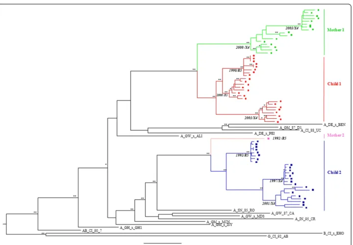
Child 1 infection was diagnosed with HIV-2 infection by PCR and virus isolation in the first month of life in 1998. To confirm the vertical transmission event and characterize the initial infecting virus population, 8 clonal full-length env gene sequences were obtained from sam-ples collected in 1998, 2000 and 2003 (in total 24env se-quences) and from his mother (mother 1-PTHCC20) in 2000 and 2003 (16env gene sequences). We were unable to obtain 1998 samples from the mother.
Child 2 infection was diagnosed in 1992 at day 39 after birth by PCR and virus isolation, and vertical transmis-sion was confirmed by phylogenetic analysis of partial
env sequences from the mother and the child [31,32].
Eight new clonal full-length env sequences were ob-tained from samples collected in 1992, 1997 and 2001 (in total 24envsequences).
segregated into one (child 2) or two (child 1) sub-clusters supported by high bootstrap values indicating that one or two virus variants were transmitted from the mothers to the children (Figure 1).
Child 1 was born with normal CD4 percentage (47%) which was sustained until age 3 without ART. The initial infecting virus was CCR5-tropic according to V3 loop se-quence analysis ofenv gene clones obtained in 1998 and to phenotypic analysis of virus isolated in 2000 (Table 1 and Figure 1). At age 5, in 2003, CD4 levels decreased to 27%, plasma viral load increased significantly and the virus changed to CXCR4-tropic as determined by phenotypic analysis [22]. Antiretroviral therapy (ART) was initiated at that time leading to a decrease in viral load to undetect-able levels and to an increase in CD4+T cells to normal
levels. Presently, this child is clinically and immunologic-ally stable and remains asymptomatic.
Child 2 was born with encephalopathy (CDC clinical stage C1) but with normal CD4+T cell percentage (52%) and undetectable viral load [31,32] (Table 1). The initial infecting virus was CCR5-tropic, as determined by our V3 loop sequence analysis, but induced syncytia forma-tion in peripheral blood mononuclear cells [31,32].
At age 5, CD4 percentage decreased to 25% and the virus changed to CXCR4-tropic, as determined by V3 loop sequence analysis. AZT therapy (1992 up to 1997) and AZT + 3TC therapy (in 2001) did not prevent in-crease in viral load and further CD4+T cell decline and the child died of AIDS at age 9.
Potent neutralizing antibodies are produced since the first year of infection especially against R5 isolates Plasma samples were available from day 27 of birth until the age of 12 for child 1, and from age 5 until age 9
(death) for child 2. Neutralizing activity of these samples was tested against autologous virus isolates obtained at age 2 (R5 isolate, CT00) and at age 5 (X4 isolate, CT03) for child 1, and at age 9 for child 2 (X4 isolate, SC01) (Figure 2). For child 1, the most notorious findings were the steep increase in Nab titers against the initial R5 iso-late (CT00) during the first 5 years of infection and the high Nab titers still present at age 12 against this isolate.
However, Nabs produced during the initial infection period and during later periods were significantly less potent against the X4 isolate that emerged in 2003 at age 5 (CT03) [median (range) of reciprocal log10 IC50
neutralization titers against CT00 and CT03 were 4.6 (3.7-5.4) and 4.1 (3.2-4.4), respectively, P = 0.0472, Mann–Whitney test]. This difference in susceptibility to neutralization of the two isolates was particularly evident
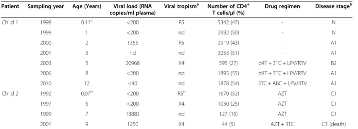
Table 1 Virological and immunological characterization of the patients
Patient Sampling year Age (Years) Viral load (RNA copies/ml plasma)
Viral tropisma Number of CD4+ T cells/μl (%)
Drug regimen Disease stageb
Child 1 1998 0.11c <200 R5 5342 (47) - N
1999 1 <200 nd 2992 (50) - N
2000 2 1355 R5 2919 (43) - A1
2001 3 nd nd 3253 (51) - A1
2003 5 20968 X4 595 (27) d4T + 3TC + LPV/RTV B2
2006 8 <200 nd 1895 (55) d4T + 3TC + LPV/RTV A1
2010 12 <40 nd 1878 (54) 3TC + ABC + LPV/RTV A1
Child 2 1992 0.07d <200 R5e 1670 (52) AZT C1
1997 5 <200 X4 1050 (25) AZT C1
1999 7 13883 nd 127 (15) AZT C1
2001 9 1250 X4 44 (5) AZT + 3TC C3 (death)
aAs determined phenotypically in TZMbl cells [22] or genotypically based on V3 loop sequence patterns [36];bAccording to the CDC revised classification system for HIV infection in children;cFirst blood collection was at day 39 after birth;dFirst blood collection was at day 27 after birth;eIsolate obtained at birth was syncytium inducing as determined in PBMCs and several cell lines [32]; d4T–stavudine, 3TC–lamivudine, LPV–lopinavir, RTV–ritonavir, ABC–abacavir, AZT–zidovudine; nd–not determined.
Figure 2Evolution of the autologous neutralizing antibody response in the children over the course of infection.The neutralizing activity present in patients plasma was analyzed against their primary virus isolates using a luciferase reporter gene assay in TZM-bl cells; plasma from child 1 from years 1998, 1999, 2000, 2001, 2003, 2006 and 2010 were tested against autologous viruses from 2000 [CT00 (R5 tropism)–pink circles] and 2003 [CT03 (X4 tropism)–green circles] and plasma from child 2 from years 1997, 1998, 1999 and 2001 were tested against
from age 5 onwards when the X4 virus emerged in the child. The close associations between rates of Nab es-cape and R5-to-X4 phenotypic switching suggests that phenotype transition in this infant is directly related with the Nab pressure.
Notably, child 1 also produced neutralizing antibodies that potently neutralized several heterologous primary HIV-2 isolates. Again, the heterologous Nabs were sig-nificantly more effective against R5 strains than against X4 strains [median (range) of reciprocal log10 IC50
neutralization titers against R5 and X4 isolates were 3.5 (1.6-4.0) and 2.5 (1.6-4.0), respectively, P = 0.0041] (Figure 3).
In child 2 we could only analyse the evolution of Nab response against the autologous X4-isolate (SC01) from age 5 onwards. Comparing Nab response at age 5 in both patients (the only age-matched data point), we found that it was significantly weaker in child 2 than in child 1 [median (range) of reciprocal log10 IC50
neutralization titer of child 2 against SC01 was 3.5 (3.4-3.6), and those of child 1 against autologous CT00 and
CT03 isolates were 5.3 (5.0-5.5) and 4.2 (4.0-4.4), re-spectively, P < 0.001] (Figure 2). Moreover, in contrast to child 1, Nab titer decreased steadily with infection time as viral load increased and disease progressed to AIDS and death at age 9 (Figure 2 and Table 1). Consid-ering all time points together, average Nab titers were lower than those of child 1 against the age-matched X4 isolate CT03 [median (range) of reciprocal log10 IC50
neutralization titer against isolates SC01 and CT03 were 3.3 (3.0-3.5) and 4.1 (3.2-4.4), respectively, P = 0.057] and against the R5 isolate CT00 [median (range) of re-ciprocal log10 IC50 neutralization titer against isolates
SC01 and CT00 were 3.3 (3.0-3.5) and 4.6 (3.7-5.4), re-spectively, P = 0.0106].
HIV-negative plasmas failed to neutralize HIV-2 strains and 2 plasmas failed to neutralize HIV-1SG3.1 or viruses pseudotyped with VSV envelope indi-cating the absence of nonspecific inhibitory activities in these samples.
Overall, the results obtained with child 1 demonstrate that potent neutralizing antibodies (autologous and
Figure 3Neutralizing antibody response against heterologous primary isolates in child 1 over the course of infection. A)A heat map of the reciprocal log-transformed IC50 value of each plasma sample from child 1 (left) against a panel of five heterologous primary virus isolates with respective tropism (top) is shown. The reciprocal log10IC50 value is colour-coded. The darkest colour indicates that neutralization above 50%
was still detected with the highest plasma dilution tested (1/5120). The lightest colour indicates that there was no detectable neutralization above 50% with the lowest plasma dilution tested (1/40). n.d.–not done (due to lack of plasma);B)Dot-plot graphic showing the mean and standard deviation of the reciprocal log10IC50 values obtained against R5 and X4 isolates indicated in A. Mann–Whitney U test was used to
heterologous) can be elicited very rapidly after HIV-2 vertical infection. Nabs are highly effective against the transmitted R5 isolates but seem to rapidly select for X4 isolates that escape neutralization. In the absence of ef-fective antiretroviral therapy, as was the case of child 2, increased replication of the Nab-resistant X4 isolates likely contributed to rapid CD4+ T cell depletion and progression to AIDS.
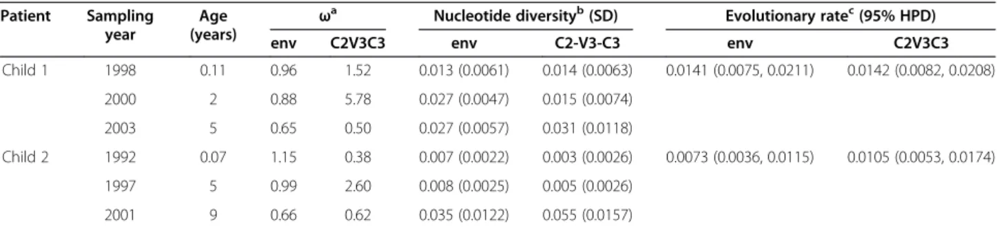
HIV-2 Env evolution in acute/early and late infection At birth, nucleotide diversity in child 1 was twice that of child 2 inenvand five times in C2V3C3 region (Table 2). At age 5, nucleotide diversity increased 2-fold inenvand
C2V3C3 in child 1 while in child 2 it only increased in C2V3C3 (2-fold) leading to an even higher difference in
env and C3V3C3 diversity (3-and 6-fold, respectively).
Interestingly, in child 2, diversity increased significantly from age 5 onward, both inenv(4-fold when compared to diversity at age 5) and C2V3C3 (11-fold) exceeding that of child 1 at age 5. However, in contrast to the first years of infection, most substitutions occurring in this period were of a synonymous nature as indicated by the sharp decrease in theωvalue both inenvand C2V3C3.
The evolutionary rate ofenv was significantly higher in child 1 than in child 2 (0.0141vs0.0073 substitutions/site/
year, posterior probability (PP) value <5%) (Table 2). When focusing on the C2V3C3 region, the evolutionary rates were not significantly different (0.0142vs0.0105
substitu-tions/site/year, PP=20%). There was a trend towards posi-tive selection in child 1 (non-synonymous rate:env, 0.0143 and V3, 0.0152; synonymous rate: env, 0.0137 and V3,
0.0124 substitutions/site/year) and purifying selection in child 2 (non-synonymous rate:env, 0.0069 and V3, 0.0092; synonymous rate: env, 0.0082 and V3, 0.0132
substitu-tions/site/year) in bothenvand V3.
We also analysed the evolution of amino acid diversity, as determined by the sum of Shannon’s entropy, in vari-able regions of gp125 which contains most of the neu-tralizing domains [19,21,26,27]. At birth, amino acid diversity was higher in child 1 than in child 2 (Table 3).
At age 5, amino acid diversity increased significantly only in V1 and V3 in both patients, this being much more pronounced in child 1. In child 2, from age 5 to age 9 (death), amino acid diversity increased in V1 (9.3-fold), V3 (2.1-fold) and V4 (1.6-(9.3-fold), though never reaching the level of diversity observed in child 1 at age 5. Amino acid changes observed after the first year in V1 and V3 are shown in Figure 4. In V1 there was no clear pattern of change except for the 2-4 amino acids deletion detected at year 5 in both patients. This dele-tion was maintained along the full course of infecdele-tion in child 2. In child 1, three mutations occurring at age 3 were fixed (were kept in year 5) and 4 mutations re-versed back to the original residue suggesting that these changes affected viral fitness; in child 2, ten mutations were fixed over the course of infection and there were no reversions suggesting that the mutations did not re-duce the fitness of the virus or that compensatory muta-tions occurred in other regions. Three of the fixed mutations in child 2 were located in a previously de-scribed neutralizing epitope [26]. Likewise, in child 1 two mutations of a potentially disruptive nature emerged in this neutralizing epitope (N to K and T to E/G).
In V3, mutations occurred almost exclusively within previously described neutralizing epitopes [26], and at residues 18, 19 and 27 that have been associated with R5 and X4 tropism [36-38]. One amino acid insertion oc-curred in the same position in both children and in-volved a hydrophobic residue (V in child 1; I in child 2). This type of insertion has also been associated to R5-to-X4 tropism switch [36,37].
Env adaptation to Nab pressure is usually associated with positive selection of specific amino acids that might be located in neutralizing domains [39]. At year 5 of in-fection there were 10 positively selected sites in Env of child 1 (seven in gp125) (Table 4). Most sites (6 out of 10) were located in confirmed neutralizing domains (V2, V3 and C5 in gp125 and MPER in gp36) [27]. In con-trast, positively selected sites were absent in child 2 at year 5 of infection and there were only 2 selected sites
Table 2 Nucleotide diversity and divergence rates in theenvgene and C2V3C3 region
Patient Sampling year
Age (years)
ωa Nucleotide diversityb(SD) Evolutionary ratec(95% HPD)
env C2V3C3 env C2-V3-C3 env C2V3C3
Child 1 1998 0.11 0.96 1.52 0.013 (0.0061) 0.014 (0.0063) 0.0141 (0.0075, 0.0211) 0.0142 (0.0082, 0.0208)
2000 2 0.88 5.78 0.027 (0.0047) 0.015 (0.0074)
2003 5 0.65 0.50 0.027 (0.0057) 0.031 (0.0118)
Child 2 1992 0.07 1.15 0.38 0.007 (0.0022) 0.003 (0.0026) 0.0073 (0.0036, 0.0115) 0.0105 (0.0053, 0.0174)
1997 5 0.99 2.60 0.008 (0.0025) 0.005 (0.0026)
2001 9 0.66 0.62 0.035 (0.0122) 0.055 (0.0157)
aRatio of nonsynonymous to synonymous substitution rates;bWithin-patient genetic distances and standard deviation (SD) as determined by averaging pairwise
in the final year of infection. These results reveal a much better adaptation to Nab pressure in child 1 compared to child 2.
In all, these results show that HIV-2 env can evolve and diversify very rapidly in the first years of infection. The positive correlation between the rate of Env evolu-tion, in terms of nucleotide divergence from the initial virus, nucleotide diversity, amino acid diversity, and positive selection, and the rate of Nab response and es-cape indicates that Nabs likely have a major impact on HIV-2 Env evolution in the first years of infection.
Tropism and susceptibility to antibody neutralization are closely associated with V3 structure
In long-term HIV-2 infected individuals the envelope V3 region adopts a significantly different structure in Nab-resistant isolates as compared to Nab-sensitive isolates, supporting a direct role of V3 conformation in the different susceptibility of these viruses to antibody neutralization [23]. To gain some insight into the struc-tural evolution of the V3 region in the first years of HIV-2 infection and try to relate it to tropism and sus-ceptibility to antibody neutralization, model structures of C2-V3-C3 regions from both children were generated by homology modelling using the three-dimensional structure of an unliganded SIV gp120 envelope glycopro-tein as template. Remarkably, the V3 loop, which was characterized by a high content of irregular secondary structure in the first year of infection, converged to an similar β-hairpin structure at year five of infection in both infants and remained in this conformation until the last year of infection in child 2 (Figure 5 and Additional file 1: Table S1). The rate of acquisition of theβ-hairpin conformation fully correlated with the rate of R5-to-X4
tropism transition and with the rate of escape from anti-body neutralization.
Discussion
There is limited knowledge on the natural history of HIV-2 infection and on the molecular and phenotypic evolutionary dynamics of HIV-2 because no study has investigated the full course of infection from the time of seroconversion. The current study is the first characterization of the Nab response and molecular and phenotypic evolution of HIV-2 followed from acute in-fection to late stage inin-fection. Our studies were based on two children infected by vertical transmission and spanned the first 12 years of infection in one case and the complete infection period in the other (9 years). We show that a potent Nab response is raised very early after infection and that the rate and pattern of molecular and phenotypic evolution of the HIV-2 Env are closely associated to the rate of Nab escape.
Child 2 was born severely ill despite the normal CD4+ T cell percentage [31,32] whereas child 1 was born asymptomatic and with normal CD4 levels. Despite the contrasting clinical conditions at birth, major CD4 de-pletion and disease progression occurred in both chil-dren in the first 5 years of infection. This fast disease course is typical of HIV-1 infected children in the ab-sence of antiretroviral treatment [40] but is highly un-usual in HIV-2 infected individuals [10,34,41,42]. Both patients were infected with R5 strains but transitions to X4 tropism occurred rapidly, being detected after only 5 years of infection. This is the first time that a full R5-to-X4 tropism switch is observed in HIV-2 infected tients and it was unexpected to find it in paediatric pa-tients. Like in some adult HIV-2 patients with advanced disease [23,37,43], the emergence of X4 viruses in our patients was associated with high viral load, marked CD4 depletion and disease progression. Hence, the rapid disease course in the two infants may have been deter-mined by the early emergence of X4 isolates.
At birth, HIV-2 nucleotide diversity in child 1 was 2-fold higher than in child 2 both inenvand C2V3C3. Nu-cleotide diversity in child 1 was also 2-fold higher than in HIV-1 infected children in the first weeks after birth [44-46] and in HIV-1 adult patients during seroconver-sion [47]. Envelope diversity also increased more signifi-cantly, both at the nucleotide and amino acid levels, in child 1 than in child 2 in the first 5 years of infection. Consistently, the evolutionary rate of the env gene in child 1 was almost two times higher than child 2 and similar to that found in chronic HIV-2 infected patients under ART (0.0102 substitutions/site/year) [24]. These results reveal a remarkably high rate of molecular evolu-tion of the HIV-2 envelope in child 1 during the first 5 years of infection and a moderate rate of evolution in
Table 3 Evolution of amino acid diversity in variable Env regions in the first five years of infection
Patient Variable regions
Sum of entropy Fold increase year of birth year 5
child 1 V1 1.324 8.657 6.5
V2 0 1.885 na
V3 0.754 3.614 4.8
V4 0.939 0.662 na
V5 0 3.402 na
V1-V5 3.017 18.22 6.0
child 2 V1 0.377 0.754 2.0
V2 1.131 0.377 na
V3 0.377 1.131 3.0
V4 0 1.316 na
V5 0.377 0 na
V1-V5 2.262 3.578 1.6
Figure 4Evolution of V1 and V3 regions.Clonal V1 and V3 amino acid sequences obtained over the course of infection from child 1
child 2. The evolutionary rate in child 2 was similar to that in HIV-1 patients who, when untreated, have a dis-ease progression generally similar to that of child 2. Thus, the higher rate in child 1 is consistent with the better immune control.
Previously, we have shown that production of gp36-specific and gp125-gp36-specific antibodies occurred during the first year of age in child 1 and that, at age 2, levels of binding antibodies to these glycoproteins were already similar to those found in HIV-2 infected patients in the chronic stage of disease [16]. In child 2, although gp36-specific antibodies were produced to near normal levels, there was a remarkably weak production of gp125-specific binding antibodies. Consistently, in this work we found that child 1 produced a much stronger Nab re-sponse than child 2. In child 1 autologous neutralizing antibodies appeared within the first year of infection, in-creased over time to levels that were similar to chronic-ally infected patients [18,19,21], and were sustained until at least the age of 12. Moreover, child 1 also produced a potent Nab response against several heterologous virus isolates. As for child 2, the autologous Nab titer was significantly lower compared to child 1 at age 5 and de-creased continuously to very low titers following the rapid decline of CD4 cells and progression to AIDS and death at age 9. Similar findings have been reported in late stage disease adult patients in whom Nab titers decrease in direct association with CD4+T cell depletion [23]. Overall, these results suggest that a potent Nab re-sponse during the acute/early phase of infection might contribute to control HIV-2 disease progression. Further investigation of the relationship between Nab response and disease progression in the acute and early stages of HIV-2 infection of adult patients is required as this might provide new insights into the benign course of most HIV-2 infections.
The differences in Nab response between the two chil-dren also correlate well with the magnitude and rate of en-velope evolution in the infants which suggest a close association between the neutralizing antibody response and the evolution of the HIV-2 envelope in these patients. Several lines of evidence further suggest that escape from Nab response is a major determinant of the evolution of the HIV-2 envelope in these infants, especially in
child 1. First, complete replacement of virus quasispecies was noted in phylogenetic analysis of envsequences pro-duced at the different time points which is compatible with a situation of ongoing viral escape from antibody neutralization [48]. Second, amino acid diversity increased significantly with infection time, especially in V1 and V3 which are major neutralizing domains in HIV-2. This is a major HIV escape mechanism as a single polymorphism can alter epitope sequence and/or conformation and reduce recognition and/or binding affinity by neutralizing antibodies [39,48-52]. Third, increase in dN/dS ratio and positive selection in the envelope were closely related to rate of Nab escape in child 1 [39,51]. Finally, the similar gain of secondary structure in V3 in both patients fully correlated with the rate of escape from antibody neutralization. This has been recently associated to HIV-2 resistance to antibody neutralization in chronic HIV-2 in-fected patients [23].
Nabs were significantly more potent against R5 isolates than against X4 isolates (autologous and heterologous) confirming the inherent resistance of X4 viruses to anti-body neutralization [23,53]. More importantly, increase in Nab resistance in child 1 preceded the emergence of X4 variant suggesting that tropism switch may have been driven by the neutralizing antibodies. Given the immuno-dominance of the V3 region in HIV-2 infected patients [54], the location of two of the three amino acid residues that are associated to R5 and X4 tropism (positions 18 and 19) [36-38] within the first neutralizing epitope in V3 [26] and the major difference in V3 conformation of R5 and X4 strains [23], the close association between HIV-2 susceptibility to antibody neutralization and tropism seen in these infants is not surprising.
The main limitations of this study are the small num-ber of patients and the inexistence of viral isolates from all time points in both children. However, worldwide it has been impossible to find individuals acutely infected with HIV-2. Moreover, due to the low or absent viral load it is often impossible to isolate virus from HIV-2 in-fected patients. Notwithstanding these limitations, we believe that our results are a major contribution to our understanding of the natural history of HIV-2 infection and of the role of the immune system in controlling and shaping HIV-2 evolution.
Table 4 Positive selective pressure on the Env glycoproteins in both children over the course of infection
Env glycoprotein
Codons under selective pressure (location)1
Child 1 Child 2
Year of birth Year 2 Year 5 Year of birth Year 5 Year 9
gp125 none 5, 7 (in SP) 178 (V2), 255, 259 (C2), 320 (V3), 459 (V5), 467, 471 (C5) none none 395 (C3)
gp36 none none 552 (HR1), 672, 673 (MPER) none none 562 (HR1)
1Codons identified as being significantly (P <0.05) under selective pressure are indicated; SP, signal peptide; V2, variable region 2; C2, conserved region 2; C3,
Conclusions
In conclusion, we show that a potent Nab response is elicited very early after HIV-2 infection and that the HIV-2 envelope evolves at a high rate in the first years of infection, this rate being directly correlated to the po-tency of the Nab response. R5-to-X4 tropism switch, in-creased nucleotide and amino acid diversity in V1 and V3, and convergence of V3 to aβ-hairpin structure were closely associated with escape from the Nab response suggesting that Nabs have a major impact on the rapid molecular and phenotypic evolution of the viral envelope in acute and early in HIV-2 infection. Our studies pro-vide further support to a model of HIV-2 pathogenesis in which Nabs play a central role.
Methods
Study subjects and ethics
Two children infected by vertical transmission were stud-ied. Blood samples were collected from child 1 (patient PT.HDE.CT), 39 days after birth in 1998, in 1999, 2000, 2001, 2003, 2006 and 2010, and from child 2 (patient PT. HDE.SC), 27 days after birth in 1992, in 1997 and 2001. Clinical and immunological characteristics of the patients are shown in Table 1. Child 1 started ART (stavudine + lamivudine + lopinavir/ritonavir) in November 2003. Pres-ently, the child is taking lamivudine + abacavir + lopinavir/ ritonavir; his viral load is undetectable and he is clinically and immunologically stable. Child 2 started ART with zidovudine immediately after birth and lamivudine was added in 2001. In 2001, viral load increased slightly and CD4+ T cells decreased sharply leading to the child’s death. Ethical approval was obtained from the Ethics Committee of Hospital Curry Cabral and written informed consent was obtained from the children's parents before entry into the study.
HIV-2 env gene PCR amplification, cloning and sequencing
Chromossomal DNA was extracted from infected PBMC’s using Wizard Genomic DNA Purification Kit (Promega) according to the manufacturer recommendations. A 2600 bp fragment encompassing the entireenv gene was amplified by nested Polymerase Chain Reaction (PCR) using the Expand Long Template PCR Systemkit (Roche) and newly designed primers (Additional file 2: Table S2). The PCR protocol consisted of denaturation at 95°C for 5 min, 35 amplification cycles of 15 sec at 94°C, 30 sec at 59°C and 3 min at 68°C with 5 sec increments and a final elongation step at 68°C for 30 min. 5 μl of PCR product was used as the template for nested PCR. The amplifica-tion profile of the nested PCR was identical to the first PCR, except for annealing temperature and extension time (61°C and 2 min respectively). PCR amplicons were purified with a JETQUICK Gel Extraction Spin Kit
Figure 5Evolution of the structure of C2-V3-C3 envelope region.Three-dimensional structures of C2-V3-C3 amino acid sequences from child 1 and 2 were generated by homology modelling using the three-dimensional structure of an unliganded SIV gp120 envelope glycoprotein as template.A)Superimposition of the structures of C2-V3-C3 of child 1 in 1998 (yellow), 2000 (blue) and 2003 (pale red);
(Genomed). For each sample, PCR products were cloned into the pcDNA3.1/V5-His-TOPO vector (Invitrogen), using the TOPO TA Expression Kit (Invitrogen) according to the manufacturer’s instructions. At least eight clones from each patient/year were sequenced using the BigDye Terminator V3.1 Cycle sequencing Kit (Applied Biosys-tems); sequencing primers are displayed in Additional file 2: Table S2. Sequencing was performed on an ABI 3100–Avant Genetic Analyzer (Applied Biosystems).
Sequence analysis
Clustal × 2.1 [55] software was used to construct align-ments of HIV-2 env sequences. Reference HIV-2
se-quences were obtained from the Los Alamos National Laboratory HIV sequence database [56]. Maximum like-lihood phylogenetic analyses were performed using the best-fit model of molecular evolution estimated in PAUP by Modeltest using likelihood ratio tests [57]. The chosen model was GTR + I + G. Tree searches were conducted in PAUP using nearest-neighbour inter-change (NNI) and tree-bisection plus reconnection (TBR) heuristic search strategies [58], and bootstrap re-sampling with 1000 replicates [59]. The genetic dis-tances between sequences were calculated by averaging pairwise tree distances using all sequences obtained for each patient at each time point, as previously described [60]. Putative recombinants were identified using the Phi-statistic [61] available in SplitsTree version 4.10 [62] by performing 10 randomized reductions of puta-tive recombinants. Putaputa-tive recombinant sequences were removed before doing the evolutionary rate ana-lyses. These were: 00PTHDECT_9, 00PTHDECT_16, 00PTHDECT_22, 00PTHDECT_6, 00PTHDECT_24, 00PTHDECT_19, 00PTHDECT_8, 00PTHDECT_12, 03PTHDECT_17, 03PTHDECT_33, 03PTHDECT_21, 01PTHDESC_13, 01PTHDESC_6 and 01PTHDESC_14. Selective pressure on the HIV-2 Env was examined with the DATAMONKEY web-server [63], after remov-ing all positions containremov-ing gaps and missremov-ing data from the dataset. All estimations were performed using the MG94 codon substitution model [64] crossed with the nucleotide substitution model GTR, previously selected with Modeltest (see above). The single-likelihood ances-tor counting (SLAC) method was used to infer the ratio of nonsynonymous to synonymous nucleotide substitu-tions (dN/dS) averaged over all codon posisubstitu-tions of the alignment. To identify individual codons under selective pressure, site-specific dN/dS rates were estimated by the relaxed-effects likelihood (REL) method, with a cut-off value for the Bayes factor of 50 [65].
The Bayesian program BEAST was used to estimate the nucleotide evolutionary rates [66]. The SRD06 model [67] of substitution was used and two different clock models were used, relaxed lognormal and strict clock. A
constant parametric demographic model as well as the non-parametric Skyline plot with 3 groups was tested. The MCMC chains were chosen so that the effective sample size for all parameters exceeded 300 and convergence was assessed by inspecting the traces in the program Tracer [68]. Appropriate demographic and molecular clock models were chosen by examining the marginal pos-terior distributions of relevant parameters.
Potential N-linked glycosylation sites were identified using the N-Glycosite software [69] and the entropy at each position in protein alignment was measured with Shannon’s entropy [70], both available at the Los Alamos National Laboratory HIV sequence database [56].
Virus isolation and tropism characterization
Primary virus isolates were obtained from both patients using the co-cultivation method as described previously [32]. Viral tropism (CCR5 and/or CXCR4 usage) was de-termined in TZM-bl cells in the presence of CCR5 or CXCR4 antagonists as described previously [22]. Trop-ism was also determined genetically using the V3 loop clonal sequences and the algorithm described by Vis-seuxet al [36] which is based in the sequence, size and charge of the V3 loop.
Neutralization assay
Structural models
Structural models of the C2-V3-C3 domain in gp125 were produced with SWISS-MODEL homology modelling ser-ver in automated mode, using PDB file 2BF1 (SIV) as tem-plate [73,74]. Accelrys Discovery Studio 2.1 (Accelrys Inc., San Diego, USA, 2008) was used to produce three dimen-sional images of the obtained models and perform the sec-ondary structure analysis of the V3 loop.
Statistical analysis
Statistical analysis was performed with GraphPad Prism 5.0 [72] with a level of significance of 5%. F test was used to compare best fit values of IC50 slopes obtained with CT00 and CT03 isolates from child 1. Non parametric Mann Whitney test was used to compare autologous Nab responses (mean IC50s) between child 1 and child 2. To compare evolutionary rates we computed the pos-terior probability (PP) that one rate exceeded the other and the probability was determined numerically by ran-domly sampling from the empirical posterior distribu-tions [24]. Kruskal-Wallis test was used to compare mean Shannon’s entropies between variable Env regions of both patients.
GenBank accession numbers
Full-length envelope sequences generated in this study are available from GenBank under the following acces-sion numbers: GU983917-GU983940 and JX219591-JX219614.
Additional files
Additional file 1: Table S1.Percentage of major secondary structure motifs present in the V3 loop of HIV-2 isolates obtained from child 1 and 2.
Additional file 2: Table S2.PCR and sequencing primers for the HIV-2 env gene.aOuter PCR primer;bInner PCR primer;cSequencing primer.
Competing interests
The authors declare that they have no competing interests.
Authors’contributions
NT and TL designed the study; CR, HS, TL, AQ and NT analysed the data and wrote the paper; CR performed the experiments; CR, HS and TL, performed the evolutionary analysis;,CR, RC, PB, JMM, IB and HB provided analytical reagents and nucleotide sequences; CR, PB and NT performed the statistical analysis; CF and AQ performed the structural analysis; PG performed the viral load assays; LR and PCS contributed clinical data from the patients. The final text was read and approved for submission by all authors.
Acknowledgements
This work was supported by grants PTDC/SAU-FAR/115290/2009 and PTDC/ SAU-EPI/122400/2010 from Fundação para a Ciência e Tecnologia (FCT) (http://www.fct.pt), Portugal, a NIH grant (R01AI087520), and by Collaborative HIV and Anti-HIV Drug Resistance Network (CHAIN), from the European Union. Cheila Rocha, Rita Calado, Pedro Borrego and Inês Bártolo were supported by PhD scholarships from Fundação para a Ciência e Tecnologia, Portugal. Helena Skar was supported by a postdoctoral fellowship from the Swedish Research Council (623-2011-1100). The following reagents were obtained through the AIDS Research and Reference Reagent Program,
Division of AIDS, NIAID, NIH: TZM-bl from Dr. John C. Kappes, Dr. Xiaoyun Wu and Tranzyme Inc.
Author details
1Unidade dos Retrovírus e Infecções Associadas, Centro de Patogénese Molecular, Faculdade de Farmácia de Lisboa, Lisboa, Portugal.2Centro de Investigação Interdisciplinar Egas Moniz (CiiEM), Instituto Superior de Ciências da Saúde Egas Moniz, Monte de Caparica, Portugal.3Unidade de
Microbiologia Médica, Instituto de Higiene e Medicina Tropical, Universidade Nova de Lisboa, Lisboa, Portugal.4Unidade de Imunohematologia, Hospital de Dona Estefânia, Lisboa, Portugal.5Laboratório de Biologia Molecular, Serviço de Medicina Transfusional, Centro Hospitalar Lisboa Ocidental–HEM, Lisboa, Portugal.6Centro de Malária e Outras Doenças Tropicais, Instituto Superior de Higiene e Medicina Tropical, Lisboa, Portugal.7Biology and Biophysics, Los Alamos National Laboratory, Los Alamos, New Mexico, U.S.A.
Received: 2 April 2013 Accepted: 15 September 2013 Published: 24 October 2013
References
1. De Silva TI, Cotten M, Rowland-Jones SL:HIV-2: the forgotten AIDS virus.
Trends Microbiol2008,16:588–595.
2. Gottlieb GS, Eholie SP, Nkengasong JN, Jallow S, Rowland-Jones S, Whittle HC, Sow PS:A call for randomized controlled trials of antiretroviral therapy for HIV-2 infection in West Africa.AIDS2008,22:2069–2072. discussion 2073-2064.
3. Yamaguchi J, Devare SG, Brennan CA:Identification of a new HIV-2 subtype based on phylogenetic analysis of full-length genomic sequence.AIDS Res Hum Retroviruses2000,16:925–930.
4. Damond F, Worobey M, Campa P, Farfara I, Colin G, Matheron S, Brun-Vezinet F, Robertson DL, Simon F:Identification of a highly divergent HIV type 2 and proposal for a change in HIV type 2 classification.AIDS Res Hum Retroviruses2004,20:666–672.
5. van der Loeff MF S, Jaffar S, Aveika AA, Sabally S, Corrah T, Harding E, Alabi A, Bayang A, Ariyoshi K, Whittle HC:Mortality of HIV-1, HIV-2 and HIV-1 /HIV-2 dually infected patients in a clinic-based cohort in The Gambia.
AIDS2002,16:1775–1783.
6. Campbell-Yesufu OT, Gandhi RT:Update on human immunodeficiency virus (HIV)-2 infection.Clin Infect Dis2011,52:780–787.
7. Matheron S, Pueyo S, Damond F, Simon F, Lepretre A, Campa P, Salamon R, Chene G, Brun-Vezinet F:Factors associated with clinical progression in HIV-2 infected-patients: the French ANRS cohort.AIDS2003,17:2593–2601. 8. van der Loeff MF, Larke N, Kaye S, Berry N, Ariyoshi K, Alabi A, Van Tienen C,
Leligdowicz A, Sarge-Njie R, Da Silva Z,et al:Undetectable plasma viral load predicts normal survival in HIV-2-infected people in a West African village.Retrovirology2010,7:46.
9. Berry N, Ariyoshi K, Jaffar S, Sabally S, Corrah T, Tedder R, Whittle H:Low peripheral blood viral HIV-2 RNA in individuals with high CD4 percentage differentiates HIV-2 from HIV-1 infection.J Hum Virol1998, 1:457–468.
10. Jaffar S, Wilkins A, Ngom PT, Sabally S, Corrah T, Bangali JE, Rolfe M, Whittle HC: Rate of decline of percentage CD4+ cells is faster in HIV-1 than in HIV-2 infection.J Acquir Immune Defic Syndr Hum Retrovirol1997,16:327–332. 11. Lisse IM, Poulsen AG, Aaby P, Knudsen K, Dias F:Serial CD4 and CD8
T-lymphocyte counts and associated mortality in an HIV-2-infected population in Guinea-Bissau.J Acquir Immune Defic Syndr Hum Retrovirol
1996,13:355–362.
12. Popper SJ, Sarr AD, Gueye-Ndiaye A, Mboup S, Essex ME, Kanki PJ:Low plasma human immunodeficiency virus type 2 viral load is independent of proviral load: low virus production in vivo.J Virol2000,74:1554–1557. 13. Andersson S, Norrgren H, Da Silva Z, Biague A, Bamba S, Kwok S,
Christopherson C, Biberfeld G, Albert J:Plasma viral load in 1 and HIV-2 singly and dually infected individuals in Guinea-Bissau, West Africa: significantly lower plasma virus set point in HIV-2 infection than in HIV-1 infection.Arch Intern Med2000,160:3286–3293.
14. Marlink R, Kanki P, Thior I, Travers K, Eisen G, Siby T, Traore I, Hsieh CC, Dia MC, Gueye EH,et al:Reduced rate of disease development after HIV-2 infection as compared to HIV-1.Science1994,265:1587–1590. 15. Drylewicz J, Matheron S, Lazaro E, Damond F, Bonnet F, Simon F, Dabis F,
marker changes between HIV-1 and HIV-2-infected patients in France.
AIDS2008,22:457–468.
16. Marcelino JM, Nilsson C, Barroso H, Gomes P, Borrego P, Maltez F, Rosado L, Doroana M, Antunes F, Taveira N:Envelope-specific antibody response in HIV-2 infection: C2V3C3-specific IgG response is associated with disease progression.AIDS2008,22:2257–2265.
17. Rodriguez SK, Sarr AD, MacNeil A, Thakore-Meloni S, Gueye-Ndiaye A, Traore I, Dia MC, Mboup S, Kanki PJ:Comparison of heterologous neutralizing antibody responses of human immunodeficiency virus type 1 (HIV-1)- and HIV-2-infected Senegalese patients: distinct patterns of breadth and magnitude distinguish HIV-1 and HIV-2 infections.J Virol2007,81:5331–5338.
18. Ozkaya Sahin G, Holmgren B, Da Silva Z, Nielsen J, Nowroozalizadeh S, Esbjornsson J, Mansson F, Andersson S, Norrgren H, Aaby P,et al:Potent intratype neutralizing activity distinguishes human immunodeficiency virus type 2 (HIV-2) from HIV-1.J Virol2012,86:961–971.
19. De Silva TI, Aasa-Chapman M, Cotten M, Hue S, Robinson J, Bibollet-Ruche F, Sarge-Njie R, Berry N, Jaye A, Aaby P,et al:Potent autologous and heterologous neutralizing antibody responses occur in HIV-2 infection across a broad range of infection outcomes.J Virol2012,86:930–946. 20. Bjorling E, Scarlatti G, Von Gegerfelt A, Albert J, Biberfeld G, Chiodi F, Norrby
E, Fenyo EM:Autologous neutralizing antibodies prevail in HIV-2 but not in HIV-1 infection.Virology1993,193:528–530.
21. Kong R, Li H, Bibollet-Ruche F, Decker JM, Zheng NN, Gottlieb GS, Kiviat NB, Sow PS, Georgiev I, Hahn BH,et al:Broad and potent neutralizing antibody responses elicited in natural HIV-2 infection.J Virol2012,86:947–960. 22. Borrego P, Calado R, Marcelino JM, Bartolo I, Rocha C, Cavaco-Silva P,
Doroana M, Antunes F, Maltez F, Caixas U,et al:Baseline susceptibility of primary HIV-2 to entry inhibitors.Antivir Ther2012,17(3):565–570. 23. Marcelino JM, Borrego P, Nilsson C, Familia C, Barroso H, Maltez F, Doroana
M, Antunes F, Quintas A, Taveira N:Resistance to antibody neutralization in HIV-2 infection occurs in late stage disease and is associated with X4 tropism.AIDS2012,26:2275–2284.
24. Skar H, Borrego P, Wallstrom TC, Mild M, Marcelino JM, Barroso H, Taveira N, Leitner T, Albert J:HIV-2 genetic evolution in patients with advanced disease is faster than that in matched HIV-1 patients.J Virol2010, 84:7412–7415.
25. Borrego P, Marcelino JM, Rocha C, Doroana M, Antunes F, Maltez F, Gomes P, Novo C, Barroso H, Taveira N:The role of the humoral immune response in the molecular evolution of the envelope C2, V3 and C3 regions in chronically HIV-2 infected patients.Retrovirology2008,5:78. 26. Bjorling E, Chiodi F, Utter G, Norrby E:Two neutralizing domains in the V3
region in the envelope glycoprotein gp125 of HIV type 2.J Immunol
1994,152:1952–1959.
27. Kong R, Li H, Georgiev I, Changela A, Bibollet-Ruche F, Decker JM, Rowland-Jones SL, Jaye A, Guan Y, Lewis GK,et al:Epitope mapping of broadly neutralizing HIV-2 human monoclonal antibodies.J Virol2012,86:12115–12128. 28. McKnight A, Shotton C, Cordell J, Jones I, Simmons G, Clapham PR:
Location, exposure, and conservation of neutralizing and
nonneutralizing epitopes on human immunodeficiency virus type 2 SU glycoprotein.J Virol1996,70:4598–4606.
29. Skott P, Achour A, Norin M, Thorstensson R, Bjorling E:Characterization of neutralizing sites in the second variable and fourth variable region in gp125 and a conserved region in gp36 of human immunodeficiency virus type 2.Viral Immunol1999,12:79–88.
30. Barroso H, Araujo F, Gomes MH, Mota-Miranda A, Taveira N:Phylogenetic demonstration of two cases of perinatal human immunodeficiency virus type 2 infection diagnosed in adulthood.AIDS Res Hum Retroviruses2004, 20:1373–1376.
31. Cavaco-Silva P, Taveira NC, Lourenco MH, Santos Ferreira MO, Daniels RS: Vertical transmission of HIV-2.Lancet1997,349:177–178.
32. Cavaco-Silva P, Taveira NC, Rosado L, Lourenco MH, Moniz-Pereira J, Douglas NW, Daniels RS, Santos-Ferreira MO:Virological and molecular demonstration of human immunodeficiency virus type 2 vertical transmission.J Virol1998,72:3418–3422.
33. Padua E, Almeida C, Nunes B, Cortes Martins H, Castela J, Neves C, Paixao MT:Assessment of mother-to-child HIV-1 and HIV-2 transmission: an AIDS reference laboratory collaborative study.HIV Med2009,10:182–190. 34. Mota-Miranda A, Gomes H, Lima-Alves C, Araujo F, Cunha-Ribeiro LM,
Taveira N:Perinatally acquired HIV-2 infection diagnosed at 15 and 24 years of age.AIDS2001,15:2460–2461.
35. Burgard M, Jasseron C, Matheron S, Damond F, Hamrene K, Blanche S, Faye A, Rouzioux C, Warszawski J, Mandelbro L:Mother-to-child transmission of HIV-2 infection from 1986 to 2007 in the ANRS French perinatal cohort EPF-CO1.Clin Infect Dis2010,51:833–843.
36. Visseaux B, Hurtado-Nedelec M, Charpentier C, Collin G, Storto A, Matheron S, Larrouy L, Damond F, Brun-Vezinet F, Descamps D:Molecular determinants of HIV-2 R5-X4 tropism in the V3 loop: development of a new genotypic tool.J Infect Dis2012,205:111–120.
37. Shi Y, Brandin E, Vincic E, Jansson M, Blaxhult A, Gyllensten K, Moberg L, Brostrom C, Fenyo EM, Albert J:Evolution of human immunodeficiency virus type 2 coreceptor usage, autologous neutralization, envelope sequence and glycosylation.J Gen Virol2005,86:3385–3396.
38. Isaka Y, Sato A, Miki S, Kawauchi S, Sakaida H, Hori T, Uchiyama T, Adachi A, Hayami M, Fujiwara T, Yoshie O:Small amino acid changes in the V3 loop of human immunodeficiency virus type 2 determines the coreceptor usage for CXCR4 and CCR5.Virology1999,264:237–243.
39. Bunnik EM, Pisas L, Van Nuenen AC, Schuitemaker H:Autologous neutralizing humoral immunity and evolution of the viral envelope in the course of subtype B human immunodeficiency virus type 1 infection.J Virol2008,82:7932–7941.
40. Rey MA, Girard PM, Harzic M, Madjar JJ, Brun-Vezinet F, Saimot AG:HIV-1 and HIV-2 double infection in French homosexual male with AIDS-related complex (Paris, 1985).Lancet1987,1:388–389.
41. Matheron S, Mendoza-Sassi G, Simon F, Olivares R, Coulaud JP, Brun-Vezinet F: HIV-1 and HIV-2 AIDS in African patients living in Paris.AIDS1997,11:934–936. 42. Norrgren H, Da Silva Z, Biague A, Andersson S, Biberfeld G:Clinical
progression in early and late stages of disease in a cohort of individuals infected with human immunodeficiency virus-2 in Guinea-Bissau.
Scand J Infect Dis2003,35:265–272.
43. Blaak H, van der Ende ME, Boers PH, Schuitemaker H, Osterhaus AD:In vitro replication capacity of HIV-2 variants from long-term aviremic individuals.Virology2006,353:144–154.
44. Wu X, Parast AB, Richardson BA, Nduati R, John-Stewart G, Mbori-Ngacha D, Rainwater SM, Overbaugh J:Neutralization escape variants of human immunodeficiency virus type 1 are transmitted from mother to infant.
J Virol2006,80:835–844.
45. Hoffmann FG, He X, West JT, Lemey P, Kankasa C, Wood C:Genetic variation in mother-child acute seroconverter pairs from Zambia.AIDS
2008,22:817–824.
46. Zhang H, Tully DC, Hoffmann FG, He J, Kankasa C, Wood C:Restricted genetic diversity of HIV-1 subtype C envelope glycoprotein from perinatally infected Zambian infants.PLoS One2010,5:e9294. 47. Salazar-Gonzalez JF, Bailes E, Pham KT, Salazar MG, Guffey MB, Keele BF,
Derdeyn CA, Farmer P, Hunter E, Allen S,et al:Deciphering human immunodeficiency virus type 1 transmission and early envelope diversification by single-genome amplification and sequencing.J Virol
2008,82:3952–3970.
48. Zhang H, Hoffmann F, He J, He X, Kankasa C, Ruprecht R, West JT, Orti G, Wood C:Evolution of subtype C HIV-1 Env in a slowly progressing Zambian infant.Retrovirology2005,2:67.
49. Zhang H, Hoffmann F, He J, He X, Kankasa C, West JT, Mitchell CD, Ruprecht RM, Orti G, Wood C:Characterization of HIV-1 subtype C envelope glycoproteins from perinatally infected children with different courses of disease.Retrovirology2006,3:73.
50. Doria-Rose NA, Georgiev I, O’Dell S, Chuang GY, Staupe RP, McLellan JS, Gorman J, Pancera M, Bonsignori M, Haynes BF,et al:A short segment of the HIV-1 gp120 V1/V2 region is a major determinant of resistance to V1/V2 neutralizing antibodies.J Virol2012,86:8319–8323.
51. Frost SD, Wrin T, Smith DM, Kosakovsky Pond SL, Liu Y, Paxinos E, Chappey C, Galovich J, Beauchaine J, Petropoulos CJ,et al:Neutralizing antibody responses drive the evolution of human immunodeficiency virus type 1 envelope during recent HIV infection.Proc Natl Acad Sci U S A2005, 102:18514–18519.
52. Rong R, Li B, Lynch RM, Haaland RE, Murphy MK, Mulenga J, Allen SA, Pinter A, Shaw GM, Hunter E,et al:Escape from autologous neutralizing antibodies in acute/early subtype C HIV-1 infection requires multiple pathways.PLoS Pathog2009,5:e1000594.
54. Soares R, Foxall R, Albuquerque A, Cortesao C, Garcia M, Victorino RM, Sousa AE:Increased frequency of circulating CCR5+ CD4+ T cells in human immunodeficiency virus type 2 infection.J Virol2006,80:12425–12429. 55. Larkin MA, Blackshields G, Brown NP, Chenna R, McGettigan PA, McWilliam
H, Valentin F, Wallace IM, Wilm A, Lopez R,et al:Clustal W and clustal X version 2.0.Bioinformatics2007,23:2947–2948.
56. SequenceDatabase:Los Alamos sequence database.Los Alamos, New Mexico: Los Alamos National Laboratory; 2009. http://www.hiv.lanl.gov/. 57. Posada D, Crandall KA:MODELTEST: testing the model of DNA
substitution.Bioinformatics1998,14:817–818.
58. Swofford DL, Waddell PJ, Huelsenbeck JP, Foster PG, Lewis PO, Rogers JS: Bias in phylogenetic estimation and its relevance to the choice between parsimony and likelihood methods.Syst Biol2001,50:525–539. 59. Felsenstein J:Confidence limits on phylogenies: an approach using the
bootstrap.Evolution1985,39:783–791.
60. Lee HY, Perelson AS, Park SC, Leitner T:Dynamic correlation between intrahost HIV-1 quasispecies evolution and disease progression.
PLoS Comput Biol2008,4:e1000240.
61. Bruen TC, Philippe H, Bryant D:A simple and robust statistical test for detecting the presence of recombination.Genetics2006,172:2665–2681. 62. Huson DH, Bryant D:Application of phylogenetic networks in
evolutionary studies.Mol Biol Evol2006,23:254–267.
63. Pond SL, Frost SD:Datamonkey: rapid detection of selective pressure on individual sites of codon alignments.Bioinformatics2005,21:2531–2533. 64. Muse SV, Gaut BS:A likelihood approach for comparing synonymous and
nonsynonymous nucleotide substitution rates, with application to the chloroplast genome.Mol Biol Evol1994,11:715–724.
65. Kosakovsky Pond SL, Frost SD:Not so different after all: a comparison of methods for detecting amino acid sites under selection.Mol Biol Evol
2005,22:1208–1222.
66. Drummond AJ, Rambaut A:BEAST: Bayesian evolutionary analysis by sampling trees.BMC Evol Biol2007,7:214.
67. Shapiro B, Rambaut A, Drummond AJ:Choosing appropriate substitution models for the phylogenetic analysis of protein-coding sequences.
Mol Biol Evol2006,23:7–9.
68. Rambaut A, Drummond AJ:Tracer v1.4.2007. Available from http://beast.bio. ed.ac.uk/Tracer.
69. Zhang M, Gaschen B, Blay W, Foley B, Haigwood N, Kuiken C, Korber B: Tracking global patterns of N-linked glycosylation site variation in highly variable viral glycoproteins: HIV, SIV, and HCV envelopes and influenza hemagglutinin.Glycobiology2004,14:1229–1246.
70. Korber BT, Kunstman KJ, Patterson BK, Furtado M, McEvilly MM, Levy R, Wolinsky SM:Genetic differences between blood-and brain-derived viral sequences from human immunodeficiency virus type 1-infected patients: evidence of conserved elements in the V3 region of the envelope protein of brain-derived sequences.J Virol1994,68:7467–7481. 71. Wei X, Decker JM, Wang S, Hui H, Kappes JC, Wu X, Salazar-Gonzalez JF,
Salazar MG, Kilby JM, Saag MS,et al:Antibody neutralization and escape by HIV-1.Nature2003,422:307–312.
72. GraphPad:Graph pad software Inc.: Prism 5, version 5.04.2010.
73. Arnold K, Bordoli L, Kopp J, Schwede T:The SWISS-MODEL workspace: a web-based environment for protein structure homology modelling.
Bioinformatics2006,22:195–201.
74. Schwede T, Kopp J, Guex N, Peitsch MC:SWISS-MODEL: an automated protein homology-modeling server.Nucleic Acids Res2003,31:3381–3385.
doi:10.1186/1742-4690-10-110
Cite this article as:Rochaet al.:Evolution of the human
immunodeficiency virus type 2 envelope in the first years of infection is associated with the dynamics of the neutralizing antibody response.
Retrovirology201310:110.
Submit your next manuscript to BioMed Central and take full advantage of:
• Convenient online submission
• Thorough peer review
• No space constraints or color figure charges
• Immediate publication on acceptance
• Inclusion in PubMed, CAS, Scopus and Google Scholar
• Research which is freely available for redistribution