Universidade do Algarve
Faculdade de Ciências e Tecnologias
Endocrine Responses of Tilapia (Oreochromis mossambicus)
females to male pheromone(s)
Ana Cláudia Nunes Rato
Dissertação para a obtenção do Grau de Mestre em Biologia Marinha
Trabalho efectuado sob a orientação de: Professor Doutor Adelino Canário Dr. Peter Hubbard
Endocrine Responses of Tilapia (Oreochromis mossambicus) females to male pheromone(s)
Declaração de autoria de trabalho
Declaro ser a autora deste trabalho, que é original e inédito. Autores e trabalhos consultados estão devidamente citados no texto e constam da listagem de referências incluída.
Copyright em nome de Ana Cláudia Nunes Rato
A Universidade do Algarve tem o direito, perpétuo e sem limites geográficos, de arquivar e publicitar este trabalho através de exemplares impressos reproduzidos em papel ou em forma digital, ou por qualquer outro meio conhecido ou que venha a ser inventado, de o divulgar através de repositórios científicos e de admitir a sua cópia e distribuição com objectivos educacionais ou de investigação, não comerciais, desde que seja dado crédito ao autor e editor.
i Agradecimentos
Primeiramente, quero agradecer ao Professor Doutor Adelino Canário por ter aceite ser o meu orientador.
Ao Dr. Peter Hubbard, muito obrigada por me ter acompanhado como co-orientador durante este longo período. Obrigada por todo o apoio e paciência.
À Elsa Couto, Tina Keller-Costa, Dr. João Saraiva e Soraia Santos pelo apoio no laboratório, pela paciência, pelos conselhos que de alguma forma me ajudaram a elaborar a minha tese e que me ajudaram a crescer profissionalmente.
Ao meu pai, à minha mãe e à minha irmã, obrigada por me terem apoiado sempre, por me terem permitido realizar este sonho que me perseguia desde a escola primária. Obrigada por terem sempre acreditado em mim, mesmo quando mais ninguém o fazia. Obrigada pelo amor, carinho, amizade, apoio que sempre me transmitiram e que fizeram de mim a pessoa que sou hoje. Muito Obrigada, Pai Carlos e Mãe Natália! Obrigada Mana Dina, por teres lido sempre as primeiras versões de todos os meus trabalhos, mesmo quando eram escritos em inglês; obrigada por me aturares, mesmo quando conseguia ser muito chata. Obrigada, “mana-metralha”! Obrigada também ao meu cunhado, Rodrigo Matos, pelo apoio e amizade.
À minha avozinha, Maria José, muito obrigada por tudo! Pelo apoio, pelas histórias que sempre me contou, as cantigas que eu tanto gostava e que me faziam adormecer à lareira! Obrigada por estes e outros momentos que nunca irei esquecer.
À minha madrinha, Elsa Fernandes, um obrigada por seres a madrinha espectacular que és e por te preocupares sempre comigo, mesmo estando longe. Obrigada Madrinha! Obrigada ao meu padrinho, Armando Costa, por tudo.
Á restante família, que por ser tão grande, é difícil nomear aqui todos, por isso, a todos um grande Obrigada por terem acreditado em mim!
Aquelas estrelinhas, que nos deixaram demasiado cedo, Obrigada por olharem por todos nós!
Aos amigos que sempre fizeram parte da minha vida, André Dias, Daniela Morgadinho, Ana Catarina Mendonça, Catarina Oliveira, Renata Galvão, Joana Rita Pereira, obrigada por fazerem parte da minha vida e por terem comprovado que, mesmo longe e sem contacto diário, é possível manter verdadeiras amizades. Obrigada pela amizade, pelas loucuras (não só aquelas de Peniche!), obrigada pelas gargalhadas constantes, obrigada por tudo!
ii Agradeço a todos aqueles que nunca me deixaram desistir durante este percurso, por me terem apoiado em todas as situações e por me terem dado força para “remar contra a maré”. Obrigada Naíde Quaresma, Andreia Cocharro, Carla Fernandes, Ana Medeiros, Verónica Rijo, Patrícia Trindade, Juliana Pacheco, Andreia Ovelheiro, Tiago Silva, Mauro Augusto, Bruno Gonçalves, Diogo Monteiro, pela amizade, por fazerem parte da minha vida, pelos momentos divertidos, pelas parvoíces ditas em longas noites de estudo, pela companhia, por serem a minha família “algarvia”. Obrigada por aturarem o meu mau- feitio e por me “darem na cabeça” sempre que necessário. Obrigada pelas longas noites de conversa na varanda e à porta de casa, obrigada por estarem lá nos bons e especialmente nos maus momentos.
Obrigada também aos “amigos do café” pelos momentos de descontracção, pelas conversas sérias, que acabavam sempre às gargalhadas. Obrigada ao Monte-Branco, por proporcionar estes momentos.
A todos aqueles que acreditaram em mim, que fizeram parte da minha vida e, que de alguma forma me ajudaram a definir tanto pessoal como profissionalmente, um muito obrigada!
iii Resumo
A comunicação química desempenha um papel fundamental na vida dos peixes, pois o ambiente que estes habitam é muitas vezes desprovido de luz, o que proporcionou a que os peixes evoluíssem de forma a detetar e responder a sinais químicos libertados por conspecificos (membros da mesma espécies). Esses sinais químicos, feromonas, são libertados para o meio, e têm a capacidade de transmitir informação entre conspecíficos, desencadeando assim uma resposta fisiológica ou comportamental. Com base na sua função, as feromonas podem-se dividir em três categorias: estímulos sociais, reprodutivos e anti-predatórios. As feromonas podem induzir alterações endócrinas e de desenvolvimento (efeito “primer”) e/ou alterações a nível comportamental (efeito “releaser”). Esteróides sexuais, ácidos biliares, prostaglandinas assim como os seus precursores e metabolitos são normalmente utilizados pelos peixes como feromonas. De forma geral, as feromonas são libertadas na água, principalmente através da urina, guelras e pela bílis. Os esteróides sexuais (androgénios, estrogénios e progestogénios) desempenham um papel ativo na reprodução. O androgénio mais comum nos teleósteos é 11-cetotestosterona, enquanto que o estradiol e a progesterona são importantes estrogénios. 17,20β-dihidróxi-4-pregnen-3-ona (17,20β-P) é um progestogénio, identificado em mais de 35 espécies de teleósteos.
A tilápia, Oreochromis mossambicus, teleósteo endémico dos rios e lagos de África oriental, devido às suas propriedades físicas e biológicas, tornou-se numa das espécies mais estudadas em laboratório. Esta espécie exibe um evidente dimorfismo sexual, no qual os machos se destacam principalmente, pelas proporções das maxilas, por apresentarem tamanhos superiores em relação às fêmeas, e pelas cores que exibem durante a época de acasalamento. Esta espécie apresenta um comportamento sexual característico, no qual, os machos formam arenas de reprodução, nas quais constroem e defendem ninhos, sendo que os machos dominantes assumem uma coloração escura. Quando estão prontas para acasalar, as fêmeas entram nas arenas, onde ocorre a desova e escolhem o macho para acasalar. Quando o ritual de acasalamento termina, a fêmea transporta o esperma e os ovos na sua boca, onde ocorre a fertilização. O.mossambicus são designados como incubadores bucais, uma vez que a fertilização e o desenvolvimento dos embriões e da fase larvar ocorre na cavidade bucal da fêmea.
iv De forma geral, sabe-se que a urina transporta feromonas que causam respostas endócrinas e comportamentais nas fêmeas. Uma dessas respostas é o aumento dos níveis de uma hormona esteróide responsável pela maturação dos oócitos, 17, 20β-P. A partir de estudos realizados anteriormente, sabe-se que existe um composto na urina dos machos que é detetado pelas fêmeas e que se encontra em concentrações superiores na urina de macho dominante quando comparado com a urina de machos subordinados. Este composto foi recentemente identificado como sendo um esteróide glucurinado e, acredita-se que é um dos componentes ativos responsável pelo aumento dos níveis da hormona esteróide responsável pela maturação dos oócitos, 17, 20β-P, nas fêmeas. Este estudo teve como principal objetivo identificar qual a fração da urina que desempenha um papel feromonal, ou seja, qual parte da urina é responsável por causar um aumento nos níveis de 17, 20β-P e determinar se o esteróide glucurinado é capaz, por si só, um aumento nos níveis de 17, 20β-P.
Para tal, primeiramente procedeu-se à recolha de urina de macho, obtendo-se posteriormente um pool de urina. Este pool foi fracionado através de um sistema de extracção “Solid-phase extraction”, que consiste na passagem da amostra por um cartucho de extração, onde os esteróides ficam retidos na matriz de sílica, sendo seguidamente obtidos por ação de um solvente, neste caso, o metanol. Assim, após extração, obteve-se o filtrado e eluato. Em laboratório, procedeu-se também à preparação do esteróide glucurinado, com concentração semelhante à presente na urina. Fêmeas de O.mossambicus foram expostas a estes estímulos, tendo-se procedido à recolha de 1l de água, uma hora antes da adição do estímulo (tempo 0h) e uma hora após a adição do estímulo (tempo 1h). A mesma experiência foi realizada em machos da mesma espécie, porém foram expostos a um único estímulo: urina de macho dominante. Após extração, as amostras foram submetidas a radioimunoensaios, para determinar a concentração de hormonas esteróides presentes. No caso das fêmeas, foram quantificadas as concentrações de 17, 20β-P e cortisol, enquanto que para as amostras dos machos, se quantificou 11-cetotestosterona e testosterona.
Uma hora após a exposição à urina, eluato, esteróide glucurinado assim como após exposição ao esteróide combinado com o filtrado e eluato juntamente com o filtrado, observou-se um aumento significativo nos níveis de 17, 20β-P. Contrariamente, uma hora após a exposição ao filtrado e metanol (usado como controlo), não se observou nenhuma mudança na taxa de libertação de 17, 20β-P. As taxas de libertação de cortisol não sofreram aumentos significativos, com exceção do cortisol glucurinado.
v Relativamente à experiência realizada nos machos, após exposição à urina de macho dominante, de forma geral, observou-se um aumento nos níveis de 11-cetotestosterona mas sem alterações nas taxas de libertação de testosterona.
Estes resultados suportam a hipótese de que, de facto, a urina desempenha um papel feromonal, e que a fração responsável pelo aumento dos níveis de 17, 20β-P, é o eluato. O facto de o filtrado não causar alterações nas taxas de libertação, ao contrário do eluato, sugere que o método de extração foi altamente eficaz, visto que o filtrado não continha esteróides. O esteróide glucurinado, é capaz, por si só de causar, um aumento significativo nos níveis da hormona esteróide responsável pela maturação dos oócitos. Os reduzidos níveis de cortisol sugerem que o método de amostragem foi o mais indicado que, apesar da recolha de amostras repetidas, causou níveis reduzidos de stress nos indivíduos. Os níveis elevados de 17, 20β-P parecem interferir com o metabolismo do cortisol, causando aumentos nos níveis de cortisol glucurinado.
Relativamente à experiência conduzida nos machos, não se pôde retirar qualquer conclusão, uma vez que foi realizada com objetivo de praticar o método de amostragem a usar posteriormente nas fêmeas, e como tal, não se obteve controlos com os quais comparar os resultados obtidos. Contudo, seria interessante repetir esta experiência, explorando também outros estímulos, à semelhança da experiência realizada nas fêmeas.
Palavras-chave:
Feromona; Esteróide glucurinado; Oreochromis mossambicus; Respostas endócrinas; 17, 20β-P.
vi Abstract
Several lines of evidence suggest that male Mozambique tilapia (Oreochromis mossambicus) release a reproductive pheromone via their urine. A recently identified steroid glucuronide, present in male urine, is probably one of the active components that increase levels of a steroid hormone responsible for oocyte maturation, 17,20β-P, in females. The aims of this study were to identify which fraction(s) of male urine is responsible for this increase and whether the steroid glucuronide is sufficient, on its own, to cause a similar increase in 17,20β-P metabolism.
Pooled male urine was passed through C18 extraction cartridges, thus obtaining the filtrate (aqueous/polar) and eluate (hydrophobic/non-polar) fractions. Females were exposed to urine, its respective fractions, and the steroid glucuronide (and in combination with each-other). One hour after exposure, water samples were collected and steroids extracted. The same experiment was conducted in males, using a urine pool from dominant males. Steroid levels (17,20β-P, cortisol, 11-ketotestosterone, and testosterone) were measured by radioimmunoassay.
Exposure to male urine, its eluate, and the steroid glucuronide (and combinations containing the eluate or steroid) evoked a dramatic increase in release rates of 17,20β-P by females. The filtrate alone had no such effect. In males, release rates of 11-ketotestosterone, but not testosterone, increased after exposure to male urine.
These results suggest that the pheromonal activity of the urine is contained wholly in the C18 eluate. Furthermore, the steroid glucuronide (originally isolated from the urine eluate) is sufficient, on its own, to cause an increase in levels of 17,20β-P metabolism. Thus, the steroid glucuronide is responsible for the pheromonal activity of male urine, at least in this endocrine effect in females. The endocrine response of males to this urinary pheromone requires further investigation.
Key-words:
Endocrine responses; Oreochromis mossambicus; pheromones; steroid glucuronide; 17, 20β-P.
vii Contents 1. Introduction... 1 1.1. Hormones ... 3 1.1.1. Sex Steroids ... 4 1.1.2. Corticosteroids ... 6 1.2. Chemical Communication ... 6 1.2.1. What is a Pheromone? ... 6
1.2.1.1 .The Goldfish (Carassius auratus)... 7
1.2.2. Functions of Pheromones in Fish Biology ... 8
1.2.3. Chemical nature of pheromones... 9
1.2.3.1. Bile Acids ... 9 1.2.3.2. Prostaglandins ... 9 1.2.3.3. Sex Steroids ... 10 1.3. Conjugated Steroids ... 10 1.4. Steroid Measurement ... 11 1.4.1. Measurement of steroids ... 12
1.4.2. Procedures for steroid determination ... 13
1.4.2.1. Radioimmunoassay ... 14
1.5. The Mozambique Tilapia, Oreochromis mossambicus ... 15
1.5.1. Sexual Behaviour and Reproduction ... 18
1.6. Role of urine among conspecifics ... 20
1.7. Aims of the study ... 22
2. Material and Methods ... 22
2.1. Experimental Animals ... 25
2.2. Urine Sample Collection... 25
2.3. Experimental Design ... 26
2.3.1. Stimuli Preparation ... 26
2.3.2. Exposure of Females to Male Urine and Derivatives ... 27
2.3.3. Exposure of Males to Male Urine ... 28
2.3.4. Hormone Analysis ... 28
2.3.4.1. Solid-Phase Extraction ... 28
2.3.4.2. Steroid Extraction ... 29
viii
2.4. Steroid Release Rates ... 33
2.5. Statistical Analysis ... 35
3. Results ... 34
3.1. Exposure of Females to Male Urine and Derivatives ... 39
3.1.1. Release Rate of 17, 20β-dihydroxypregnen-4-en-3-one (17,20β-P) ... 39
3.1.2. Comparisons of responses to different stimuli ... 42
3.1.3. Release Rate of Cortisol... 42
3.2. Exposure of Males to Male Urine ... 44
4. Discussion ... 43
5. Conclusion ... 55
6. Future Research ... 55
3 Chemical communication is important in several aspects of fish biology, such as migration, the alarm response and – particularly - in reproduction.
Previous studies have shown that females and males release pheromones that affect the sexual behaviour and reproductive physiology of conspecifics. However, few fish pheromones have been chemically identified and fully characterized.
The Mozambique tilapia (Oreochromis mossambicus) is an African cichlid of immense scientific interest and economic value; it is also a prolific invasive species in countries far from its normal geographical range (e.g. South America and Australasia). Therefore, knowledge of its reproductive pheromones may not only increase scientific knowledge, but may also help in the management of farmed and invasive stocks.
Several studies suggest the importance of male urine during courtship and spawning in this species; male urination dramatically increases in the presence of ripe females, the urine contains potent odorants (the concentration of some depends on social status), and the urinary bladder is larger and more muscular in dominant males (Barata et al., 2007, 2008; Keller-Costa et al., 2012). Recent studies have shown that exposure to male urine dramatically increases the metabolism of 17,20-P, the maturation-inducing steroid, in females (Huertas et al., unpublished) and at least one of the active compounds is a steroid glucuronide (Keller-Costa et al., unpublished). The current study was therefore designed to test the pheromonal activity of this steroid glucuronide on female 17,20-P metabolism; does it act on its own, or are other urinary components necessary?
The following literature review intends to introduce and clarify some concepts required to understand the world of chemical communication, as well as the objective of the current study.
1.1. Hormones
The classical definition of a hormone is a specific messenger molecule synthesized and secreted by a group of specialized cells (endocrine gland) which is carried by the circulatory system to a distant target organ or tissue, where it evokes its effect(s). In vertebrates, hormones can be classified, according to chemical structure, as steroids (including sex steroids) which are formed in the adrenal glands and gonads;
4
A
B
C
D
6 2 3 4 5 7 1 8 9 10 11 12 13 14 15 16 17A
B
C
D
6 2 3 4 5 7 1 8 9 10 11 12 13 14 15 16 17amino acid derivatives, peptide hormones and prostaglandins (Crisp et al., 1998; Cornish, 1998).
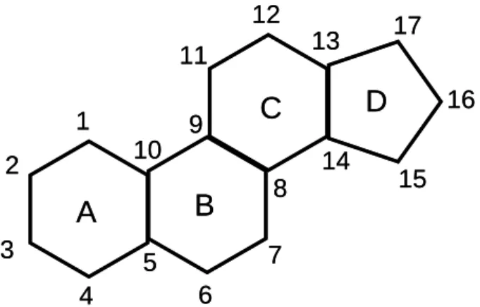
Steroids constitute a vast group of natural and synthetic organic compounds with a characteristic chemical structure consisting of a four-ring system (figure 1.1), with functional groups attached (Stacey & Sorensen, 2011; Gomes et al., 2009). The important roles played by steroids include regulating body functions, such as growth, digestion, development, reproduction and functioning of the sexual organs (Gomes et al., 2009; Crisp et al., 1998).
Figure 1.1 – Basic structure of a steroid, showing the four ring structure (A, B, C and D) and the numbering of the carbons (Based on Kime, 1993).
This group is composed of sterols, corticosteroids (mineralcorticoids and glucocorticoids), bile acids and sex steroids (Gomes et al., 2009). Some steroids can act as hormones, such as 11-ketotestosterone and 17,20β-P (Stacey & Sorensen, 2011).
1.1.1. Sex Steroids
Sex steroids refer to a group of hormones which perform an active role in reproduction and in controlling secondary sexual characters (Sunny et al., 2002). These hormones are classified as estrogens, androgens and progestogens (Gomes et al., 2009). According to Kime (1993), the definition of androgens is associated with C19 steroids, estrogens with C18 steroids and progestogens have a C21 structure (figure 1.2).
5
Figure 1.2- Structure of the androgens (C19), estrogens (C18) and progestogens (C21), showing the numbering of carbons.
Androgens can be characterized as “any natural steroid hormone”, involved mainly in growth, development, maintenance of the reproductive system (spermiation, spermatogenesis, gonadal differentiation) of the male and are also responsible for secondary sexual characteristics and regulation of sexual behaviour (Stacey & Sorensen, 2011; Rocha & Henriques, 1996; Oliveira, 1995). In teleost males, testosterone and 11-ketotestosterone are the androgens more commonly found in plasma (Oliveira, 1995); 11-ketotestosterone is the more physiologically active (Kime, 1993). However, testosterone is also an important androgen, not only in males, but also in females‟ reproductive cycle, since this is the only androgen synthesized by the ovary (Kime, 1993).
Stacey and Sorensen (2011) have defined estrogens as “any natural steroid hormone that controls female sexual development, secondary sexual characteristics, and stimulates egg production”. Estradiol and progesterone are the major estrogens regulating female reproductive cycle (Kime, 1993; Stacey & Sorensen, 2011). Progesterone is designated as the mammalian pregnancy hormone, thus and according to Kime (1993), does not play a role in the majority of teleost fishes. A well-studied progestogen is 17,20β-dihydroxypregn-4-en-3-one (17,20β-P), was first isolated in the plasma of Pacific salmon (Oncorhynchus nerka) (Kime, 1993; Nagahama, 1987), and
6 since then it was measured in females of more than 35 teleost species (Kime, 1993). However, progestogens are not exclusive to females; they also play a role during final gamete maturation in reproductive cycle of males in at least some species (Kime, 1993).
1.1.2. Corticosteroids
In teleost fish, corticosteroids are mainly synthesized in the interrenal tissue (Milla et al., 2009; Galhardo, 2010). Cortisol and cortisone are the main corticosteroids isolated from fish blood (Milla et al., 2009). Stress hormones, such as glucocorticoids, are released when the individual is exposed to a stress factor, such as threat of predation, confinement, social conflicts or pollution (Gabor & Contreras, 2012).
Cortisol is the major glucocorticoid in fishes and, as such, is often used as indicator of stress (Gabor & Contreras, 2012; Huang et al., 2007; Galhardo, 2010). According to Fox et al (1997), its plasma concentration depends on reproductive and social status of an individual and on social situation stability. Cortisol is also an important osmoregulatory hormone in fish (Dharmamba, 1979).
An increase in glucocorticoid release can decrease rates of the sex steroids production, since energetic costs of stress affect reproductive processes by altering the production of other hormones (Gabor & Contreras, 2012; Scott et al., 2008).
1.2. Chemical Communication
1.2.1. What is a Pheromone?
Although the environment surrounding fishes – water – is an excellent solvent, it is often turbid or devoid of light. Thus, fish have evolved systems to detect and respond to chemical cues released by individuals of the same species, rather than rely solely on visual and/or auditory cues (Sorensen & Stacey, 2004). To coordinate various aspects of their reproductive biology and non-reproductive functions (Cole & Stacey, 2006), and to mediate social behaviours (Sorensen & Stacey, 2004), fish use pheromones, chemical signals that pass between members of the same species (Stacey & Sorensen, 2006).
Pheromones were first described by Karlson & Luscher (1959) as “substances that are excreted to the outside by an individual and received by a second individual in which they release, for example, a definite behaviour or development process” (Sorensen & Stacey, 2004). According to Stacey and Sorensen (2006, 2011), a pheromone is a “chemical or a mixture of chemicals released into the environment by an
7 individual (the donor), capable of evoking a specific and adaptive response in conspecifics (the receivers), the expression of which does not require learning” (Stacey & Sorensen, 2006). Although pheromone detection involves specialisation within the chemo-sensory system(s) of the receiver, no specialization of the donor is required (Sorensen & Stacey, 2004). A pheromone might consist of a single compound, which does not need to be a specialized compound; however, pheromones are usually mixtures of chemicals (Sorensen & Stacey, 2004). Some fish employ reproductive hormones and their precursors and metabolites as “hormonal pheromones”, to induce important physiological and behavioural effects in conspecifics (Stacey, 2010).
Stacey and Sorensen (2006) characterize a reproductive pheromone as a “pheromone that induces any behavioural or physiological response associated with reproductive activity”, while a hormonal pheromone is any reproductive pheromone containing at least one derived compound from a chemical pathway that produces hormones, i.e., internal chemical signals. According to the same authors, hormonal pheromones can contain unmodified hormones, hormonal precursors and/or their metabolites (Stacey & Sorensen, 2006).
Evidence that hormones can function as pheromones is provided by 17,20β-P, an oocyte maturation inducing steroid that is released by female goldfish (Carassius auratus) and generates strong endocrinological and behavioural effects in conspecific males. Thus, conspecific behaviour and/ or physiology in several species are influenced by hormones and related compounds, which act as potent odorants (Stacey & Sorensen, 2006).
In 1963, Wilson and Bossert proposed that rapid behavioural and slower physiological effects induced by pheromones could be termed “releaser” and “primer” respectively (Sorensen & Stacey, 2004). However, these terms led to some confusion since the same pheromone can be both a primer and a releaser, and have since fallen out of favour (Stacey & Sorensen, 2006).
1.2.1.1 .The Goldfish (Carassius auratus)
A well-known sex pheromonal system in teleosts is that of the goldfish, in which hormonal pheromones from females primarily function to synchronize both male and female spawning physiology and behaviour (Kobayashi et al., 2002). Male goldfish also respond to the presence of male conspecifics, by increasing sperm stores either in
8 response to nearby males with greater levels of sex steroids “or in response to isolation from a group of conspecifics males in a basal endocrine condition”, whilst in females, there is some evidence that the released steroids are likewise detected and used as a priming pheromones, suggesting that female goldfish synchronize their ovulations (Kobayashi et al., 2002; Sorensen & Stacey, 2004; Stacey & Sorensen, 2006). In summary, male goldfish enhance reproductive success through a set of physiological and behavioural strategies in response to the changing odour of peri-ovulatory females (Stacey & Sorensen, 2006).
1.2.2. Functions of Pheromones in Fish Biology
Chemical communication is important in several aspects of fish biology, such as migration, the alarm response and reproduction (Sorensen & Caprio, 1998; Smith, 1992; Selset & Døving, 1980; Bjerselius et al., 2000 cited in Frade et al., 2002), particularly during reproduction (Miranda et al., 2005). Based on their function, pheromones can be divided into three categories: social, anti-predator and reproductive cues. Each of these categories includes „primer‟ pheromones (that induce endocrine, physiological and developmental changes) and/or „releaser‟ pheromones (strong behavioural changes) (Sorensen & Stacey, 2004). For example, several teleosts respond to pheromones, immediately before and during spawning, by increasing gonadal development and hormonal changes, capable of inducing final gamete maturation (priming response before spawning) (Sorensen & Stacey, 2004).
Pheromones include anti-predation and alarm cues: fishes display several chemically-mediated responses in order to reduce predation risk; kin and individual recognition in dominance hierarchies. Some fishes show a complex social system that helps to determine relatedness of conspecifics (including the establishment of dominance hierarchies and kin recognition) through the evolution of chemosensory mechanisms (Sorensen & Stacey, 2004).
Species recognition and aggregation, as well as migration, can be also mediated by pheromone release. Concerning migratory attraction, some species of migratory fishes seem to find spawning and feeding habitats through conspecific odours (Sorensen & Stacey, 2004).
9
1.2.3. Chemical nature of pheromones
Pheromones play an important role in the life histories of many fish species (Sorensen & Stacey, 2004), particularly in reproduction. However, few have been chemically identified and characterized to date: bile acids, sex steroids and F prostaglandins (Stacey & Sorensen, 2006).
Several electrophysiological studies, mostly using the electro-olfactogram (EOG), have shown that many species have high olfactory sensitivity to these substances (Stacey & Sorensen, 2006).Steroid and prostaglandin hormones, and metabolites, are capable of transmitting information to conspecifics, since their synthesis is predictably linked with reproductive events (Stacey & Sorensen, 2006). Therefore, such steroids and prostaglandins or their metabolites, once released into the water, have the potential to be used as hormonal pheromones (Barata et al., 2007).
Pheromones may be released into the water through several routes, namely via the urine, gills, skin, faeces or gonadal fluids (Barata et al., 2007; Stacey & Sorensen, 2006). Routes and rates of steroid release are both associated with conjugation of the steroids; free steroids are rapidly released across the gills, while sulphated and glucuronidated steroids are released in the urine and bile, respectively, although over a longer time-scale (Stacey & Sorensen, 2006; Vermeirssen & Scott, 1996).
1.2.3.1. Bile Acids
Bile acids are sterols produced by the liver and released in the bile and play a well know role in digestive system. Many fishes produce, release and detect bile acids: in the sea lamprey (Petromyzon marinus) a mixture of bile acids act as attractants for migratory adults and as male sex pheromone (Stacey & Sorensen, 2006); while in the European eel (Anguilla anguilla), Huertas et al (2010) suggested that bile acids could act as sex pheromones since the odour of bile depends on gender and changes with sexual maturity (Huertas et al., 2007).
1.2.3.2. Prostaglandins
Prostaglandins belong to a group of lipid compounds, derivatives of 20-carbon fatty acid, structurally characterized by a five-carbon ring. This class of compounds mediates several functions in fishes, such as female sexual behaviour, ovulation
10 (follicular rupture); they also perform pheromonal functions in some species (Stacey & Sorensen, 2011). In preovulatory female goldfish, levels of circulating prostaglandins F2α increase considerably at ovulation (Sorensen et al., 1995 cited in Sorensen & Stacey, 2006); thus being released as a postovulatory pheromone, that attracts male and it can increases milt volume (Kobayashi et al., 2002; Fraser & Stacey, 2002). According to Appelt and Sorensen (2007), goldfish control the release of urinary prostaglandin pheromones to advertise about their location and physiological condition.
As demonstrated by Moore and Waring (1996) in Atlantic salmon parr (Salmo salar), F-type prostaglandins may function as priming pheromones, released by ovulated females, via the urine (Moore & Waring, 1996).
1.2.3.3. Sex Steroids
The oocyte maturation-inducing steroid (17,20β-P) released by female goldfish acts as a pheromone inducing strong behavioural and endocrinological changes in males, such as increase in concentration of male plasma gonadotropin II (GtH II), in volume of sperm and seminal fluid, as well as in sexual activity (Zheng et al., 1997; Stacey & Sorensen, 2006).
In tench (Tinca tinca), males demonstrated high olfactory sensitivity to some classic teleost sex steroids, in particular, highly sensitive to glucuronidated 17,20β-P while 100 times less sensitive to the sulphated form (Pinillos et al, 2002). Nevertheless, tilapia are neither sensitive to androgens found in male urine (testosterone and 11-ketotestosterone) nor to steroids that act as pheromones in other species (17,20β-P and its conjugated forms; Frade et al., 2002).
1.3. Conjugated Steroids
In endocrine studies, it is important to consider that the initial “steroid will be excreted in a modified and inactivated form” (Gomes et al., 2009). Therefore, steroid metabolism needs to be taken into account. Catabolic pathways are divided into phase I and phase II reactions, being the principal goal to transform steroid substrate into a more polar and less active form (Kuuranne, 2010).
During phase I reactions, polarity increases by reduction, oxidation or hydrolysis reactions, which introduce new functional groups for the following phase II reactions, also designated as conjugation, where the biological activity of the free steroid is
11 O S OH O O O S OH O O
reduced, thus converting the non-polar compounds to a more easily excreted form, to be subsequently released in the urine (Gomes et al., 2009; Kuuranne, 2010).
Thus, conjugation implies the addition of sulphate or glucuronide groups to the free steroid, thereby promoting its excretion from the body (Gomes et al., 2009). Glucurone and sulphate conjugation are catalyzed by enzymes: uridine-5‟-diphosphoglucuronic acid (UDPGA), leading to the addition of a polar glucuronic acid to the structure of the steroid (glucuronidation) (figure 1.3); sulphotransferase enzymes which transfer sulphate group (SO3), from a co-substrate, 3‟-phosphoadenosine-5‟-phosphosulphate, to the steroid (sulphation) (figure 1.4) (Kuuranne, 2010).
Figure 1.3- Basic structure of a steroid with a glucuronic acid attached at carbon 17 (A), however this acid can also be attached at carbon 3 in B.
Figure 1.4- Basic structure of a steroid, with a sulphate group attached.
1.4. Steroid Measurement
Usually, the endocrine status of an individual is assessed by measurement of the concentration of hormones and their metabolites in the blood (Scott et al., 2008). Nevertheless, blood sampling may be disadvantageous; in order to bleed the fish, it needs to be caught, handled and/or anaesthetised, all of which can modify behaviour and physiological condition, and therefore plasma concentrations of the hormones under
A OH O O OH O OH HO OH O O OH O OH HO O OH O O OH OH HO O OH O O OH OH HO OH O O OH OH HO B
12 study. Furthermore, some fish may be too small, rare or valuable to be bled (Scott et al., 2008). Measuring fish steroids in water enables the study of reproductive physiology of the fish through a non-invasive technique, an alternative to measurement in samples of blood (Scott & Ellis, 2007; Scott et al., 2008). This technique offers minimal intervention (no anaesthetic, bleeding or handling stress), repeated measurements on the same individual and given that it involves measurements over time, this integration may reduce the short-term fluctuations in hormone levels that may occur in plasma (Scott & Ellis, 2007).
To understand the hormonal condition of the fish, it is necessary to know how much steroid has been released by the fish, over a given time, i.e. steroid release rate (Scott et al., 2008). There are two ways of measuring steroid release rate: “static sampling procedure” and “dynamic sampling procedure”. The first method has been used essentially for behavioural studies and consists of removing the fish from its tank and temporarily placing it in a tank with clean water for a given period. While the “dynamic sampling procedure” implies keeping the fish in controlled flow-through water conditions, and performing at least two water samples to estimate steroid release over time (Scott et al., 2008). With only a single measurement, it is not possible to determine the amount of steroid that has been released over time and how much has been lost by degradation, water replacement or reabsorption by fish (Scott et al., 2008).
In theory, since free steroids are released in the water mainly by passive diffusion across the gills, their release rate should be directly proportional to their concentration in plasma (Scott et al., 2008). However, this correlation between plasma steroid and water steroid concentrations is not always direct and is dependent on a variety of factors, such as gill surface area, affinity for specific steroid binding proteins in plasma, fish size, salinity, water temperature, among others (Scott & Ellis, 2007; Scott et al., 2008). Apart from these factors, steroid concentrations in water will also be affected by bacterial degradation, reabsorption by fish and adsorption to surfaces, as well as steroid instability during storage (Scott & Ellis, 2007).
1.4.1. Measurement of steroids
Glucuronidated and sulphated steroid concentrations are therefore heavily influenced by urination and defecation rates, and by the time that taken for the conjugation process (Scott & Ellis, 2007).
13 Nevertheless, the measurement of conjugated steroids is followed by advantages and disadvantages. It is advantageous to measure conjugated steroids when the goal is to reveal physiological and behavioural strategies that would not be seen by only measuring free steroids (Scott et al., 2008). It is also advantageous because fishes are capable of using some conjugated steroids as pheromones and thus in order to understand both physiological and behavioural reactions of conspecifics and to study when and how they are released, conjugated steroids need to be measured (Scott et al., 2008). However, measuring conjugated steroids can also be disadvantageous, since pathways from synthesis to release are more complicated than in free steroids and according to Scott et al (2008) “conjugated steroids can be temporarily “stored” in the bile and urine and their release is, thus, subject to factors such as glomerular filtration rate, urination frequency, feeding, gut passage time and defecation”.
1.4.2. Procedures for steroid determination
A specific, relatively cheap and rapid method of steroid measurement is immunoassay, such as radio or enzyme-immunoassay (Scott et al., 2008). Immunoassay methods, were first described by Yalow and Berson (1960), and are characterized by high sensitivity and precision, that allow the measurement of hormone levels at low concentrations in biological fluids (saliva, urine, blood) or, in this case, water (Wheeler, 2006).
Immunoassays rely on reaction of an antigen (ligand) with a specific antibody (binder). These methods are also referred to as “binding assays”, since quantification of a substance depends on progressive saturation of the specific antibody by that substance and the subsequent determination of its distribution between free and “bound” phases, which is achieved by the incorporation of a “tracer” (Chard, 1990). The tracer consists of a small quantity of the ligand or the binder labelled with a material that can be precisely measured in very small amounts, as radioactive isotopes in radioimmunoassay (RIA), fluorescent compounds in fluoroimmunoassay (FIA) or enzymes in enzymoimmunoassay (EIA) (Chard, 1990).
To quantify the amount of a particular steroid present in a given sample, initially and before performing immunoassay procedures, steroids need to be extracted from the water and subsequently concentrated, since steroid concentrations in water are, in general, too low to allow direct determination (Scott & Ellis, 2007).
14 For steroid extraction from aqueous solutions, Solid-phase Extraction (SPE) is the preferred approach. This well-established technique allows the isolation of organic compounds present in aqueous samples (Junk et al., 1988; Gomes et al., 2009).
The water sample is pumped through a solid-phase cartridge, where steroid molecules are retained in a silica matrix; when using C18 cartridges, for example, the silica matrix consists of 18 carbons. Since steroids are non-polar compounds, they are retained by the silica matrix, whereas polar compounds pass through. In order to release the retained steroids, they eluted by using an organic solvent such as ethanol or methanol.
1.4.2.1. Radioimmunoassay
Radioimmunoassay (RIA) is one of the most sensitive methods for quantitative analysis of antigen-antibody reactions, in which radioisotopes are used as tracers and are attached to antibodies or antigens (Voller et al., 1976). It involves competition between a radioactive labelled antigen (Ag*), an unlabeled antigen (Ag) with a specific antibody (Ab) with fixed and limiting concentration. After an incubation period, Ag* will be quantified in the free fraction or in binding one (Ag-Ab complex), once antigen distribution in both phases is positively related with the amount of antigen in the sample (Chard, 1990).
In competitive immunoassays, where the bound fraction is used to determine antigen concentration in the sample, the amount of antigen and the quantity of radioactive labelled antigen that bound to antibody are inversely proportional, i.e., the higher the antigen concentration in the sample, the lower the amount of Ag*-Ab (Andrade, 2006). The amount of antibody to be used in the assay is assessed through a dilution curve, which involves the incubation of a fixed amount of labelled antigen with different antibody concentrations. Generally, the concentration of antibody to be used is the one for which there is a 50% binding of the tracer ligand (Chard, 1990).
A standard curve is the basic requirement for quantification of the ligand in unknown samples, since it allows quantification of the antigen (hormones) present in the samples through incubation of fixed amounts of labelled hormone and specific antibody with different concentrations of unlabelled antigen (Chard, 1990).
Radioimmunoassay is advantageous since it can be applied to several types of compounds to which there are antibodies available, also because it is extremely
15 sensitive, stable, specific and precise. On the other hand, it can be also disadvantageous, because it needs purified antigens for Ag* preparation and it requires special attention regarding to handling due to radioactivity (Chard, 1990). The utility of the assay is also dependent on the affinity and specificity of the antibody.
1.5. The Mozambique Tilapia, Oreochromis mossambicus
Tilapia are a large group of teleost fishes belonging to the family Cichlidae and order Perciformes, the most evolutionarily advanced and the largest order of teleosts (Cruz, 2006; Huang et al., 2007, Mulero et al., 2007). Due to the exhibition of complex social behaviour together with high reproductive rates and nutritional value, tilapia became one of the most studied fishes (Cruz, 2006). The Mozambique tilapia, a lek-breeding maternal mouth-brooding African cichlid, Oreochromis mossambicus (Amorim & Almada, 2005; Almeida et al., 2005) was firstly described as Chromis (Tilapia) mossambicus by Peters in 1852, from specimens collected in the Mozambique region (Oliveira, 1995). The taxonomy of the species is shown below according to the Integrated Taxonomic Information System (ITIS):
Kingdom: Animalia Phylum: Chordata Subphylum: Vertebrata Class: Actinopterygii Order: Perciformes Suborder: Labroidei Family: Cichlidae Genus: Oreochromis
Species: Oreochromis mossambicus (Peters, 1852)
Figure 1.5 - Reaction between radiolabelled antigen, antigen to quantify and the antibody. Source: Wheeler, 2006
16 The Mozambique tilapia is a euryhaline cichlid fish, endemic to the lakes and rivers of the east coast of Africa, though it has been introduced from its native habitat to tropical freshwater and marine environments around the world (Oliveira & Almada, 1998a; Barata et al., 2007; Morgan et al., 1997). Its physical robustness, combined with resistance to variation in physical and chemical factors, has allowed the occupation of a vast diversity of habitats in a wide geographical distribution that comprises equatorial rivers, tropical and subtropical lakes, estuaries and irrigation channels, for instance (Oliveira, 1995); tilapia have become a major invasive species in several countries, in all five continents (Russell et al., 2012).
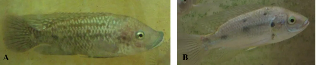
In its natural distribution, this eurythermal species can support temperatures ranging from 17ºC to 35ºC (Oliveira, 1995); its optimal temperature is around 27ºC (±1ºC). From the perspective of feeding, O.mossambicus is an opportunistic species, consuming phytoplankton, zooplankton, benthos and detritus (Caskey et al., 2007; Oliveira & Almada, 1995). O. mossambicus, as well as other tilapine fishes, exhibits a clear sexual dimorphism, including the height of the dorsal and anal fins, the proportions of the jaw and in the shape and size of the genital papilla, which is emphasized during the breeding season (Oliveira & Canário, 2000) (figure 1.6). Male O. mossambicus exhibit characteristic breeding colours and generally they grow faster and reach larger sizes than females. Males also present one urogenital opening while females have two, and mature males have a thick lip in upper jaw (Oliveira & Almada, 1995). Females have a larger oral cavity due to a greater development of the pre-opercular and inter-pre-opercular bones; although males have a larger and stronger mouth, they do not mouth-brood (Oliveira & Almada, 1995).
The highest expression of morphological (dorsal and anal fin height, mandible width and pre-maxilla length) and behavioural (nuptial coloration, time defending a territory, spawning pit volume and courtship rate) characters are showed by dominant fishes (Oliveira & Canário, 2000).
Figure 1.6 – Individuals of Oreochromis mossambicus: it is possible to observe the dimorphism in sexual characteristics of the species. Figure 5A shows a male while in Figure 5B shows a female.
17 This species displays a pronounced dichromatism, which is more evident during breeding season; breeding males exhibit an intense black coloration over the entire body, only with the lower jaw region displaying white and the edges of dorsal and anal fins showing a red coloration. Non-breeding males and females exhibit a grey coloration (Cruz, 2006) (figure 1.7).
Figure 1.7- Male tilapia exhibiting characteristic breeding colours.
In their classic study on cichlid behaviour, Baerends and Baerends van Roon (1950) distinguished three basic patterns of social relations: dominance hierarchies, territoriality and shoals. According to Oliveira (1995), males tend to be dominant over females, while large and medium sized fish tend to be dominant over smaller ones. Thus, females of O.mossambicus occupy the lowest places in the hierarchies (Oliveira, 1995).
As in many cichlids, in Mozambique tilapia, social status is revealed by the colour pattern of the skin, with dominant males having a darker pigmentation (Van der Salm et al., 2005), larger gonadosomatic indices, longer dorsal fins, larger genital papilla and larger testes than subordinate males (Oliveira et al., 1996; Oliveira & Canário, 2000). Social dominance allows dominant individuals to have priority in access to limited resources; either food or reproductive females (Oliveira, 1995). In teleosts, a correlation between hierarchical position of the males and reproductive success has been observed, where dominant individuals show a higher rate of mating and more access to females (Oliveira, 1995). From studies carried out in captivity, it has been shown that male tilapia form stable linear hierarchies, with the largest males (alpha males) receiving more visits of spawning females (Oliveira & Almada, 1996; Oliveira & Almada, 1998a).
In captivity, males are commonly divided into „territorial‟ (dominant), „floater‟ and „sneaker‟. Dominant males usually dig nests, assume a dark coloration, defend a centred territory in the nest and dynamically court females. Floater males typically live
18 in the water column, display a light dark coloration and occupy territories for a short period (from seconds to minutes), when the owners are away to court females. On the other hand, sneaker males usually invade nests during spawning and try to remain near the female while exhibiting trembling behaviour, which is normally related to sperm release (Oliveira & Canário, 2000).
1.5.1. Sexual Behaviour and Reproduction
In this species, during the breeding season, males gather in breeding arenas or leks, forming dense aggregations over sandy or muddy substrates (Barata et al., 2007; Oliveira & Almada, 1998b). After males‟ aggregation, dominance hierarchies are established, where pheromones may play an important role in modulating aggressive interactions (Barata et al., 2007, Keller-Costa et al., 2012).
Within the arenas, males establish individual territories where they dig spawning nests (pits) in the substratum, which they defend, and the associated area, from possible invaders, and assume a characteristic black colouration (Barata et al., 2007; Barata et al., 2008; Frade et al., 2002; Miranda et al., 2005; Oliveira et al., 1996; Oliveira & Almada, 1998b; Galhardo et al., 2008). From a few seconds to a few minutes after the formation of the group, some individuals begin to darken and participate in symmetrical fights (i.e. when an individual retaliates to an agonistic act with another aggressive action), essentially involving circle fights, mouth-to-mouth fighting and mutual displays (Oliveira & Almada, 1998c; Oliveira & Canário, 2000; Barata et al., 2007).
Circle fights consists of two individuals in antiparallel position, exhibiting lateral display, moving around a central spot, and as they attempt to bite, both individuals attack alternately (figure 1.7) (Oliveira, 1995). In mouth-to-mouth fighting (figure 1.8), fishes display frontally to each other and attack with open mouths (Baerends & Baerends van Roon, 1950; Oliveira, 1995). Both of these behaviours, as well as mutual displays of individuals (fish remains motionless in front of its opponent), are considered as symmetrical agonistic interactions (Oliveira, 1995).
19
Figure 1.5 – Agonistic interactions between tilapia males: circle fights (from Oliveira, 1995).
Figure 1.6- Agonistic behaviour exhibit by tilapia males: mouth-to-mouth fighting (from Oliveira, 1995).
Females are attracted to spawning pits, which are usually located in shallow waters, where males display to attract females for mating, exhibiting a series of behaviours, such as nest digging, trembling and circling the female (Amorim & Almada, 2005; Almeida et al., 2005).
Although Mozambique tilapia can spawn repeatedly throughout the year, with females having a regular ovulatory cycle of 15–20 days, only when the females are ready to spawn do they visit the breeding areas (Barata et al., 2008; Barata et al., 2007).
In lekking cichlids, with a breeding system analogous to O.mossambicus, dominant fish occupy a central position in the nest, dig larger nests, are more effective at defending territories, also court at a higher ratio and have a higher breeding succession (Amorim & Almada, 2005; Oliveira et al., 1996), which can be explained by the fact that females prefer males with larger nests (Amorim et al., 2003).
20 Mature females enter the lekking area, where spawning takes place, and choose one or more males with which to mate (Frade et al., 2002). When the mating sequence ends, males tremble while circling the nest followed by female, which in turn, takes both eggs and sperm into her mouth, where fertilization occurs (Oliveira & Almada, 1998b; Amorim et al., 2003). After spawning, the female leaves the arena and during the next 20-22 days carries the embryos and the offspring in her mouth, to brood in a nursery area separate from males (Frade et al., 2002). Hence, parental care is restricted to females, which delay their next ovulatory cycle until the brood is released (Oliveira et al., 1996; Miranda et al., 2005).
Throughout the mouth-brooding cycle, females become progressively more aggressive to other conspecifics. However, contrary to males, they defend mobile space around themselves, instead of defending territories on the substrate (Oliveira & Almada, 1998b; Oliveira, 1995).
In this species, maternal aggression is a well-developed phenomenon; however, female agonistic behaviour differs from that of territorial males in that aggressive acts are limited to charges, chases and butting (Oliveira & Almada, 1998b). Female pigmentation pattern also changes progressively during the mouth-brooding cycle; the body becomes light grey with an overlaid pattern of dark stripes, the eyes show horizontal bars in the irises, the lips get darken and display a characteristic mandibular spot (Oliveira & Almada, 1998b; Oliveira, 1995).
Maternal aggression seems to function principally to defend the brood against predators, conspecifics included. This is different from the aggression directed against the brood as a way to impose control over their behaviour (Oliveira & Almada, 1998b). Throughout the oral incubation cycle, Oliveira (1995) observed that females suppressed almost totally their feeding activity; however, there was no decrease in the condition factor of the incubating females, whilst a decrease in the number of eggs/juveniles during the incubation cycle was observed. This suggests the existence of a partial cannibalism of the eggs as a reproductive strategy of the females (Oliveira, 1995).
1.6. Role of urine among conspecifics
Urine is used as a chemical signal, at least for conspecific females (Almeida et al., 2005; Barata et al., 2007). Several studies have shown that Mozambique tilapia males regulate urine release depending on the social context; territorial males (males
21 from a higher social rank) may signal their status and aggressiveness to other males, potential rivals, through the release of urine (Almeida et al., 2005), which may contain male-male pheromones (Barata et al., 2007), as well as by changing behaviour and coloration (Almeida et al., 2005). The females‟ olfactory system is extremely sensitive to substances released by territorial males into the water as well as to male body fluids, demonstrating that urine has a special importance (Barata et al., 2008; Almeida et al., 2005).
O. mossambicus males are capable of storing urine: the bladder of a fish weighting 100g may contain up to 2 ml of urine (Almeida et al., 2005). According to Barata et al (2007), dominant males dynamically release chemical information through increasing urination rate during aggression, and in the presence of females, dominant males dramatically increase their release of urine (Almeida et al., 2005). Urination rate remains high during courtship, being considerably higher in the presence of pre-ovulatory females, and their urine has higher olfactory potency than that of subordinate males (Almeida et al., 2005; Barata et al., 2007). On the other hand, females release urine at higher frequency and smaller pulses, and this seems to be unaffected by the presence of dominant males (Almeida et al., 2005).
In a study carried out by Barata et al. (2008), using a liquid chromatography linked to mass spectrometry and recording the electroolfactogram (EOG) it was demonstrated that female tilapia detect a potent odorant in the non-polar fraction of male urine suggested to be a sulphated amino-sterol. This odorant is present at higher concentrations in urine from dominant males than in subordinate males. However, further work has shown that this compound is, in fact, a steroid glucuronide (Keller-Costa et al., unpublished).
These authors suggested that the urine of dominant males has a higher concentration of this compound and it is related to a higher olfactory potency. The same authors also suggested that social dominance, instead of reproductive capability, is reflected by both urine volume stored in the bladder and concentration of the odorant (Barata et al., 2008). However, according to the same study, the most active and important odorant found in male urine is non-polar. Consequently, this study suggested that this urinary odorant “is may be detected as part of mixture of odorants and that females may use the ratio, rather than the absolute amount, to discriminate between dominant and subordinate males” (Barata et al., 2008). In turn, subordinate males are able to store less urine in their samller bladder, so they are less capable of increasing
22 their urination rate and are consequently less able to stimulate females (Barata et al., 2008, Keller-Costa et al., 2012).
As mentioned before, male urine may act as a pheromone signal, causing responses in female endocrine status and behaviour. One such response is the increasing the synthesis for of the oocyte maturation steroid. Frade et al (2002) suggested that males release a signal in their urine in way to attract females (“releaser” effect), but this signal may be also capable of inducing ovulation (“primer” effect). This possibility is supported by a recent study where females were exposed to male urine in way to explore “primer” effects on females‟ endocrine system. Less than one hour after exposure, an increase in the levels of 17,20β-P, responsible for oocyte maturation (Huertas et al., unpublished).
1.7. Aims of the study
Pheromones present in male urine, once released, affect steroid levels in females. In the case of the Mozambique tilapia, exposure to male urine elevates the release rate of the maturation-inducing steroid hormone 17,20β-P by females.
The main objective of this study was to identify which constituent(s) of male urine (filtrate, eluate or both) contain(s) the pheromonal components. In other words, which fraction of urine is responsible for causing an increase in the oocyte maturation-inducing steroid (17,20β-P) in females.
Specifically, the study aimed to determine whether the steroid glucuronide recently identified as the main component in the eluate of male urine is sufficient - on its own - to cause the increase in 17,20β-P metabolism in females, or whether other urinary components are necessary.
25 2.1. Experimental Animals
Mozambique tilapia (Oreochromis mossambicus) used in the current study were obtained from a stock population previously established at the University of the Algarve. This stock originated from wild specimens caught in Mozambique, from the River Incomati (early 1970s), kept and raised at Aquário Vasco da Gama (Lisbon) from which some individuals were bred at Instituto Superior de Psicologia Aplicada (Lisbon); subsequently, individuals were donated to the University of the Algarve (Frade et al., 2002).
In order to form families, animals were grouped (one male and four females) in fibre-glass tanks (250 l) with sand substratum and kept at 27ºC (±1ºC), where they were maintained and fed once a day with an appropriate diet. Under these semi-natural conditions, individuals exhibited the normal mating behaviour and, as such, spawning occurred in each tank naturally. Nevertheless, after each spawning, eggs were removed from the mother‟s mouth, by applying a slight pressure in the posterior area of the opercula, to maintain the females‟ ovulatory cycle and to predict the next ovulation. The removal date of the eggs was recorded, so that a given female could be designated as „pre-ovulatory‟ (predicted to ovulate in the next three days or „post-ovulatory‟ (having ovulated during the past three days).
2.2. Urine Sample Collection
Urine samples were collected daily (except at weekends) by application of slight pressure at the terminal part of the abdomen, immediately above and anterior to the urogenital opening (Frade et al., 2002), and avoiding any possible contamination with faeces, from each male (n=7) to 1.5 ml Eppendorf tubes. Males were then replaced into their tank of origin. Urine samples were clearly identified (number of the male and collection date) and frozen (-20ºC) until a given volume per male was collected. Then a pool was made, using an equal volume of urine (4.9 ml) from each male, and this pool was subsequently aliquoted, extracted and frozen until use (see below).
26 1 2 3 4 5 6 7 8 9 10 19 11 12 13 14 15 16 17 18 20 21 O OH OH H O OH O H O H -OOC H H H 22 23 24 25 26 27 5β-Pregnan-3α,17α,20β-triol-3α-glucuronide 1 2 3 4 5 6 7 8 9 10 19 11 12 13 14 15 16 17 18 20 21 O OH OH H O OH O H O H -OOC H H H 22 23 24 25 26 27 5β-Pregnan-3α,17α,20α-triol-3α-glucuronide 1 2 3 4 5 6 7 8 9 10 19 11 12 13 14 15 16 17 18 20 21 O OH OH H O OH O H O H -OOC H H H 22 23 24 25 26 27 5β-Pregnan-3α,17α,20β-triol-3α-glucuronide 1 2 3 4 5 6 7 8 9 10 19 11 12 13 14 15 16 17 18 20 21 O OH OH H O OH O H O H -OOC H H H 22 23 24 25 26 27 5β-Pregnan-3α,17α,20α-triol-3α-glucuronide 2.3. Experimental Design 2.3.1. Stimuli Preparation
Of the total volume of the urine pool (34.3 ml), 15 ml was aliquoted into 1.5 ml Eppendorf tubes, each with 0.5 ml of urine, designated as “urine pool”. Another 15 ml of urine pool was extracted (see Solid-Phase Extraction), to obtain 15 ml of filtrate (aqueous fraction), which was aliquotted into Eppendorf tubes, each with 0.5 ml; and 5 ml of eluate (hydrophobic fraction), which was stored in glass vials containing 1 ml each.
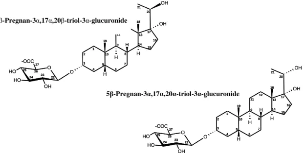
Two glucuronidated steroids present at high concentration in dominant male urine and previously identified as the most potent olfactory stimuli in the eluate, [pregnane-3α,17α,20β-triol-3α-glucuronide (20β-P-Gluc) (main compound); 5β-pregnane-3α,17α,20α-triol-3α-glucuronide (20α-P-Gluc) (minor compound)] (Keller-Costa et al., unpublished) were mixed in a proportion of 4:1, respectively (figure 2.1).
Figure 2.1- Structures of steroid present in high concentrations in urine of dominant male, identified by Keller-Costa et al (unpublished), consisting of two steroid glucuronides: 5β-Pregnan-3α, 17α, 20β-triol-3α-glucuronide (main compound); 5β-Pregnan-5β-Pregnan-3α, 17α, 20α-triol-3α-glucuronide (minor compound).
Each steroid has a molecular weight of 534.6 g mol-1 and to achieve a steroid concentration of 5x10⁻³M (2.673 mg/ml, see table 1), it was added 1.407 ml and 0.449 ml of methanol to 3.76 mg of steroid 20β-P-Gluc and 1.2 mg of 20α-P-Gluc, respectively (see table 1). From these stock solutions, 1.2 ml of 20β-P-Gluc, corresponding to 4-fold of 20β-P-Gluc (5x10⁻³M), and 0.3 ml of 20α-P-Gluc,
27 equivalent to 1-fold of 20α-P-Gluc (5x10⁻³M) was then taken and mixed together in glass vial. This 1.5 ml of 4:1 20β-P-Gluc/20α-P-Gluc (5x10⁻³M) mixture was diluted with 13.5 ml of ethanol to a total volume of 15 ml, thus achieving a total mixture concentration of 5x10⁻⁴M, approximately the same concentration as in urine. This stimulus was then divided into glass vials (with a capacity of 1.5 ml), each containing 1ml of the stimulus and stored at -20ºC.
Table 2.1: Preparation of the stimulus "Steroid" in order to achieve a concentration of 5x10⁻⁴ M
Preparation of the stimulus:
20--P-Gluc-sodium salt (charge III) in mg mg 3.76
20--P-Gluc-sodium salt (charge III) in mg mg 1.2
molecular weight (each) in g /mol g/mol 534.6
concentration of the stock solution 5x10⁻³M: mmol/ml mmol/ ml 0.005
steroid concentration_ 5x10⁻³M: mg/ml mg/ml 2.673
Methanol volume (ml) to 20--Gluc to obtain stock solution ml 1.407
volume (ml) to 20--Gluc to obtain stock solution ml 0.449
4x 20--P-Gluc 5x10⁻³M ml 1.2
1x 20--P-Gluc 5x10⁻³M ml 0.3
4:1 20-/20- MIX total: 5x10⁻³M ml 1.5
Ethanol
4:1 20-/20- MIX total: 5x10⁻⁴M ml 15
All the four stimuli obtained (urine pool, filtrate, eluate, “steroid”) were stored at -20ºC until further use. Some of the stimuli were combined during the experiment: eluate plus filtrate and filtrate plus “steroid”. Methanol (Sigma-Aldrich or VWR) was also used as control stimulus.
2.3.2. Exposure of Females to Male Urine and Derivatives
Firstly, females (n=8) were selected from the family tanks according to their ovulatory stage; pre-ovulatory females with a regular ovulatory cycles were preferred. The weight and length of each female were recorded, and the female was then isolated in a glass aquarium with 6l of de-chlorinated tap-water at 27ºC (±1ºC) and equipped with an air supply, and maintained over-night. The next day, females were transferred to an identical aquarium with a volume of clean de-chlorinated tap-water normalized to the weight of the fish (1 litre of water per 10 g of fish; Huertas et al., unpublished). An hour after the transfer, one litre of water was collected (control sample, at time 0h), through siphoning with a tube previously placed in the tank. This sample was extracted (see Solid-Phase Extraction).