Catarina Sofia Rodrigues do Carmo
Role of sirtuin 3 on mitochondrial dynamics
in Huntington’s disease striatal cells
Outubro 2015
Tese de Mestrado
Mestrado em Genética Molecular
Trabalho efectuado sob a orientação de
Professora Doutora Ana Cristina Rego
Professora Doutora Olga Coutinho
Catarina Sofia Rodrigues do Carmo
Role of sirtuin 3 on mitochondrial dynamics
in Huntington’s disease striatal cells
ii
DECLARAÇÃO
Nome: Catarina Sofia Rodrigues do Carmo
Endereço electrónico: [email protected] Telefone: +351 964 321 097 Número do Bilhete de Identidade: 14187861
Título da dissertação de Mestrado:
Role of sirtuin 3 on mitochondrial dynamics in Huntington’s disease striatal cells
Orientadores:
Professora Doutora Ana Cristina Rego Professora Doutora Olga Coutinho
Ano de conclusão: 2015
Designação do Mestrado: Mestrado em Genética Molecular
DE ACORDO COM A LEGISLAÇÃO EM VIGOR, NÃO É PERMITIDA A REPRODUÇÃO DE QUALQUER PARTE DESTA TESE/TRABALHO.
Universidade do Minho, 30 / 10 / 2015 _________________________________ ( Catarina Sofia Rodrigues do Carmo )
iii Depois de um ano intensivo mas, sem dúvida, recompensador, esta será uma das secções mais difíceis de concluir. Embora seja um trabalho apresentado por uma pessoa, não seria de todo possível sem o contínuo apoio e ajuda de um vasto número de pessoas. Não poderia assim passar a oportunidade de expressar o meu profundo agradecimento e reconhecimento a todos os que de alguma forma contribuíram para a sua realização e concretização.
Às minha orientadoras: à Professora Doutora Ana Cristina Rego pela oportunidade de poder integrar no seu grupo de investigação, pelo enorme voto de confiança que depositou em mim e por ajudar continuamente no meu crescimento científico; à Professora Doutora Olga Coutinho pelo apoio e ajuda desde o início deste trabalho e pela constante presença e disponibilidade. Quero ainda agradecer a revisão crítica desta dissertação. O meu obrigada por tudo.
Às minhas “supervisoras” não oficiais: Luana Naia e Ana Oliveira, que enalteceram grandemente o meu crescimento no laboratório, pelo apoio contínuo ao longo de todo este ano e por todo o tempo e atenção que disponibilizaram para mim. Este trabalho é tanto meu como é vosso.
Aos colegas (e amigos) do grupo Mitochondrial Dysfunction and Signaling in
Neurodegeneration (António, Carla, Elisabete, Inês, Jorge, Luísa, Mário, Maura e
Sandra), que sempre se mostraram disponíveis e prestáveis mas acima de tudo por permitirem a fácil integração no grupo e a vossa amizade, que sei que vou levar comigo.
Aos amigos de todo o país (de Braga a Lisboa, passando por Viseu), que sempre me acompanharam e partilharam comigo os sucessos e insucessos.
À minha família, em especial, aos meus pais, pelo apoio incondicional e por acreditarem em mim muito mais que o que eu acredito.
iv
This work was funded by FEDER funds through the Operational Programme Competitiveness Factors - COMPETE, national funds by FCT - Foundation for Science and Technology (project reference: EXPL/BIM-MEC/2220/2013), ‘Fundação Luso-Americana para o Desenvolvimento’ (FLAD) and by Gabinete de Apoio à Investigação, funded by FMUC and Santander Totta Bank. Center for Neuroscience and Cell Biology (CNC) is supported by projects PEst-C/SAU/LA0001/2013-2014 and UID/NEU/04539/2013.
v ABSTRACT
Altered mitochondrial dynamics has been implicated in the pathogenesis of several neurodegenerative disorders, including Huntington’s disease (HD). Sirtuins, NAD+-dependent lysine deacetylases, have emerged as important cellular targets that can interfere with mitochondrial biogenesis, fission/fusion, motility and mitophagy. Among them, sirtuin 3 (SIRT3) is particularly relevant, being the main deacetylase located in mitochondria. Here we evaluated the influence of SIRT3 on mitochondrial dynamics using striatal cells derived from HD knock-in mice (STHdhQ111/Q111) versus wild-type cells (STHdhQ7/Q7).
Increased mitochondrial fragmentation was observed in untransfected HD cells. Indeed, STHdhQ111/Q111 cells exhibited an overall decrease in the levels of mitochondrial fusion proteins (Mfn2, OPA1) and an increase in fission-related Fis1. Drp1 (also involved in mitochondrial fission) was preferentially accumulated in the mitochondrial fraction of HD cells. Increased LC3-II/I ratio, which evaluates autophagosome formation, was observed in STHdhQ111/Q111 cells. Moreover, the autophagy adaptor p62 was found to be decreased in mutant cells. Parkin and PINK1, two markers of mitophagy, were also assessed. Untransfected HD cells exhibited lower levels of both proteins. No significant changes were detected in phosphorylated Parkin (required for its enzymatic activation and mitochondrial translocation). These data suggest that PINK1/Parkin-dependent mitophagy is impaired in HD striatal cells.
Overexpression (OE) of SIRT3 reduced the unbalance between fission/fusion by decreasing the protein levels of Fis1 in STHdhQ7/Q7 and STHdhQ111/Q111 cells, and Drp1 accumulation in mitochondria in STHdhQ111/Q111 cells. Concordantly, an increased number of mutant cells presenting tubular mitochondria was observed after SIRT3OE. An additional significant increase in LC3-II/I ratio was observed in STHdhQ111/Q111-SIRT3 cells, indicative of macroautophagy activation.
Data suggest that enhanced SIRT3 levels restore mitochondrial morphology in mutant cells by reducing mitochondrial fission, with additional activation of macroautophagy.1
KEYWORDS: HUNTINGTON’S DISEASE, MITOCHONDRIAL DYSFUNCTION, MITOCHONDRIAL DYNAMICS,
vii RESUMO
Alterações na dinâmica mitocondrial têm sido relacionadas com diversas doenças neurodegenerativas, incluindo a doença de Huntington (DH). As sirtuínas são deacetilases de lisinas dependentes de NAD+ que demonstraram ter um papel importante no re-estabelecimento do equilíbrio entre biogénese e fissão/fusão mitocondrial, e mitofagia. De todas, a sirtuína 3 (SIRT3) destaca-se por ser a deacetilase predominantemente localizada na mitocôndria com maior número de alvos proteícos. Neste trabalho avaliou-se o efeito da SIRT3 na dinâmica mitocondrial recorrendo ao uso de células estriatais derivadas de murganhos knock-in para a DH (STHdhQ111/Q111) versus células ‘wild-type’ (STHdhQ7/Q7).
As células mutantes não transfetadas apresentaram um aumento da fragmentação mitocondrial. De facto, as células STHdhQ111/Q111 apresentaram um decréscimo dos níveis proteícos de Mfn2 e OPA1, duas proteínas envolvidas na fusão mitocondrial, e um aumento de Fis1, uma proteína relacionada com a fissão mitocondrial. Verificou-se ainda uma acumulação preferencial da Drp1 (também envolvida na fissão mitocondrial) na fração mitocondrial das células STHdhQ111/Q111. Embora se tenha observado um aumento do rácio LC3-II/I (que avalia a formação de autofagossomas) nas células STHdhQ111/Q111, os níveis do adaptador autofágico p62 encontraram-se diminuídos. Células mutantes não transfetadas apresentaram ainda uma redução dos níveis de Parkina e PINK1, dois marcadores do processo mitofágico. Contudo, não se observaram diferenças significativas nos níveis da forma fosforilada da Parkina (indicador da sua ativação enzimática e translocação para a mitocôndria). Estas evidências sugerem alterações deste processo mitofágico nas células mutantes. A sobre-expressão de SIRT3 reduziu o desequilíbrio entre fissão/fusão ao diminuir os níveis de Fis1 nas células STHdhQ7/Q7 e STHdhQ111/Q111, e a acumulação da Drp1 na mitocôndria nas células STHdhQ111/Q111. Consequentemente, observou-se um aumentou do número de células mutantes com mitocôndrias tubulares. Verificou-se ainda um aumento significativo do rácio LC3-II/I nas células STHdhQ111/Q111- -SIRT3, indicativo de uma ativação da macroautofagia.
Em conclusão, o aumento dos níveis de SIRT3 permite restaurar a morfologia mitochondrial em células mutantes ao reduzir a fissão mitocondrial, conduzindo ainda à ativação da macroautofagia.
ix
TABLE OF CONTENTS
DECLARAÇÃO ...ii AGRADECIMENTOS ... iii ABSTRACT ... v RESUMO ... vii LIST OF FIGURES ... xi LIST OF TABLES ... xi ABBREVIATIONS ... xiii CHAPTER I – INTRODUCTION ... 1 1.1. HUNTINGTON’S DISEASE ... 31.1.1. Huntingtin: structure, function and post-translational modifications ... 4
1.1.2. Mutant huntingtin and mechanisms of cytotoxicity ... 7
1.2. CHANGES IN MITOCHONDRIAL FUNCTION AND DYNAMICS IN HD ... 9
1.2.1. Mitochondrial dysfunction – from transcription deregulation to altered calcium handling 10 1.2.2. Alterations in mitochondrial dynamics ... 13
1.3. LYSINE DEACETYLASES AND THEIR ROLE IN NEURODEGENERATION ... 19
1.3.1. Lysine deacetylases: what are they? ... 20
1.3.2. Role of KDACs in Huntington’s disease ... 24
1.4. MAIN GOALS ... 27
CHAPTER II – MATERIAL & METHODS ... 29
2.1. MATERIALS ... 31
2.2. CELL CULTURE ... 32
2.3. CONSTRUCTS, TRANSFECTION AND INCUBATIONS ... 32
2.3.1. Constructs ... 32
2.3.2. Bacteria transformation ... 33
2.3.3. Plasmid DNA extraction ... 33
2.3.4. Transfection of STHdh(Q111/Q111) and STHdh(Q7/Q7) striatal cells ... 33
2.3.5. Analysis of Autophagy flux in untransfected STHdh(Q111/Q111) and STHdh(Q7/Q7) striatal
x
2.4. SAMPLE PREPARATION AND WESTERN BLOTTING ... 34
2.5.1. Subcellular fractionation ... 34
2.5.2. Total protein extracts ... 35
2.5.3. Western Blotting ... 35
2.5. IMMUNOCYTOCHEMISTRY ... 35
2.6. IMAGE ANALYSIS ... 36
2.7. STATISTICAL ANALYSIS ... 38
CHAPTER III – RESULTS ... 39
3.1. HD STRIATAL CELLS SHOW INCREASED SIRT3-GFP ACCUMULATION IN MITOCHONDRIA ... 41
3.2. FISSION IS REDUCED UPON SIRT3 OVEREXPRESSION IN HD STRATAL CELLS THROUGH DECREASED DRP1 ACCUMULATION IN MITOCHONDRIA AND FIS1 TOTAL PROTEIN LEVELS ... 43
3.3. MITOCHONDRIAL FUSION-RELATED PROTEIN LEVELS ARE DECREASED IN HD STRIATAL CELLS AND REMAIN UNALTERED AFTER SIRT3 OVEREXPRESSION ... 46
3.4. SIRT3 OVEREXPRESSION APPEARS TO RESTORE MITOCHONDRIAL MORPHOLOGY IN MUTANT CELLS... 49
3.5. SIRT3 OVEREXPRESSION MIGHT ACTIVATE MACROAUTOPHAGY BUT NOT PARKIN-DEPENDENT MITOPHAGY IN MUTANT CELLS ... 54
CHAPTER IV – DISCUSSION & CONCLUSIONS ... 61
4.1. DISCUSSION ... 63
4.2. CONCLUSIONS ... 71
REFERENCES ... 73
ATTACHMENTS ... xvii
1. SUPPLEMENTARY METHODS ... xvii
1.1. Macro used to design ROIs ... xvii
1.2. Macro used to analyze mitochondrial morphology and protein colocalization ... xxi
xi
LIST OF FIGURES
Fig. 1 | A Schematic diagram of the HTT amino acid sequence. ... 5 Fig. 2 | Visual representation of mitochondrial morphology analysis done in Image J. ... 37 Fig. 3 | Overexpressed SIRT3-GFP accumulates more in the mitochondria of STHdhQ111/Q111 cells.
... 42 Fig. 4 | Drp1 accumulation in mitochondria and increased Fis1 protein levels in HD striatal cells are rescued by SIRT3 overexpression... 45 Fig. 5 | STHdhQ111/Q111 cells show decreased levels of fusion proteins, Mfn2 and OPA1, that are
not recovered after SIRT3 overexpression. ... 48 Fig. 6 | Categorization of mitochondrial morphology. ... 50 Fig. 7 | Mutant cells display more fragmented mitochondria with decreased percentage of the cellular area occupied by the mitochondrial network than wild-type cells and the former is counteracted upon SIRT3 overexpression. ... 53 Fig. 8 | Analysis of mitophagy in HD striatal cells – Decreased levels of Parkin were maintained after SIRT3OE, with no additional changes in Parkin phosphorylation (at S65) and PINK1. ... 56 Fig. 9 | Apparent activation of macroautophagy upon SIRT3 overexpression in STHdhQ111/Q111
cells. ... 60 Fig. 10 | Schematic representation of fission/fusion-related protein localization in untransfected and in STHdhQ111/Q111 cells overexpressing SIRT3. ... 72
Fig. S 1 | Study of the variables Aspect Ratio and Roundness and their adequacy towards the categorization of mitochondrial morphology. ... xxxiii
LIST OF TABLES
Table 1 | KDACs classification and subcellular localization. ... 21 Table 2 | Antibody information used in this study. ... 31 Table 3 | Variables used in the study of mitochondrial morphology and protein quantification by immunocytochemistry. ... 37 Table 4 | Thresholds used for mitochondrial morphology evaluation. ... 51
xiii
ABBREVIATIONS
ADP, Adenosine diphosphate ATP, Adenosine triphosphate
BDNF, Brain-derived neurotrophic factor BSA, Bovine serum albumin
cAMP, Cyclic adenosine monophosphate CBP, CREB binding protein
CCCP, Carbonyl cyanide m-chlorophenyl hydrazone CREB, cAMP response element-binding protein DMEM, Dulbecco’s Modified Eagle’s Medium Drp1, Dynamin-related protein 1
DTT, Dithiothreitol
ER, Endoplasmic reticulum FBS, Fetal bovine serum Fis1, Mitochondria fission 1 GABA, γ-aminobutyric acid
GAPDH, Glyceraldehyde-3-phosphate dehydrogenase HAP1, Huntingtin-associated protein 1
HD, Huntington’s disease HDAC, Histone deacetylase
HEAT, Huntingtin, Elongation factor 3, protein phosphatase 2A and yeast kinase TOR1
HIP, Huntingtin interacting protein HTT, Huntingtin
H2O2, Hydrogen peroxide
IMM, Inner mitochondrial membrane K, Lysine
KAT, Lysine acetyltransferase KDAC, Lysine deacetylases LC3, Light chain 3
xiv
mHTT, Mutant huntingtin
MPP, Mitochondrial processing peptidase MPT, Mitochondrial permeability transition MSN, Medium spiny neurons
mtDNA, Mitochondrial DNA
NAD+, β-nicotinamide adenine dinucleotide NAM, Nicotinamide
NO, Nitric oxide
Nrf2, Nuclear factor-erythroid 2-related factor-2 NRF, Nuclear respiratory factor
OE, Overexpression
OMM, Outer mitochondrial membrane OPA1, Optic atrophy 1
OXPHOS, Oxidative phosphorylation O2• -, Superoxide anion radical
PGC-1α , PPARγ – coactivator-1α
PARL, Presenilin-associated rhomboid-like protease PBS, Phosphate-buffered saline
PE, Phosphatidylethanolamine
PINK1, PTEN-induced putative kinase 1 PI3K, Phosphatidylinositol-3-kinase PMSF, Phenylmethylsulfonyl fluoride polyQ, Polyglutamines
PPARγ, Peroxisome proliferator-activated receptor γ PTEN, Phosphatase and tensin homolog
ROI, Region of Interest ROS, Reactive oxygen species SIRT, Sirtuin
Sir2, Silent information regulator 2 SNO, S-nitrosylation
SOD2, Superoxide dismutase 2 TAF, TBP-associated factor
xv TBS-T, Tris Buffered Saline with Tween-20
TBP, TATA-binding protein TCA, Trichloroacetic acid TOR, Target of rapamycin TSA, Trichostatin A
UPS, Ubiquitin-Proteasome System Viniferin, Trans-(–)-ε-viniferin YAC, Yeast artificial chromosome
1
3 1.1. HUNTINGTON’S DISEASE
Huntington’s disease (HD) is an inherited autosomal dominant neurodegenerative disorder, with a prevalence of 2-5 per 100 000 individuals in Portugal (Costa et al, 2003). Unlike other neurodegenerative diseases, HD is known to be caused by an unstable expansion of CAG trinucleotide repeats in the exon 1 of
HTT gene, located on the short arm of chromosome 4 (4p16.3) (Walker, 2007). The
normal allele is transmitted according to Mendelian laws, while the mutant one shows instability during meiosis, changing in length with either slight increases or decreases (1-4 or 1-2 units, respectively) (Gil & Rego, 2008). In 73% of the cases, instability accounts for expansion, with contraction taking about 23%, occurring mainly through paternal transmission (Rosas et al, 2008).
The HTT gene contains less than 27 repeats in the general population, and although 27-35 repeats still remains under a non-pathological condition, expansion and anticipation may be manifested in offsprings (Morreale, 2015). Disease is manifested with over 39 repeats, causing long stretches of polyglutamines (polyQ) at the N-terminal of the encoded protein huntingtin (HTT). An intermediate number of repeats (36-39) is associated with a slower progression of HD, due to incomplete penetrance of the mutant allele (Bano et al, 2011). Disease onset and progression display an inverse correlation with the number of polyQ repeats, which is evident for CAG repeats higher than about 50. The majority of HD patients exhibit the first symptons at middle-age between 35-50 years; younger occurrences have been documented for CAG repeats higher than 60 (Gil & Rego, 2008). With a progressive decline over time, HD ultimately leads to the patient’s death 15-20 years after the onset (Ross & Tabrizi, 2011).
HD is widely perceived as a movement disorder, still patients exhibit significant cognitive, behavioral and psychiatric symptoms that might precede motor abnormalities. Affected individuals demonstrate changes in behavior and personality, ranging from lack of inhibition with impulsivity and irritability to apathy and indifference. Cognitive decline is manifested with altered emotional recognition, working and learning memory with overall memory impairment, although not as pronounced as in other neurodegenerative disorders associated with dementia. The
4
most characteristic symptom of HD is chorea, often being the initial indication of motor illness that can be mistaken for clumsiness in early stages. It starts distally but progresses to the proximal, axial and facial musculature. Dystonia and bradykinesia develop in later stages of HD. As the disease progresses, the initial uncontrolled movements lead to impairment of nearly all movement-associated functions and cognitive deficits become more severe. HD ultimately culminates in the patient’s death from complications of falls, inanition, dysphagia or aspiration pneumonia (Morreale, 2015; Ross & Tabrizi, 2011).
Neurodegeneration related with HD is specific for striatum (caudate nucleus and putamen) and in later stages for cerebral cortex (Quintanilla & Johnson, 2009). Striatal medium spiny neurons (MSN) containing γ-aminobutyric acid (GABA) are particularly vulnerable and are the reason for the characteristic involuntary movements (Mochel & Haller, 2011). The reason behind the specific neurodegeneration remains unclear to date, with several hypotheses suggested. Subramaniam and colleagues for instance, showed that Rhes, a protein that localizes particularly in striatum, has the ability to bind to mutant huntingtin (mHTT), thereby inducing its small ubiquitin-like modifier (SUMO)ylation in such a way that may result in neurotoxicity (Subramaniam et al, 2009). Neuronal intranuclear inclusions are also a characteristic of HD, along with protein aggregation in dystrophic neurites in striatal and cortical neurons. The number of cortical inclusions also seems to correlate with the length of CAG repeats and inversely with disease onset (Gil & Rego, 2008).
1.1.1. Huntingtin: structure, function and post-translational modifications
HTT (OMIM:613004) is naturally expressed among all human and mammalian cells, with a higher expression in brain and testes although it can also be found in the liver, heart and lungs (Walker, 2007).
Wild-type HTT is a ~350 kDa protein with a polymorphic stretch of 6-35 glutamine residues localized in its N-terminus (Borrell-Pagès et al, 2006). Longer polyQ stretches induce conformational changes, resulting in a form of HTT causative
5 of disease (Gil & Rego, 2008). It is mainly a cytosolic protein, where it can associate with multiple organelles (endoplasmic reticulum (ER), Golgi complex, mitochondria, among others), but can also be present in nucleus. HTT can also be found in neurites and at synapses (Mochel & Haller, 2011; Cattaneo et al, 2005).
The N-terminus of HTT contains an amphipatic alpha helical membrane- -binding domain that can help in targeting vesicles, such as late endosomes and autophagic vesicles and ER. Due to the presence of an active nuclear localization signal in the same terminus, HTT can translocate to the nucleus in response to several stimuli, such as ER stress (Desmond et al, 2012; Atwal et al, 2007). In addition, it possesses a putative nuclear export signal near the C-terminus, regulating HTT localization towards the cytoplasm (Maiuri et al, 2013; Zheng et al, 2013; Xia, 2003). The polyQ expansion can impair the normal nuclear export and maintain the affected protein in the nucleus (Cornett et al, 2005). A number of HEAT (Huntingtin, Elongation factor 3, protein phosphatase 2A and the yeast kinase Target of rapamycin (TOR) 1) repeats (~40 amino acids forming two hydrophobic α-helices) downstream of the glutamine/proline-rich domain at the N-terminus (see Fig. 1) is also present (Andrade et al, 2001). This domain is highly conserved among eukaryotic proteins involved in cytoplasmic/nuclear transport-related processes, microtubule dynamics, and thus may confer the same functions to HTT (Neuwald & Hirano, 2000).
Fig. 1 | A Schematic diagram of the HTT amino acid sequence.
(Q)n indicates the polyQ tract, followed by the polyproline sequence, (P)n, and the red squares indicate the three main clusters of HEAT repeats. The arrows indicate the caspase cleavage sites and their amino acid positions. B identifies the regions cleaved preferentially in the cerebral cortex, C indicates those cleaved mainly in the striatum, and A indicates regions cleaved in both. Green and orange arrowheads point to the approximate amino acid regions for protease cleavage. NES is the nuclear export signal. The red and blue circles indicate post-translational modifications: ubiquitination (UBI) and/or SUMOylation (SUMO) (red), and phosphorylation at serine 421 and serine 434 (blue). The glutamic acid (Glu)-, serine (Ser)- and proline (Pro)-rich regions are indicated (serine-rich regions encircled in green). AA indicates number of amino acids. In E. Cattaneo, C. Zuccato, and M. Tartari, “Normal huntingtin function: an alternative approach to Huntington’s disease.,” Nat. Rev. Neurosci., vol. 6, no. 12, pp. 919–30, Dec. 2005.
6
HTT is subjected to extensive post-translational modifications (as seen in Fig. 1). It may be subjected to SUMOylation and ubiquitination at the N-terminal lysines (K6, K9, K15), with the former reducing the ability of HTT to form aggregates (Steffan, 2004; Kalchman et al, 1996). Phosphorylation has been reported at serines (S13, S16, S421, S434) and appears to play a protective role, influencing cleavage and toxicity, with lower levels associated with HD (Gu et al, 2009; Warby et al, 2009; Luo
et al, 2005; Humbert et al, 2002). HTT can also be palmitoylated by huntingtin
interacting protein (HIP)14, a palmitoyl transferase, at cysteine 214 and has been correlated with its trafficking and function. In the case of HD, there is a reduction in palmitoylation that can lead to increased toxic effects generated by the mutant protein due to enhanced formation of inclusion bodies (Fukata & Fukata, 2010; Yanai
et al, 2006).
A possible role for HTT in early embryonic development was one of the first processes to be related with its function, since embryos of homozygous knockout mice do not survive after gestation (Cattaneo, 2003). Since then, it has been implicated in hippocampal neurogenesis by increasing axonal transport of brain- -derived neurotrophic factor (BDNF), related with differentiation and maintenance of neurons, and in neural induction as well as in early stages of embryogenesis (Ismailoglu et al, 2014; Ben M’Barek et al, 2013; Nguyen et al, 2013). On the other hand, wild-type HTT appears to have an anti-apoptotic function, associated with the promoted expression of BDNF and with interaction with HIP1, a pro-apoptotic protein, preventing the later from activating caspase 8 (Gervais et al, 2002; Zuccato, 2001; Rigamonti et al, 2000).
Given its subcellular localization, ubiquitous expression and the fact that no homologues are known, a precise function of this protein is yet to be elucidated. Meanwhile, it has been proven that HTT interacts with a number of proteins involved in numerous functions – gene expression, intracellular transport, signaling and trafficking (Gil & Rego, 2008). In fact, HTT appears to be involved in endocytosis, microtubule-dependent transport of organelles (including mitochondria) or even recycling at plasma membrane by interacting with microtubules, β-tubulin, clathrin, to name a few (Brandstaetter et al, 2014; Li & Li, 2004). Such may implicate that HTT
7 may act as a scaffold protein and act to coordinate complexes composed by other proteins, as it was recently proposed by Cuervo’s lab in relation to selective autophagy (Rui et al, 2015).
1.1.2. Mutant huntingtin and mechanisms of cytotoxicity
The mutation characteristic of HD leads to long polyQ stretches in HTT N- -terminal domain, altering its conformation and protein-protein interactions combined with decreased levels of the wild-type protein. Since wild-type HTT is involved in numerous cellular functions, mHTT ultimately induces profound alterations in several signaling pathways, including transcription, apoptosis, vesicular transport and/or mitochondrial function, among others (Caviston & Holzbaur, 2009; Harjes & Wanker, 2003).
mHTT suffers a proteolytic caspase-dependent cleavage generating toxic N- -terminal fragments. Both forms are prone to aggregation and its propensity is directly correlated with the polyQ tract length (Rubinsztein, 2006; Wellington et al, 2000). N-terminal fragments due to their smaller size can easily translocate towards the nucleus and promote apoptosis and toxicity (Wellington et al, 2000). One of the hallmarks of HD is, in fact, the formation of insoluble mHTT aggregates that can be found in the nucleus as neuronal intranuclear inclusions, as well as in other cellular compartments – cytoplasm, dendrites and axon terminals (Gil & Rego, 2008).
These aggregates can interfere with cell metabolism and cause cytotoxicity, though the associated mechanisms are still unclear. One possibility can be related to ER stress and another to the sequestration of proteins that have glutamine-rich domains, such as cyclic adenosine monophosphate (cAMP) response element- -binding protein (CREB) and Sp1, making them unable to achieve their transcriptional function (Leitman et al, 2014; Schaffar et al, 2004; Sakahira et al, 2002). However, growing evidence suggests that the toxic role is not associated with the insoluble aggregates of mHTT, but rather with the soluble oligomeric mHTT, which could result in a change in therapeutic approaches (Kumar et al, 2014; Leitman et al, 2013).
8
When localizing in the nucleus, abnormal protein-protein interactions may occur between mHTT and nuclear proteins and transcription factors by including them into the protein aggregates or inhibiting their normal transcriptional activity. The later could occur either through chromatin modification or direct interaction with genomic DNA. Abnormal interactions of mHTT were described with TATA- -binding protein (TBP)/TFIID, p53 or CREB binding protein (CBP), resulting in a wide transcription deregulation (Kumar et al, 2014; Moumné et al, 2013).
In fact, mHTT interaction with p53 not only highlights nuclear dysfunction but also mitochondrial dysfunction (see section 1.2.1.). Additionally, mitochondrial dysfunction can aggravate cellular homeostasis by increasing oxidative stress. Mitochondria constitue the major sources for reactive oxygen species (ROS) production as a byproduct of oxidative phosphorylation (OXPHOS). Increased ROS levels can also induce oxidative DNA damage (nuclear and mitochondrial) that, if not repaired, will result in DNA instability and further pathogenesis (Ayala-Peña, 2013).
Expression of mHTT leads to protein aggregation, recruiting other proteins than just the modified protein. It can overcome the ability of the cell’s protein quality control/degradation pathways to successfully degrade protein aggregates, resulting in a greater accumulation of the mHTT (Rubinsztein, 2006). It was initially thought that soluble mHTT would not be successfully degraded by physically blocking the channel of the 20S proteolytic chamber, leading to further Ubiquitin-Proteasome System (UPS) dysfunction with accumulation of 26S proteasome (McKinnon & Tabrizi, 2014). Hipp and colleagues studied the relation between aggregation of N-terminal fragments and UPS function, elegantly demonstrating that such fragments, aggregated or not, could not block the 26S proteasome (Hipp et al, 2012). Meanwhile, the 26S proteasome was also reported to be sequestered into mHTT- -derived aggregates which could explain protein accumulation (Jana, 2001). Consequently, with general decreased protein degradation, inhibition of ER- -associated protein degradation (ERAD) pathway occurs. Ultimately, it culminates in reduced protein load in the ER, with accumulation of unfolded/misfolded proteins in ER and activation of the unfolded protein response (UPR) (Leitman et al, 2013). As a result, Bax incorporates in the ER membrane leading to caspase 7 activation and cell death (Ueda et al, 2014).
9 Autophagy impairment has long been implicated in HD, preventing an efficient starvation response and nutrient recycling (Tan et al, 2014; Levine & Kroemer, 2008). Alterations in autophagy can also increase the susceptibility to apoptosis and formation of ubiquitinated inclusions (Ghavami et al, 2014). mHTT contributes not only to sequestration of mTOR, thereby inducing autophagy, but also with autophagosome motility and impedes cytosolic cargo from being recognized (Wong & Holzbaur, 2014; Ghavami et al, 2014; Martinez-Vicente et al, 2010). It results in an increased number of empty autophagosomes, with aggregated mHTT as well as damaged organelles kept from degradation, which then accumulate in the cytoplasm and increase cytotoxcicity (Martin et al, 2015).
mHTT has been studied as a target for treatment of HD and a great effort has been made in exploring new techniques that culminate in mHTT clearance or silencing (Appl et al, 2012; Lu & Yang, 2012; Carroll et al, 2011). Meanwhile, recent findings demonstrate the presence of mHTT in genetically normal cells from unrelated neural tissue grafts that were transplanted in the brain of affected HD patients. The authors suggest several hypotheses for their intriguing findings including cell-to-cell transport/transmission (Cicchetti et al, 2014). This has yet to be further explored to determine the therapeutic, and even scientific, implications for patients and the complete elucidation of HD pathogenesis.
1.2. CHANGES IN MITOCHONDRIAL FUNCTION AND DYNAMICS IN HD
The presence of mHTT alters profoundly the cellular homeostasis further leading (directly or not) to excitotoxicity, oxidative stress, nucleolar and mitochondrial dysfunction and overall metabolic impairment (Sepers & Raymond, 2014; Radi et al, 2014; Lee et al, 2014; Naia et al, 2012).
Mitochondria are essential organelles that control the production of energy via adenosine triphosphate (ATP) through OXPHOS, intracellular Ca2+ homeostasis, cell metabolism, apoptosis and overall cellular homeostasis (Rosenstock et al, 2010). Thus, mitochondrial dysfunction is considered a hallmark of HD pathogenesis since they are major contributors for the increase in generation of ROS, excitotoxicity and
10
neuronal cell death (Ribeiro et al, 2014; Federico et al, 2012; Gil & Rego, 2008). Additionally, HD patients demonstrate a marked weight loss in spite of unchanged calorie uptake, decreased brain glucose consumption with consequent elevated lactate production in early stages, prior to pronounced striatal atrophy (Mochel & Haller, 2011; Berent et al, 1988). Moreover, ROS production by mitochondria has also been related with increased mitochondrial DNA (mtDNA) damage (Siddiqui et al, 2012). As such, mitochondrial function stands for a hallmark of HD pathogenesis and a vast effort has been continuously made to improve it.
1.2.1. Mitochondrial dysfunction – from transcription deregulation to altered calcium handling
Interference of mHTT with nuclear gene transcription may mediate the mitochondrial dysfunction observed in HD. In its soluble form, mHTT interacts with several transcriptional regulators such as CBP, TBP-associated factor (TAF)4/ TAFII130 and peroxisome proliferator-activated receptor γ – PPARγ – coactivator-1α (PGC-1α) (Jin & Johnson, 2010).
mHTT interferes with CREB/TAF4 signaling pathway that regulates various mitochondrial genes, such as β-nicotinamide adenine dinucleotide (NADH) dehydrogenase subunit 5 (ND5) that codes for a subunit of complex I (Steffan et al, 2000). PGC-1α is also found repressed in HD in in vitro and in vivo models, partially due to the direct interaction of mHTT with the signaling pathway mentioned above that regulates its expression, but also by direct binding to its promoter (Cui et al, 2006). PGC-1α is a major regulator of mitochondrial function, mediating mitochondrial biogenesis and respiration. Being a transcriptional coactivator, it regulates the expression of nuclear-encoded subunits of each of the electron transport-chain complexes, along with genes involved in antioxidant response (Johri
et al, 2013). PGC-1α also regulates the nuclear respiratory factor-1/2 (NRF-1/2) and
PPARα, PPARδ and PPARγ by forming heteromeric complexes, sharing a role in the expression of genes such as cytochrome c, complexes I-V and the mitochondrial
11 transcription factor A (Tfam) (Jin & Johnson, 2010). The foremost contribution of PGC1-α to HD can be assigned from the neuroprotective effects that come from its restoration in HD transgenic mice (Tsunemi et al, 2012).
Mitochondrial function can also be affected by the regulation of nuclear p53 function. p53 has the ability to bind to HTT and regulate it at transcriptional level, by inducing HTT expression (Feng et al, 2006). Since p53 responds to different stress stimuli, we can anticipate that many environmental factors would then increase mHTT protein expression levels, by altering p53 activity. Besides this, p53 also regulates the expression of genes involved in metabolism: from glycolysis (e.g.: TP53- -induced glycolysis and apoptosis regulator (TIGAR)) to oxidative phosphorylation (e.g.: SCO2 cytochrome c oxidase assembly protein), and so it may participate in mitochondrial dysfunction (Wickramasekera & Das, 2014; Bae et al, 2005; Nakaso et
al, 2004). Furthermore, impairment of mitochondrial energy metabolism, with a
consequent decrease in ATP levels, can result in induction of p53 expression in the striatum. This will consequently lead to induction of autophagy and neuronal cell death (Zhang et al, 2009). In this way, suppression of p53 function can promote stabilization of mitochondria against dysfunction (Lau & Bading, 2009).
Additionally, biochemical studies showed reduced activities of enzymes involved in metabolism such as aconitase – of the tricarboxylic acid cycle and that can be used as an indirect indicator of ROS generation and as thus oxidative stress –, succinate dehydrogenase (complex II), cytochrome c oxidase (complex IV), pyruvate dehydrogenase complex, α-ketoglutarate dehydrogenase complex, as detected in caudate and putamen (striatum) tissue derived from symptomatic/advanced HD patients (Solans et al, 2006; Benchoua et al, 2006; Tabrizi et al, 1999). Studies done using mitochondria isolated from human platelets demonstrate that while pre- -symptomatic HD patients displayed reduced complex I and citrate synthase activities, symptomatic HD patients presented decreased complex I and increased complex IV activities (Silva et al, 2013). Still, there is no relevant deficiency of the respiratory chain complexes in symptomatic patient-derived cybrids or in transgenic mice when expressing full-length mHTT, and thus, these defects can be a secondary feature in the HD pathogenesis (Ferreira et al, 2010; Guidetti et al, 2001). In accordance, Milakovic and colleagues did not find impairment of mitochondrial
12
complexes I-IV, but related the presence of mHTT to impaired mitochondrial ATP production (Milakovic & Johnson, 2005). Concordantly, Pickrell and co-authors published some interesting results by showing that the striatum appears to be particularly sensitive to defects in OXPHOS, as they may depend largely on this mechanism and have a higher membrane potential. The authors further suggested that such distinct mitochondrial properties could be due to differential expression of PGC-1α/β (Pickrell et al, 2011).
In the presence of mHTT mitochondria have abnormal conformations and morphology as shown in postmortem patient’s brain tissue, as well as in human HD lymphoblasts (Napoli et al, 2013; Squitieri et al, 2006; Goebel et al, 1978). Evidence from human HD lymphoblasts, transgenic yeast artificial chromosome (YAC) mice expressing full-length HTT with 72 glutamines, R6/2 and R6/1 mice (expressing exon 1 of human mutant HTT), knock-in Hdh150 mice (expressing full-length HTT with an expansion of 150 glutamines) and STHdhQ111/Q111 cells that produce N-terminal
fragments of mHTT containing the polyQ expansion associate with mitochondria, accumulating at the outer mitochondrial membrane (OMM) (Yu et al, 2003; Panov et
al, 2002). Recently, it was described by Yano and co-authors that the N-terminal of
mHTT interacts with mitochondrial import machinery, namely translocase of the inner membrane 23 (TIM23) complex (Yano et al, 2014). Furthermore, it was established that this association resulted in the organelle’s uncoupling from microtubule-based transport proteins, mitochondrial depolarization with additional impacts on Ca2+ homeostasis (Orr et al, 2008; Rockabrand et al, 2007). Apparently, mHTT interaction with the OMM can destabilize it and in that way increase mitochondrial permeability transition (MPT) pore sensitivity to Ca2+ or other stimuli, resulting in apoptosis through the release of cytochrome c (Milakovic et al, 2006; Panov et al, 2005; Choo et al, 2004). Meanwhile, recent data obtained with isolated brain synaptic and non-synaptic mitochondria from YAC128 mice suggest that mHTT increases mitochondrial Ca2+ uptake, contradictory to the detrimental effect so far documented not only in YAC128 mice, but also in R6/2 and knock-in Hdh150 mice (Pellman et al, 2015; Zhang et al, 2008; Oliveira et al, 2007, 2006).
13
1.2.2. Alterations in mitochondrial dynamics
Mitochondria are dynamic organelles with frequent changes in size, shape, number and even cellular distribution directly related with its function in response to cellular need or to diverse stimuli (Detmer & Chan, 2007). The control of length, shape, size and number of mitochondria is controlled by a wide range of processes – biogenesis, fission/fusion balance, trafficking and mitophagy.
Increasing evidence suggests that unbalanced mitochondrial dynamics take an important role in neurodegeneration, as is the case with HD (Rosenstock & Rego, 2012). The presence of mHTT appears to reduce the number of mitochondria and lead to its fragmentation, with defects in anterograde and retrograde transport and velocity, ultimately causing neuronal cell death (Shirendeb et al, 2011).
1.2.2.1. Biogenesis
Mitochondria biogenesis compromises a multistep process, where mtDNA transcription and translation, along with translation of nuclear-encoded mitochondria-related transcripts, mitochondrial protein import and overall assembly into a mitochondrial reticulum must proceed correctly (Zhu et al, 2013). NRF1 and NRF2, which are regulated by PGC-1α and PGC-1α itself are considered the major transcriptional regulator of organelle’s de novo generation (Scarpulla, 2008). NRF (1 and 2) regulate transcription of Tfam, the major transcriptional regulator of mtDNA, while PGC-1α can also command NRFs activity and regulate enzymes such as ATP synthase or superoxide dismutase 2 (SOD2) (Palikaras & Tavernarakis, 2014).
HD patients show reduced levels of Tfam and PGC-1α as disease severity increases, combined with mitochondrial loss (Kim et al, 2010b). This was seen not only in brain tissue from HD patients, but also in several animal models and muscle of HD patients and transgenic mice (Shirendeb et al, 2012; Chaturvedi et al, 2009; Cui et al, 2006; Weydt et al, 2006).
14
1.2.2.2. Fission/fusion balance
Both processes of fusion (joining of different mitochondria) and fission (division of a mother mitochondria resulting in two daughter ones) are regulated by members of the dynamin family. In the case of fission, dynamin-related protein 1 (Drp1) takes control of the process. It is largely cytosolic, but can transit towards the OMM upon a fission stimulus, having an effector guanosine triphosphate (GTP)ase domain. Drp1 undergoes complex and numerous post-translational modifications on two main serines (in human, S616 and S637). Phosphorylation at S637 by protein kinase A (PKA) causes a decrease in Drp1 GTPase activity, whilst phosphorylation at S616 by cyclin-dependent kinase 1 (Cdk1)/cyclin B results in translocation of the mitochondrial fission modulator to the effector site. SUMOylation has also been suggested to stabilize and enhance Drp1 binding to the mitochondria (Knott et al, 2008).
Drp1 assembles into punctuate spots on mitochondrial tubules, assembling into rings that will constrict the mitochondrial tubule. For the recruitment of Drp1 to the effector site, mitochondrial fission 1 (Fis1), an integral protein of the OMM, is fundamental, binding directly to Drp1 (Chen & Chan, 2004).
While fission modulators are only associated with the OMM, fusion counts with machinery in both the inner mitochondrial membrane (IMM) and OMM. Mitofusins (Mfn) 1 and 2 are also GTPases, located on the OMM, being responsible for the fusion of OMMs of the juxtaposing mitochondria. They form homo- and hetero-oligomeric complexes in the sites that are close together of the opposing mitochondria. Optic atrophy 1 (OPA1) is the regulator for the IMM fusion process, found in the intermembrane space and showing association with IMM. Maintenance of mitochondrial membrane potential (Δѱm) is required for mitochondrial fusion. As
such, when there is a dissipation of Δѱm, fusion is inhibited but fission can still occur
and mitochondrial fragmentation can become a dominant morphology (van der Bliek
et al, 2013; Griffin et al, 2006; Legros, 2002). In addition to its role in IMM fusion,
OPA1 is also connected with maintaining and remodeling cristae junctions and the release of cytochrome c (Costa & Scorrano, 2012).
15 In normal conditions both processes are balanced. Cells that show increased fusion over fission have fewer mitochondria, being long and connected, while cells that show the reversed case have numerous mitochondria, with small and spherical shape, also referred as fragmented mitochondria (Otera & Mihara, 2012; Detmer & Chan, 2007). Fusion and fission control the shape, length and number of mitochondria with functional consequences. They permit the exchange of lipid membranes and intramitochondrial content, mobility of the organelle itself to specific subcellular locations; also, fission facilitates apoptosis by regulating the release of intermembrane space proteins into the cytosol (Detmer & Chan, 2007). In addition, the internal structures also show a dynamic behavior, being linked to the metabolic state of mitochondria: when in low adenosine diphosphate (ADP) conditions, there is limited respiration, with fewer and narrower cristae present; in high ADP and substrate conditions, the inverse situation is seen, with condensed and large cristae (Mannella, 2006).
Fission/fusion balance has been reported to be altered in HD. Altered expression of genes involved in these processes culminates in abnormal mitochondria and consequently in neuronal dysfunction (Reddy & Shirendeb, 2012). Kim and colleagues assessed for the first time altered mitochondrial dynamics in HD. They started by analyzing neostriatal tissues from HD patients by 3D deconvolutional digital imaging using cytochrome c oxidase subunit 2 (COX2) for mitochondrial labeling. They observed a visible decrease in the number of mitochondria in HD striatal spiny neurons that appeared to directly correlate with disease severity. Moreover, alterations in size were apparent, with a higher loss of larger and medium-sized mitochondria in the mutant cells. A significant increase in Drp1 expression with a decrease in Mfn1 expression (correlating with overall transcriptional deregulation) showed a preference for fission in HD (Kim et al, 2010b). Besides the alterations seen in striatum, increased levels of Drp1 and Fis1 and decreased levels of Mfn1, Mfn2 and OPA1 were also visible in cortex, being specific to disease affected areas (Shirendeb et al, 2011).
In addition to the increased expression, Drp1 appears to display an increased GTPase activity due to interaction with mHTT (Song et al, 2011). mHTT-induced fragmented mitochondria are found to be localized mainly in the cell body, not being
16
able to transport to dendrites, axons and synapses, which consequently results in low ATP levels at these sites and in overall synaptic degeneration (Shirendeb et al, 2012). On the other hand, along with oxidative stress there is a significant production of nitric oxide (NO), a reactive nitrogen species (RNS), that leads to increasing S-nitrosylation (SNO) of Drp1. SNO-Drp1 was associated with neurotoxic events related with excessive mitochondrial fragmentation, that could be abrogated using NO inhibitors, further suggesting that this mechanism may be a key mediator of mHTT associated toxicity (Haun et al, 2013). Additionally, the Ca2+-dependent phosphatase calcineurin displays a higher activity in HD and has been related with activating Drp1 dephosphorylation at S637, resulting in the translocation of the fission modulator to the mitochondria (Ermak et al, 2009; Cereghetti et al, 2008).
Nuclear factor-erythroid 2-related factor-2 (Nrf2) signaling has a prominent role in the antioxidant response along with regulation of mitochondrial biogenesis (by inducing NRF-1 transcription), and it is found altered in HD. It was reported that this particular signaling pathway can contribute to altered mitochondrial morphology, namely to fragmented mitochondria related with oxidative stress (Jin et
al, 2013).
Data from lymphoblasts from HD patients, knock-in Hdh111 mice and transgenic YAC128 mice show an excessive mitochondrial fragmentation in HD, in accordance with the aforementioned increase in expression of fission-related genes and a decrease in expression of fusion-related genes (Costa et al, 2010).
1.2.2.3. Motility
Mitochondria trafficking along the cell allows for the organelle to be present in subcellular compartments that are in need of a higher energy demand. This process is critically important when considering polarized cells, as is the case of neurons, that need energy outside the regular bioenergetic requirements, such as for synaptic transmission. The processes of mitochondrial fusion and fission can be directly related to their motility. Fission allows for smaller mitochondria to be separated from the rest of the network and to be transported along the cell’s cytoskeleton –
17 microtubules and actin filaments – with the aid of dynein, dynactin (retrograde transport) and kinesins motors (anterograde transport) (Zinsmaier et al, 2009). Mitochondria enlists motor adaptors such as TRAK1 and TRAK2 that bind Miro (OMM protein) to kynesin motors and ensures targeted and precise trafficking in response to neuronal activity (Lin & Sheng, 2015).
If impaired, mitochondria transport from the cell body to dendrites, axons and synapses will not occur, and damaged mitochondria will be accumulated at these sites (Chen & Chan, 2009).
Impairment in mitochondrial transport along neuronal processes, with slower translocation of the organelle has been associated to HD. Both N-terminal fragments and full-length mHTT can directly affect mitochondria motility in both anterograde and retrograde movement, leading to accumulation of the organelle in close location to mHTT aggregates through destabilization of microtubules in a polyQ expansion- -dependent manner. This cytoplasmic dysfunction was suggested to precede transcriptional dysfunction (Shirendeb et al, 2012; Orr et al, 2008; Trushina et al, 2004). Sequestration of mitochondrial transport machinery and blockage by the presence of aggregates may also take place in making impossible for mitochondria to move through narrow neuronal projections, as seen in cortical neurons overexpressing mHTT and in HD striatal neurons (Chang et al, 2006; Trushina et al, 2004).
1.2.2.4. Mitophagy
Accumulation of damaged mitochondria due to several means – loss of Δѱm,
oxidative stress, impaired OXPHOS, excessive fragmentation, decreased biogenesis – occurs in HD cells and can induce apoptosis by cytochrome c release and additional neuronal damage (Whitworth & Pallanck, 2009). In these conditions, cells are equipped with specific mechanisms to degrade damaged organelles. Selective mitochondrial degradation by autophagy (hereafter termed mitophagy) ensures mitochondrial quality control and recycling, but must be balanced with organelle’s
18
mitophagy has been extensively studied, but its complex dynamics remain to be fully understood. Trying to make some sense of the numerous findings on a wide range of cell and animal models, Lemasters (2014) proposed that the mechanism has several and distinct variants: type 1 mitophagy – phosphatidylinositol-3-kinase (PI3K)- -dependent and occurs during nutrient deprivation; type 2 mitophagy – stimulated by mitochondrial damage, counting with autophagic light chain 3 (LC3)-containing vesicles; and type 3 mitophagy – formation of mitochondria-derived vesicles containing oxidized mitochondrial proteins that travel into multivesicular bodies (Lemasters, 2014).
Type 2 mitophagy selectively targets depolarized damaged mitochondria, generated via fission events (Buhlman et al, 2014; Gomes & Scorrano, 2013; Palikaras & Tavernarakis, 2012; Twig & Shirihai, 2011). Phosphatase and tensin homolog (PTEN)-induced putative kinase 1 (PINK1)/Parkin-dependent mitophagy pathway is the most well characterized type 2 mitophagy pathway, although PINK1/Parkin-independent mitophagy can also occur (Strappazzon et al, 2015; Allen
et al, 2013). PINK1 is a serine/threonine kinase that localizes in the cytosol and, due
to its N-terminus, it is also imported through translocase of the outer membrane 40 (TOM40) into IMM where it is degraded by mitochondrial proteases, namely the presenilin-associated rhomboid-like protease (PARL) (cleaves in the transmembrane domain) and mitochondrial processing peptidase (MPP) (cleaves in the N-terminus and mitochondrial targeting sequence) (Okatsu et al, 2015; Greene et al, 2012; Deas
et al, 2011a). PINK1 is needed to suppress fusion and autophagy. This kinase appears
to sense damage in the mitochondria that results from lesions to mtDNA, oxidative stress and others (Matsuda et al, 2013; Gautier et al, 2008). When in the presence of damaged mitochondria with loss of Δѱm, PINK1 is stabilized in the OMM, where it
induces Parkin translocation to mitochondria and causes phosphorylation of both Parkin and ubiquitin, at serine 65 (S65) (Kazlauskaite et al, 2015; Caulfield et al, 2014; Narendra et al, 2010; Matsuda et al, 2010). Parkin on the other hand is a E3 ubiquitin ligase, that ligates ubiquitin chains on OMM proteins, that are recognized by autophagy receptors such as p62 (Narendra et al, 2008).
Mitophagy impairment has been associated with several neurodegenerative disorders, including Alzheimer’s disease and Parkinson’s disease, where mutations in
19 the multiple genes involved were connected to familial form (Deas et al, 2011b; Moreira et al, 2007). Meanwhile, a lot was left unsaid in HD and only recently started to change. Although not directly associated with mitophagy, the work by Wong and Holzbaur elegantly proposed that HTT, along with huntingtin-associated protein 1 (HAP1), controlled autophagosome dynamics through regulation of dynein and kinesin, promoting their transport. When considering the expanded polyQ version of HTT, axonal transport of autophagosomes was found impaired. It ultimately ended in inefficient degradation of internalized mitochondria probably due to inhibition of autophagosome/lysosome fusion (Wong & Holzbaur, 2014). Furthermore, wild-type HTT was recently proposed to function in selective autophagy, not just regarding mitochondria, aiding autophagic adaptor p62 to associate with LC3 (present in the autophagosome membrane) and lysine 63 (K63)- -linked ubiquitinated substrates (Rui et al, 2015).
Using immortalized striatal cells derived from HdhQ111 knock-in mice, it was recently assigned a neuroprotective effect in HD through mitophagy following PINK1 overexpression (Khalil et al, 2015). On that same note, Mochly-Rosen group cleverly assayed mitophagy in the same HD cell model and in R6/2 mice. Knowing that in HD, glyceraldehyde-3-phosphate dehydrogenase (GAPDH) is found inactive and associated with damaged mitochondria in a selective way, they used the inactive form to assess mitophagic flux. In the presence of mHTT, GAPDH association with mitochondria became impaired with increased cell death. Interestingly, this effect was counteracted by GAPDH overexpression (Hwang et al, 2015). As such, improving mitophagy in HD could prove a successful therapeutic option.
1.3. LYSINE DEACETYLASES AND THEIR ROLE IN
NEURODEGENERATION
Lysine (Lys, K) acetylation is a reversible post-translational modification known to target a broad number of proteins in order to manage diverse cellular processes from nutrient adaptation to metabolite homeostasis. It is rapidly reversible, being regulated by lysine acetyltransferases (KATs) and lysine deacetylases (KDACs)
20
(Karabulut & Frishman, 2015). It has recently emerged as possible therapeutics due to its association with several disorders, from cancer to neurodegenerative diseases (Lu et al, 2015b; Xu et al, 2014; Yuan et al, 2013).
1.3.1. Lysine deacetylases: what are they?
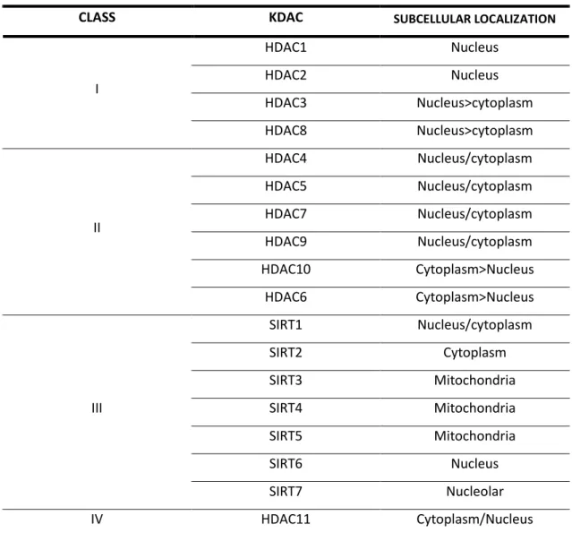
KDACs are present in all organisms, from yeast to mammals. They are mostly known as epigenetic modulators of gene expression by removing acetyl groups from Lys residues found in the N-terminal tails of nucleosomal histone proteins. In this way, chromatin compaction is favored followed by decreased levels of gene transcription. They can be divided in two major families: those with a bound Zn2+ ion (histone deacetylases, HDACs) and those dependent on NAD+ cofactor (sirtuins, SIRTs). Complementary, when regarding their structural homology, KDACs can also be divided into classes I-IV (see Table 1.) (Van Dyke, 2014).
21
Table 1 | KDACs classification and subcellular localization. (Adapted from Van Dyke M.W., 2014)
CLASS KDAC SUBCELLULAR LOCALIZATION
I HDAC1 Nucleus HDAC2 Nucleus HDAC3 Nucleus>cytoplasm HDAC8 Nucleus>cytoplasm II HDAC4 Nucleus/cytoplasm HDAC5 Nucleus/cytoplasm HDAC7 Nucleus/cytoplasm HDAC9 Nucleus/cytoplasm HDAC10 Cytoplasm>Nucleus HDAC6 Cytoplasm>Nucleus III SIRT1 Nucleus/cytoplasm SIRT2 Cytoplasm SIRT3 Mitochondria SIRT4 Mitochondria SIRT5 Mitochondria SIRT6 Nucleus SIRT7 Nucleolar IV HDAC11 Cytoplasm/Nucleus
HDACS are known to function in transcriptional repression through deacetylation of acetyl-L-lysine side chains in histone proteins, although non-histone targets have also been reported (Lombardi et al, 2011). Class I KDACs are predominantly nuclear and are ubiquitous, except for HDAC8 that is confined to smooth muscle. Meanwhile, class II KDACs show tissue specificity, being expressed mostly in the brain, heart and muscle. HDAC4, 5 and 7 translocate between nucleus and cytoplasm depending on a phosphorylation stimuli (whilst for HDAC9 this is only true for its splice variant) as HDAC6 and 10 are mostly cytosolic. HDAC11, the only member of class IV, it is largely found in the nucleus, being involved in regulation of immune tolerance (Guedes-Dias & Oliveira, 2013).
22
HDAC1 can deacetylate all four core histones (H2A, H2B, H3 and H4) but with varying efficiency. HDAC8 was reported to preferentially deacetylate histone H3 and H4, while HDAC11 might deacetylate H3 specifically at K9 and K14. Further studies are still required though to fully comprehend HDACs histone substrate specificity (Seto & Yoshida, 2014). Considering non-histone targets, HDAC8 interacts with α- -actin, increasing contractile action (Waltregny, 2005) and HDAC6 is involved in cell motility, cell adhesion and even activation of protein kinases by deacetylation of targets such as α-tubulin and Hsp90, but also participates in clearance of misfolded proteins (Liu et al, 2012).
For SIRTs, the case becomes less ambiguous. SIRTs, homologous of yeast silent information regulator 2 (Sir2) that was proven to extend replicative lifespan, are NAD+-dependent KDACs, resulting in the generation of nicotinamide and –O-acetyl- -ADP ribose after substrate deacetylation (Haigis & Guarente, 2006; Tanner et al, 2000). Mammals have 7 Sir2 homologs with a highly conserved NAD+- -dependent SIRT core domain, whilst being functionally nonredunctant. They are found in different subcellular locations – SIRT1, 6 and 7 are located mainly in the nucleus, SIRT3, 4 and 5 are mitochondrial and SIRT2 is cytoplasmic –, each containg signal sequences that explain their intracellular localization (Haigis & Sinclair, 2010; Michan & Sinclair, 2007).
SIRT1 and SIRT2 have been reported to deacetylate histones. This may seem contradictory in terms of SIRT2, being mainly cytosolic, but it was observed to localize to chromatin during cell cycle. SIRT2, in the same manner of HDAC6, can deacetylate α-tubulin. SIRT6 has a very low deacetylase activity but has a key role in telomere maintenance and DNA repair, whilst SIRT7 histone deacetylation is involved in cellular transformation in tumorigenesis (Seto & Yoshida, 2014). SIRT1 was found to regulate its own expression, and may also regulate SIRT3 expression, indirectly through regulation of PGC-1α (Bell & Guarente, 2011).
Among the large number of proteins that are known to be acetylated, a high percentage of them are of mitochondrial nature (Kim et al, 2006). Although the source for protein acetylation in mitochondria remains relatively unknown, SIRT3 acquired a prominent role in deacetylation of mitochondrial proteins in comparison to SIRT4 and SIRT5 that only show a weak deacetylase activity (Lombard et al, 2007).
23 Mitochondrial SIRTs have not been studied as extensively as SIRT1, but there is a growing interest in them and it has been suggested that they may regulate energy production, signaling and apoptosis (Verdin et al, 2010).
Deacetylation of manganese superoxide dismutase (SOD2) on K68 by SIRT3 leads to its activation, generating an antioxidant response and as such, conferring to SIRT3 an antioxidant function (Lu et al, 2015a; He et al, 2012). Besides this, SIRT3 also facilitates OXPHOS by activating complexes I and II (Finley et al, 2011). While SIRT5 and SIRT4 show weak deacetylase activity, demalonylase/desuccinylase and lipoamidase activities have been attributed, respectively (Mathias et al, 2014; Guedes-Dias & Oliveira, 2013).
As the most striking characteristic, calorie restriction was shown to correlate with extension of life span through the action of SIRTs. There is an up-regulation of oxidative metabolism, concomitant with a lowering of glycolysis (Qiu et al, 2010). This then leads to an increase in the available NAD+ levels as well as the levels of SIRT1 and SIRT3 (Guarente, 2011). Deacetylation of target proteins by SIRTs can lead to the control of metabolism and to a response against oxidative stress (Guarente, 2011; Rodgers et al, 2005). Deacetylation of components of the DNA repair machinery has also been implicated in stress responses. For instance, p53 is a target of SIRT1, keeping the balance between repair and apoptosis (Finkel et al, 2009; Haigis & Guarente, 2006).
During nutrient stress there is a shift in metabolism by action of SIRT3, promoting catabolism of acetate and fatty acids via deacetylation of several enzymes, for which, glutamate dehydrogenase (GDH) is one example (Hirschey et al, 2010). It has also been suggested that by moving away from the normal carbohydrate metabolism, there might be a decrease in ROS production, decreasing in this way oxidative stress and ameliorating aging (Guarente, 2008). In accordance to its importance, SIRT3-/- mouse embryonic fibroblasts show abnormal mitochondrial morphology, with increases in ROS levels and genomic instability (Kim
24
1.3.2. Role of KDACs in Huntington’s disease
Epigenetic dysregulation can underline cognition disorders, and thus epigenetics may play an important role in learning and memory processing. As such, epigenetic modulation through KDACs modulation seems promising as a therapy in neurodegeneration (Coppedè, 2014; Jakovcevski & Akbarian, 2012).
Morever, evidence indicates hypoacetylation of H3 at K9 and K14 in HD transgenic mice with a polyQ expansion of 82 glutamines and R6/2 mice (Valor & Guiretti, 2014; McFarland et al, 2012). Oal administration of a HDAC inhibitor in R6/2 mice after onset of motor symptoms reduced the marked H3 hypoacetylation, resulting in correction of mRNA expression levels. Additionally, treated mice displayed improved motor performance and body weight (Thomas et al, 2008). Further studies showed additional beneficial effects connected to modulation of the ubiquitin-proteasomal and autophagy (Jia et al, 2012a). Such effect was later associated by the same group to HDAC1 and HDAC3 preferential inhibition and confirmed on additional HD animal models (Jia et al, 2015, 2012b). However, knockdown of HDAC3 in R6/2 mice did not generate physiological or behavior changes with no additional transcriptional effects. As such, the beneficial effects derived from the HDAC1 and HDAC3 inhibitor may not be due to HDAC3 itself (Moumné et al, 2012).
Unselective HDAC inhibitors (trichostatin A, TSA and sodium butyrate, SB) improved Ca2+ handling after an excitotoxic-like stimuli in primary striatal neuron cultures from YAC128 mice and immortalized HD striatal cells derived from knock-in
HdhQ111 mice (Oliveira et al, 2006). Additionally, TSA was seen to increase α-tubulin
acetylation in HD cell models, further resulting in enhanced vesicle transport and BDNF release (Dompierre et al, 2007). Although this effect was justified as HDAC6- -derived, its genetic knock-out in R6/2 mice also cause α-tubulin deacetylation, while it did not in increased BDNF transport, or additional changes on behavior or physiology assessments (Bobrowska et al, 2011).
mHTT was described to interact with HDAC4. Interestingly, when HDAC4 knockdown was achieved in R6/2 and HdhQ150 mice, mHTT aggregation was delayed in cytoplasm, with no changes occurring in the nucleus. Meanwhile, reduced
25 HDAC4 levels led to restoration of membrane electrophysiological properties of MSN and corticostriatal synaptic transmission (Mielcarek et al, 2013).
When it comes to SIRTs modulation in HD, the data becomes more puzzling. Overall, SIRTs pharmacological inhibition using nicotinamide (NAM) in B6.HDR6/1 transgenic mice (that display human HTT exon 1 with 1 kB of the endogenous promoter) resulted in increased expression of BDNF and PGC-1α, with additional beneficial motor effects (Hathorn et al, 2011). Meanwhile, beneficial effects were also reported after SIRTs activation. Using Caenorhabditis elegans as an early phase HD model with 128Q, Parker and colleagues correlated increased Sir2 levels with neuronal rescue and reduced axonal aggregation. The authors went further, and tested SIRTs activator resveratrol on STHdhQ111/Q111 cells, achieving decreased cell death (Parker et al, 2005). The use of resveratrol was also seen to improve gene expression of genes involved in OXPHOS and mitochondrial biogenesis, the latter through an increase in PGC-1α activity subsequent of decreased acetylation, thus promoting overall mitochondrial function (Lagouge et al, 2006).
Resveratrol effects have been associated with SIRT1 activation, although it lacks complete specificity. A large focus has been made in regards to the possible neuroprotection derived from increased SIRT1 activation (Naia & Rego, 2015). Overexpression of SIRT1 in cortical neurons overexpressing 120Q HTT prevented mitochondrial loss, although the effect was achieved after co-transfection with PGC- -1α/β (Wareski et al, 2009). Furthermore, it appeared to cause autophagy activation in SH-SY5Y cells transfected with HTT exon 1 with 97Q, resulting in reduced polyQ aggregation (Shin et al, 2013). The observed neuroprotection was proposed to occur at the transcriptional level, namely through the activation of transcriptional factor forkhead box O3A (FOXO3a) and CREB-regulated transcription coactivator 1 (TORC1), and subsequent modulation of CREB activity (Jiang et al, 2011; Jeong et al, 2011).
Meanwhile, inhibition of SIRT2 was reported as a possible neuroprotective strategy for HD. Inhibition of SIRT2 in HD Drosophila melanogaster, C. elegans and R6/2 mice increased neuronal viability, which was accounted for by alterations in sterol biosynthesis pathway (Luthi-Carter et al, 2010). This was quickly challenged by Cattaneo’s group as the previous authors did not show data regarding sterol biosynthesis in all models and in equivalent conditions, with their hypothesis being