Electroacupuncture inhibits apoptosis in annulus fibrosis cells through
suppression of the mitochondria-dependent pathway in a rat model of cervical
intervertebral disc degradation
Jun Liao
1*, Meigui Ke
1*, Teng Xu
2and Lili Lin
11
Department of Acupuncture and Moxa and Tuina, Fujian University of Traditional Chinese Medicine,
Fujian, P.R. China.
2
Fujian Academy of Integrative Medicine, Fujian University of Traditional Chinese Medicine, Fujian,
P.R. China.
Abstract
The purpose of this study was to investigate whether treatment with electroacupuncture (EA) inhibited mitochon-dria-dependent apoptosis in annulus fibrosis (AF) cells in a rat model of cervical intervertebral disc degradation in-duced by unbalanced dynamic and static forces. Forty Sprague-Dawley rats were used in this study, of which 30 underwent surgery to induce cervical intervertebral disc degradation, 10 rats received EA at acupoints Dazhui (DU 14) and Shousanli (LI 10). TUNEL staining was measured to assess apoptosis in AF cells, immunohistochemistry was used to examine Bcl-2 and Bax expression, colorimetric assays were used to determine caspase 9 and caspase 3 activities and RT-PCR and western blotting were used to assess the mRNA and protein expression of Crk and ERK2. Treatment with EA reduced the number of AF-positive cells in TUNEL staining, increased Bcl-2-positive cells and decreased Bax-positive cells in immunohistochemical staining, significantly inhibited the activation of caspa-ses-9 and -3, and enhanced the mRNA and protein expression of Crk and ERK2. Our data show that EA inhibits AF cell apoptosis via the mitochondria-dependent pathway and up-regulates Crk and ERK2 expression. These results suggest that treatment with may be a good alternative therapy for preventing cervical spondylosis.
Key words:annulus fibrosis cells, apoptosis, electroacupuncture, mitochondria.
Received: January 13, 2012; Accepted: May 9, 2012.
Introduction
Intervertebral disc (IVD) degeneration is an impor-tant phenomenon in pathological conditions commonly seen in orthopedic practice, such as disc herniation, spinal instability and radiculopathy (Gruberet al., 2005). How-ever, the pathogenesis of IVD degeneration remains un-clear (Kasraet al., 2006; Aliet al., 2008). Cell apoptosis is one of the crucial pathological changes in disc degeneration (Zhaoet al., 2006; Joneset al., 2008; Tschoekeet al., 2008; Weiet al., 2008). The existence of an apoptotic signal path-way in annulus fibrosis (AF) cells remains controversial. Previous studies of AF focused on the period after AF had ruptured, i.e, when IVD degeneration had occurred, and the methods used to produce AF degeneration cincluded surgi-cal ablation of IVD and biologisurgi-cal approaches such as mo-lecular therapy (Kimet al., 2003; Neidlinger-Wilkeet al., 2009), gene therapy (Moonet al., 2000, 2008; Yoonet al.,
2004), cell transplantation (Sakaiet al., 2005), tissue engi-neering (Mizunoet al., 2004; O’Halloran DM and Pandit, 2007) and traditional Chinese medicine such as acupunc-ture.
Acupuncture is recognized by the World Health Or-ganization as an effective treatment for pain relieve and for cervical spondylosis. Dazhui (DU 14) and Shousanli (LI 10), which belong to the DU channel and Large Intestine channel, respectively, can dredge meridians and free collaterals, free Qi and stop pain, promote blood circulation and eliminate blood stasis, and are often used for neck stiff-ness, shoulder pain, upper limb paralysis and lumbar pain. However, the precise mechanism involved in these benefi-cial effects remains to be elucidated. To date, no study has investigated apoptosis in AF before IVD degeneration. The aims of this study were therefore to investigate the occur-rence of apoptosis in AF before IVD degeneration, to deter-mine the apoptotic pathway involved, to exadeter-mine the relationship between apoptosis and Crk and ERK2 expres-sion and to assess the usefulness of electroacupuncture (EA) for treating cervical intervertebral disc degeneration in rats.
Send correspondence to Jun Liao. Department of Acupuncture and Moxa and Tuina, Fujian University of Traditional Chinese Medicine, 1 Huatuo Road, Minhou Shangjie, 350108 Fuzhou, Fujian, P.R. China. E-mail: [email protected].
Materials and Methods
Forty Sprague-Dawley rats (one month old) were pro-vided by the Shanghai Laboratory Animal Center and all animal procedures were approved by the Ethical Commit-tee of Fujian University of Traditional Chinese Medicine. The rats were maintained on a 12 h light/dark cycle at 21±2 °C with free access to food and tap water.
Prior to use, the rats were randomly assigned to two groups: a sham group (n = 10; 5 males and 5 females) in which the rats did not undergo any surgery and a surgical group (n = 30; 15 males and 15 females) that underwent surgery to induce cervical IVD degradation through unbal-anced dynamic and static forces, as described by Wanget al.(2006). After observation for seven days, the 30 rats that have accepted surgery were randomly allocated into three groups of 10 rats (5 males and 5 females): a control group that was handled identically to the other groups but without acupuncture or electrical treatment, a group treated with meloxicam tablets (MT; Boehringer Ingelheim Corpora-tion, Germany) that served as a positive control and a group treated with EA. For the EA protocol, rats were kept in spe-cially designed holders with their necks and limbs exposed. Acupuncture needles were inserted in turn to depths of ap-proximately 3 mm at acupoint Dazhui (DU 14) and approx-imately 1 mm at acupoint Shousanli (LI 10) bilaterally (Zhongren Li, 2003) and the rats then stimulated electri-cally (1 mA in intensity at 2/100 Hz) using a HANS EA In-strument (Model No. 100A, Shijiazhuang Fusai Medical Devices Ltd., China). The EA treatment was applied for 30 min once a day over 14 days (a complete course) with a two-day interval between two courses. In the MT group, meloxicam (0.75 mg/kg) was administered intragastrically for 30 days. All of these rats were euthanized with pento-barbitone sodium (Nembutal®; 100 mg/kg, i.p.; Boehringer Ingelheim, Artarmon, NSW, Australia) and the cervical spines were harvested for analysis.
TUNEL assay for apoptosis
For the quantitative analyses of apoptosis, sections from paraffin-embedded AFs were processed for terminal deoxynucleotidyl transferase-mediated dUTPFITC nick end-labeling (TUNEL) by using anin situapoptosis detec-tion kit (Wako Pure Chemical Industries, Ltd. Osaka, Ja-pan). The assay was done according to the manufacturer’s instructions, with minor modifications. TUNEL-positive cells were scored in viable regions peripheral to areas of ne-crosis in AF sections. The number of TUNEL-positive cells was counted in five random high-power (x400) fields in AF sections from each rat.
Immunohistochemical staining for Bcl-2 and Bax
The slides were processed using standard protocols for deparaffinization and rehydration. Endogenous pero-xidase activity was blocked by incubating the sections with
3% H2O2for 10 min followed by digestion with 0.01%
pro-tease K for 10 min. Non-specific binding sites were blocked by incubation with confining liquid for 10 min after which the sections were incubated with rat polyclonal antibody to Bcl-2 or Bax (Cell Signaling Inc., Danvers, MA) at 4 °C for 12 h. After thorough washing, the sections were incubated with biotinylated goat anti-rabbit IgG at 4 °C for 60 min and then in Streptavidin-HRP for 10 min. The final color reac-tion was developed by incubareac-tion with the chromogenic substrate 3,3’-diaminobenzidine (0.5 mg/mL in Tris). The sections were counterstained with hematoxylin and mounted for examination with an Olympus BX50 micro-scope coupled to an Image Analysis System (Olympus).
Caspase activities
The activities of caspases 3 and 9 were determined by a colorimetric assay using caspase 3 and 9 activation kits (Invitrogen), according to the manufacturer’s instructions. Briefly, AF samples were lysed in lysis buffer for 30 min on ice. The lysed cells were centrifuged at 16,000 x g for 10 min and 100mg of protein was incubated with 50mL of the colorimetric tetrapeptide Asp-Glu-Val-Asp (DEAD)-p-nitroaniline (pNA) (specific substrate of caspase 3) or Leu-Glu-His-Asp (LEHD)-pNA (specific substrate for cas-pase 9) at 37 °C in the dark for 2 h after which the plates were read at 405 nm in an ELISA reader (Model EXL800, BioTek, USA). The data were normalized to the caspase ac-tivities in control cells (treated with 0.5% DMSO vehicle) and expressed as the fold increase.
RNA extraction and RT-PCR analysis
Total RNA from AF samples was extracted with TRIzol reagent (Sigma, St. Louis, MO) according to the manufacturer’s protocol. Oligo(dT)-primed RNA (1 mg) was reverse-transcribed with SuperScript II reverse tran-scriptase (Promega) according to the manufacturer’s in-structions. The resulting cDNA was used to determine the amount of ERK2 or Crk mRNA by PCR withTaq DNA polymerase (Fermentas). GAPDH was used as an internal control. The primers used for amplification were: ERK2 forward 5’-TCCAACCTGCTGCTCAACACCAC-3’ and reverse 5’-CACTCGGGTCGTAATACTGCTCC-3’; CRK forward 5-ACTATGTGCTCAGCGTCTCA-3’ and reverse 5’-ATTCCACCACTGCTCTTCA-3’, and GAPDH forward 5’-GTCACCATGACAACTTTGG -3’ and reverse 5’-GAGCTTGACAAAGTGGTCGT-3’.
Western blotting
pro-tein assay kit (Bio-Rad). 30mg of protein exact were sepa-rated by SDS-PAGE and then transferred to a PVDF membrane (NEN Life Science Products Inc., Boston, MA). The blots were probed overnight at 4 °C with either rabbit anti-rat Crk or ERK2 (Abcam, Cambridge, MA). After washing, the membranes were incubated for 1 h at 20 °C with goat anti-rabbit IgG (Oncogen, Boston, MA). Immune complexes were detected using ECL (Amersham, Pisca-taway, NJ). Membranes were reprobed for GAPDH to con-firm equal protein loading.
Results
TUNEL staining
The effect of EA treatment on apoptosis in AF cells was assessed by TUNEL staining. This procedure allows the detection and quantification of apoptosis at a cellular level based on the labeling of free 3-OH terminals created during double-strand and single-strand cleavage of ge-nomic DNA. Figure 1A-D shows the TUNEL staining in the four experimental groups. Qualitatively, TUNEL stain-ing was more intense in the cell nuclei of the control group than in the sham group. In the former group, the nuclei were denser, there was disruption of the nuclear membrane and
apoptotic bodies were seen in the cytosol; cells in the EA group showed a relatively intact nuclear membrane. The number of TUNEL-positive cells in the control group was significantly higher than in the sham group, and the number of TUNEL-positive cells in the EA group was significantly lower than the control group but similar to the sham and MT groups (Figure 1E).
Immunohistochemical staining for Bcl-2 and Bax
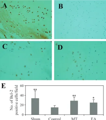
Bcl-2 family proteins are key regulators of mitochon-dria-mediated apoptosis and include anti-apoptotic mem-bers such as Bcl-2 and pro-apoptotic memmem-bers such as Bax. Bcl-2 has been implicated in the inhibition of apoptosis. In the present study, Bcl-2-positive cells were detected mainly in the sham, EA and MT groups, although positively stained cells were also seen in the control group (Figure 2A-D). There was a significant increase in the number of Bcl-2-positive cells in the EA group compared to the con-trol group (Figure 2E) and this could explain the decreased apoptosis seen in the former group (Figure 1E).
Unlike Bcl-2, which is anti-apoptotic, Bax induces apoptosis in several cell lines. Bax immunoreactivity was detected mainly in the control group (Figure 3B). Quantita-tive analysis revealed a significant difference in the number of Bax-positive cells in the control group compared to the other groups (Figure 3E).
Figure 1- Effect of EA on TUNEL staining of AF cells. (A) Sham group, (B) Control group, (C) MT group and (D) EA group. (E) Number of TUNEL-positive cells/field in each experimental group. Note the decrease in the number of TUNEL-positive cells in the EA group compared to the control group. The columns represent the mean±SD (n = 8). **p < 0.01 compared to the control group. Magnification: x400.
Assay for caspases 3 and 9
To identify the downstream effectors in the apoptotic signaling pathway, the activation of caspases 9 and 3 was examined by using specific chromophores, i.e., DEVD-pNA (a specific substrate for caspase 3) and LEHD-DEVD-pNA (a specific substrate for caspase 9). The mitochondria- de-pendent pathway is the most common apoptotic pathway in vertebrate cells. Permeabilization of the mitochondrial membrane, accompanied by the collapse of the electro-chemical gradient across the mitochondrial membrane, is one of the key events during cellular apoptosis. This event leads to the release of numerous apoptogenic proteins, such as cytochrome c, from mitochondria, thereby triggering the activation of caspases 3 and 9 and eventually inducing apoptosis. As shown in Figure 4A,B, treatment with EA significantly inhibited the activation of caspases 3 and 9 in AF cells. These data suggest that EA inhibits apoptosis in AF cells, probably by blocking the mitochondria-depen-dent pathway.
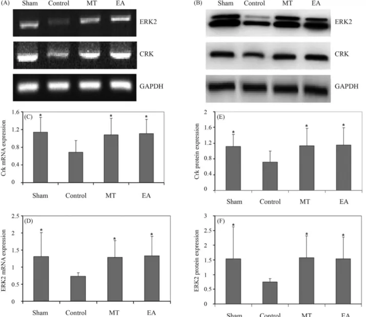
EA modulates the expression of Crk and ERK2
To enhance our understanding of the anti-apoptotic activity of EA, we used RT-PCR and western blotting to
examine the mRNA and protein expression of Crk and ERK2 in EA-treated AF cells. Figure 5A,C and D shows that Crk and ERK2 gene expression in the control group was lower than in the sham group and that EA treatment in-creased the mRNA expression of these two proteins; the pattern of Crk and ERK2 protein expression was similar to that of their respective mRNA levels (Figure 5B,E and F).
Discussion
Previous studies have shown that apoptosis in AF cells plays a key role in IVD degeneration. Degenerative changes in the IVD can lead to nerve root compression, re-sulting in radiculopathy (Hacker and Miller, 2003; Chenet al., 2005). To investigate whether electroacupuncture pro-tected AF cells from apoptosis caused by cervical IVD de-generation, we established a rat model of cervical inter-vertebral disc degradation induced by the application of unbalanced dynamic and static forces, as described in the literature (Wanget al., 2006). TUNEL staining showed that EA treatment could reduce the signs of apoptosis after sur-gery.
Apoptosis is a complex process by which individual cells undergo self-destruction without inducing an inflam-matory response. Caspases, which belong to a family of cysteine proteases, are the key proteins that modulate the apoptotic response. These enzymes occur in a latent form in the cytoplasm and are activated late in the apoptotic
pro-Figure 3- Effect of EA on immunohistochemical staining for Bax in AF cells. (A) Sham group, (B) Control group, (C) MT group and (D) EA group. AF cells were stained with anti-Bax antibody. Note the decrease in the number of Bax-positive cells in the EA group compared to the control group. The columns represent the mean±SD (n = 8). *p < 0.05 and **p < 0.01 compared to the control group. Magnification: x400.
cess. Caspases are organized as a cascade with two major pathways for activation, namely, the mitochondria-me-diated pathway and the fas-memitochondria-me-diated pathway (Tsujimoto and Shimizu, 2000; Pedersenet al., 2002). Caspase 3 is a key mediator of apoptosis that is activated by an initiator caspase such as caspase 9 during mitochondria-mediated apoptosis. As shown here, EA suppressed the activation of caspases 9 and 3 in AF cells and lead to a reduction in the activities of these two enzymes in these cells.
Mitochondria-dependent apoptosis is regulated mainly by Bcl-2 family proteins, a group of evolutionarily conserved pro- and anti-apoptotic proteins. Bcl-2 is an anti-apoptotic protein that forms channels that stabilize the mitochondrial membrane, thereby preventing the release of cytochrome c, a second mitochondria-derived activator of
caspases. Bax is a pro-apoptotic protein that forms heterodimers with Bcl-2, thereby inactivating the latter. Mitochondrial outer membrane permeabilization (MOMP) is thought to involve the formation of pores in the mito-chondrial membrane by pro-apoptotic Bax-like proteins; the action of these proteins can be inhibited by anti-apoptotic Bcl-2-like members (Reed,2000; Cory and Adams,2002). The ratio of Bax to Bcl-2 is therefore critical in determining the fate of cells. The results described here show that EA treatment increased the number of Bcl-2-positive cells and reduced the number of Bax-positive AF cells. This finding indicates that EA inhib-its apoptosis by affecting the Bax/Bcl-2 ratio.
We examined whether changes in the expression of Crk and ERK2 were related to the suppression of apoptosis.
Integrins are adhesion receptors that transmit signals from the extracellular matrix to cells. Of the numerous signals emanating from integrin receptors, Crk-associated sub-strate and its recruitment of and binding to the adaptor pro-tein Crk are critical events in controlling integrin-dependent processes (Schwartzet al., 1995; Schlaepfer and Hunter, 1998). The amplification of Crk signaling in epi-thelial cells and fibroblasts suppresses apoptotic mecha-nisms and may itself be a transforming factor (Cho and Klemke,2000; Iwaharaet al., 2003). Activation of mem-bers of the mitogen-activated protein (MAP) kinase family by Crk plays a key role in regulating chondrocyte gene ex-pression.
The Ras-dependent extracellular signal-regulated ki-nase 1/2 (ERK1/2) pathway also plays a central role in con-trolling cell proliferation (Meloche and Pouyssegur,2007). The ERK pathway promotes the transcription of cyclin D (Albanese et al., 1995) and c-Myc, and induces protea-somal degradation of FOXO3a (Yang et al., 2008) and p21Cip1 (Hwang et al., 2009) through direct phosphoryl-ation, thereby resulting in cellular proliferation. As shown here for the first time, EA at DU 14 and LI 10 increased the gene and protein expression of Crk and ERK2 in cervical IVD degeneration; the pattern of protein expression agreed with that seen for mRNA. These data indicate that Crk and ERK2 signaling may be associated with AF cell apoptosis during cervical IVD degeneration. However, it is unclear whether these molecules are involved in the signaling cas-cade of programmed cell death or whether their enhanced expression is simply a non-specific response to apoptosis. Further studies are needed to elucidate the specific role of these molecules in the apoptotic pathway (Nurminskayaet al., 1998; Koikeet al., 2003).
Recent work has suggested that EA stimulation can suppress apoptosis induced by surgical trauma stress, pos-sibly by modulating Fas protein expression (Wanget al., 2005), and improve ulcerative colitis in rats, perhaps by promoting neutrophil apoptosis and down-regulating cyto-kine production by monocytes (Wu et al., 2007). Some studies have also shown that pretreatment with EA can sig-nificantly attenuate neuronal apoptosis, preserve neuronal morphology and inhibit caspase 3 activity in the hippo-campal CA1 region after exposure to +Gz (Wanget al., 2010). EA may also stimulate endogenousxPKC-mediated anti-apoptosis pathways to protect against ischemic dam-age after focal cerebral ischemia caused by the activation of cannabinoid receptor type 1 (Wanget al., 2011).
In conclusion, our data demonstrate that EA treat-ment at DU 14 and LI 10 inhibits AF cell apoptosis via the mitochondria-dependent pathway and up-regulates Crk and ERK2. These results suggest that EA may be a good alter-native therapy for preventing cervical spondylosis.
Acknowledgments
The study was supported by the National Natural Sci-ence Foundation of China (grant nos. 30901934 and 81001554), the New Teachers’ Fund for Doctor Stations, Ministry of Education (grant no. 20113519120003) and the Educational Office of Fujian Province (grant no. JK2010028).
References
Albanese C, Johnson J, Watanabe G, Eklund N, Vu D, Arnold A and Pestell RG (1995) Transforming p21ras mutants and c-Ets-2 activate the cyclin D1 promoter through distinguish-able regions. J Biol Chem 270:23589-23597.
Ali R, Le Maitre CL, Richardson SM, Hoyland JA and Freemont AJ (2008) Connective tissue growth factor expression in hu-man intervertebral disc: Implications for angiogenesis in intervertebral disc degeneration. Biotech Histochem 83:239-245.
Chen B, Fellenberg J, Wang H, Carstens C and Richter W (2005) Occurrence and regional distribution of apoptosis in sco-liotic discs. Spine 30:519-524.
Cho SY and Klemke RL (2000) Extracellular-regulated kinase ac-tivation and CAS/Crk coupling regulate cell migration and suppress apoptosis during invasion of the extracellular ma-trix. J Cell Biol 149:223-236.
Cory S and Adams JM (2002) The Bcl-2 family: Regulators of the cellular life-of-death switch. Nat Rev Cancer 2:647-656. Gruber HE, Norton HJ, Ingram JA and Hanley Jr EN (2005) The
SOX9 transcription factor in the human disc: Decreased immunolocalization with age and disc degeneration. Spine 30:625-630.
Hacker RJ and Miller CG (2003) Failed anterior cervical fora-minotomy. J Neurosurg Spine 98:126-130.
Hwang CY, Lee C and Kwon KS (2009) Extracellular signal-regulated kinase 2-dependent phosphorylation induces cyto-plasmic localization and degradation of p21Cip1. Mol Cell Biol 29:3379-3389.
Iwahara T, Akagi T, Shishido T and Hanafusa H (2003) CrkII in-duces serum response factor activation and cellular transfor-mation through its function in Rho activation. Oncogene 22:2946-5957.
Jones P, Gardner L, Menage J, Williams GT and Roberts S (2008) Intervertebral disc cells as competent phagocytesin vitro: Implications for cell death in disc degeneration. Arthritis Res Ther 10:R86.
Kasra M, Merryman WD, Loveless KN, Goel VK, Martin JD and Buckwalter JA (2006) Frequency response of pig inter-vertebral disc cells subjected to dynamic hydrostatic pres-sure. J Orthop Res 24:1967-1973.
Meloche S and Pouyssegur J (2007) The ERK1/2 mitogen-acti-vated protein kinase pathway as a master regulator of the G1-to S-phase transition. Oncogene 26:3227-3239. Mizuno H, Roy AK, Vacanti CA, Kojima K, Ueda M and
Bonas-sar LJ (2004) Tissue-engineered composites of anulus fibrosus and nucleus pulposus for intervertebral disc re-placement. Spine 29:1290-1297.
Moon SH, Gilbertson LG, Nishida K, Knaub M, Muzzonigro T, Robbins PD, Evans CH and Kang JD (2000) Human inter-vertebral disc cells are genetically modifiable by adeno-virus-mediated gene transfer: Implications for the clinical management of intervertebral disc disorders. Spine 25:2573-2579.
Moon SH, Nishida K, Gilbertson LG, Lee HM, Kim H, Hall RA, Robbins PD and Kang JD (2008) Biologic response of hu-man intervertebral disc cells to gene therapy cocktail. Spine 33:1850-1855.
Neidlinger-Wilke C, Liedert A, Wuertz K, Buser Z, Rinkler C, Käfer W, Ignatius A, Claes L, Roberts S and Johnson WE (2009) Mechanical stimulation alters pleiotrophin and aggrecan expression by human intervertebral disc cells and influences their capacity to stimulate endothelial migration. Spine 34:663-669.
Nurminskaya M, Magee C, Nurminsky D and Linsenmayer TF (1998) Plasma transglutaminase in hypertrophic chondro-cytes: Expression and cell-specific intracellular activation produce cell death and externalization. J Cell Biol 142:1135-1144.
O’Halloran DM and Pandit AS (2007) Tissue-engineering ap-proach to regenerating the intervertebral disc. Tissue Eng 13:1927-1945.
Pedersen IM, Kitada S, Schimmer A, Kim Y, Zapata JM, Char-boneau L, Rassenti L, Andreeff M, Bennett F, Sporn MB,et al.(2002) The triterpenoid CDDO induces apoptosis in re-fractory CLL B cells. Blood 100:3541-3553.
Reed JC (2000) Mechanisms of apoptosis. Am J Pathol 157:1415-1430.
Sakai D, Mochida J, Iwashina T, Watanabe T, Nakai T, Ando K and Hotta T (2005) Differentiation of mesenchymal stem cells transplanted to a rabbit degenerative disc model: Po-tential and limitations for stem cell therapy in disc regenera-tion. Spine 30:2379-2387.
Schlaepfer DD and Hunter T (1998) Integrin signalling and tyro-sine phosphorylation: Just the FAKs? Trends Cell Biol 8:151-157.
Schwartz MA, Schaller MD and Ginsberg MH (1995) Integrins: Emerging paradigms of signal transduction. Annu Rev Cell Dev Biol 11:549-599.
Tschoeke SK, Hellmuth M, Hostmann A, Robinson Y, Ertel W, Oberholzer A and Heyde CE (2008) Apoptosis of human intervertebral discs after trauma compares to degenerated discs involving both receptor-mediated and mitochondrial-dependent pathways. J Orthop Res 26:999-1006.
Tsujimoto Y and Shimizu S (2000) Bcl-2 family: Life-or-death switch. FEBS Lett 466:6-10.
Wang H, Peng Y, Wang L, Lu Y, Shi T and Xiong L (2010) Electroacupuncture pretreatment ameliorates hypergravity-induced impairment of learning and memory and apoptosis of hippocampal neurons in rats. Neurosci Lett 478:150-155. Wang J, Wang YQ, Yu J, Cao XD and Wu GC (2005)
Elec-troacupuncture suppresses surgical trauma stress-induced lymphocyte apoptosis in rats. Neurosci Lett 383:68-72. Wang Q, Li X, Chen Y, Wang F, Yang Q, Chen S, Min Y, Li X
and Xiong L (2011) Activation of epsilon protein kinase C-mediated anti-apoptosis is involved in rapid tolerance in-duced by electroacupuncture pretreatment through canna-binoid receptor type 1. Stroke 42:389-396.
Wang YJ, Shi Q, Lu WW, Cheung KC, Darowish M, Li TF, Dong YF, Zhou CJ, Zhou Q, Hu ZJ,et al.(2006) Cervical inter-vertebral disc degeneration induced by unbalanced dynamic and static forces: A novel in vivo rat model. Spine 31:1532-1538.
Wei A, Brisby H, Chung SA and Diwan AD (2008) Bone mor-phogenetic protein-7 protects human intervertebral disc cellsin vitrofrom apoptosis. Spine 8:466-474.
Wu HG, Liu HR, Tan LY, Gong YJ, Shi Y, Zhao TP, Yi Y and Yang Y (2007) Electroacupuncture and moxibustion pro-mote neutrophil apoptosis and improve ulcerative colitis in rats. Dig Dis Sci 52:379-384.
Yang JY, Zong CS and Xia W (2008) ERK promotes tumo-rigenesis by inhibiting FOXO3a via MDM2-mediated deg-radation. Nat Cell Biol 10:138-148.
Yoon ST, Park JS, Kim KS, Li J, Attallah-Wasif ES, Hutton WC and Boden SD (2004) ISSLS prize winner: LMP-1 upre-gulates intervertebral disc cell production of proteoglycans and BMPsin vitroandin vivo. Spine 29:2603-2611. Zhao CQ, Jiang LS and Dai LY (2006) Programmed cell death in
intervertebral disc degeneration. Apoptosis 11:2079-2088. Zhongren Li (2003) Experimental Acupuncture and Moxibustion
Science. China Press of Traditional Chinese Medicine, Beijing, pp 327-329.
Associate Editor: Carlos R. Machado