Received 8 Feb 2015|Accepted 7 Apr 2015|Published 21 May 2015
Major histocompatibility complex associations
of ankylosing spondylitis are complex and involve
further epistasis with
ERAP1
Adrian Cortes1, Sara L. Pulit2, Paul J. Leo1, Jenny J. Pointon3, Philip C. Robinson1, Michael H. Weisman4, Michael Ward5, Lianne S. Gensler6, Xiaodong Zhou7, Henri-Jean Garchon8,9, Gilles Chiocchia8, Johannes Nossent10,11, Benedicte A. Lie12,13, Øystein Førre14, Jaakko Tuomilehto15,16,17, Kari Laiho18, Linda A. Bradbury1, Dirk Elewaut19,20, Ruben Burgos-Vargas21, Simon Stebbings22, Louise Appleton3, Claire Farrah3, Jonathan Lau3, Nigil Haroon23, Juan Mulero24, Francisco J. Blanco25, Miguel A. Gonzalez-Gay26, C. Lopez-Larrea27,28, Paul Bowness3, Karl Gaffney29, Hill Gaston30, Dafna D. Gladman31,32,33, Proton Rahman34, Walter P. Maksymowych35, J. Bart A. Crusius36, Irene E. van der Horst-Bruinsma37, Raphael Valle-On˜ate38, Consuelo Romero-Sa´nchez38, Inger Myrnes Hansen39, Fernando M. Pimentel-Santos40, Robert D. Inman23, Javier Martin41, Maxime Breban8,42, Bryan Paul Wordsworth3, John D. Reveille7, David M. Evans1,43,44, Paul I.W. de Bakker2,45
& Matthew A. Brown1
Ankylosing spondylitis (AS) is a common, highly heritable, inflammatory arthritis for which
HLA-B*27is the major genetic risk factor, although its role in the aetiology of AS remains elusive.
To better understand the genetic basis of the MHC susceptibility loci, we genotyped 7,264 MHC SNPs in 22,647 AS cases and controls of European descent. We impute SNPs, classical HLA alleles and amino-acid residues within HLA proteins, and tested these for association to AS status. Here we show that in addition to effects due toHLA-B*27alleles, several otherHLA-B
alleles also affect susceptibility. After controlling for the associated haplotypes in HLA-B, we observe independent associations with variants in theHLA-A,HLA-DPB1andHLA-DRB1loci. We also demonstrate that theERAP1SNP rs30187 association is not restricted only to carriers of
HLA-B*27but also found inHLA-B*40:01carriers independently of HLA-B*27 genotype.
DOI: 10.1038/ncomms8146 OPEN
A
nkylosing spondylitis (AS) is a common, highly heritable1,inflammatory arthritis for whichHLA-B*27is the major
genetic risk factor. To better understand the genetic basis of the major histocompatibility complex (MHC) susceptibility loci, we genotyped 7,264 MHC single-nucleotide polymorphisms (SNPs) in 9,069 AS cases and 13,578 population controls of European descent using the Illumina Immunochip microarray. In
addition to extremely strong effects due to HLA-B*27:02 and
B*27:05, several otherHLA-B alleles (B*07:02, B*13:02, B*40:01,
B*40:02,B*47:01,B*51:01andB*57:01) also affect susceptibility to AS. HLA-B-independent associations were demonstrated with
variants in theHLA-A,HLA-DPB1andHLA-DRB1loci. We also
demonstrate that the ERAP1 SNP rs30187 association is not
restricted only to carriers ofHLA-B*27 but also found in
HLA-B*40:01 carriers independently of theHLA-B*27 genotype. The presence of associations in both HLA class I and II loci might
reflect effects on antigen presentation to both CD4þ and CD8þ
T lymphocytes in the pathogenesis of AS.
While the classicalHLA-B*27allele is found in over 85% of AS patients2–4, it is clearly not sufficient alone to cause disease, with
only 1–5% of HLA-B*27 carriers developing the disease. From
epidemiological data, it is evident that susceptibility to AS is affected by other genes within and outside the MHC1. Indeed, 26 risk loci outside the MHC have now been identified by genome-wide association studies5–8.
The biological mechanism(s) by which HLA-B27 confers risk of disease remains elusive. The main hypotheses regarding this mechanism can be divided into canonical mechanisms based on the known function of HLA-B27 within the adaptive immune system, and non-canonical mechanisms related to unusual properties of HLA-B27, notably its propensity to dimerise or misfold. Suggested canonical mechanisms propose either that HLA-B27 is uniquely capable of presenting particular peptide(s) found at sites of inflammation in AS to cytotoxic T lymphocytes
(the arthritogenic peptide hypothesis)9 or that HLA-B27 is
associated with reduced gut mucosal immunity, leading to migration of enteric bacteria across the intestinal mucosa, driving the production of the pro-inflammatory cytokine interleukin (IL)-23 and development of AS (the mucosal
immunodeficiency hypothesis)10,11. Both these theories place
antigenic peptide presentation and handling as critical steps in the pathogenesis of AS. One of the first non-MHC susceptibility loci to be identified in AS was endoplasmic reticulum aminopeptidase 1 (ERAP1)5, the main function of which is to trim peptides in the endoplasmic reticulum (ER) to optimal length for binding to MHC class I molecules on antigen-presenting cells for subsequent interaction with CD8þ T cells12,13. Moreover, this association is so far uniquely found inHLA-B*27-positive disease7. HLA-B27 has an unusual property of forming homodimers through disulphide bonding of the unpaired cysteine residue at position 67 (ref. 14). It has been proposed that these homodimers may cause AS through abnormal presentation of peptides or by facilitating ‘abnormal’ interaction with natural killer cells15. Apart fromHLA-B*27, the subtypes of the allelesHLA-B*14,HLA-B*15,
HLA-B*38,HLA-B*39andHLA-B*75encode a cysteine residue at position 67 but of these there is only evidence thatHLA-B*14may be AS associated16,17. It is also unclear if these other non-HLA-B27 Cys67 variants can form homodimers. In addition, Cys67 is found on all B27 subtypes, including the subtypes
HLA-B*27:06 and HLA-B*27:09, which are not AS associated18,19. A
further hypothesis suggests that abnormal folding of the HLA-B27 molecule during assembly results in ER stress and activation of the unfolded protein response20,21. ER stress is evident in the
HLA-B*27-transgenic rat model of AS and correlates with production of IL-23 (ref. 21), but has not been demonstrated in
HLA-B*27-positive patients22–24.
While non-B27 HLA associations have been reported, notably withHLA-B40(refs 25–27) andHLA-A*02(ref. 8), most have not been definitive or replicated in independent studies. In this study, we analyse the associations of AS across the MHC aiming to identify functional and potentially causal variants using a large, previously reported, panel of cases and controls of European ancestry8. Here we extend on our primary analysis of this cohort by fine mapping the MHC region with imputation of SNPs, MHC class I and II classical alleles, and amino-acid residues within the classical HLA proteins28. In addition to HLA-B27, we identify further HLA-B and other HLA class I and II alleles associated with AS, and demonstrate that HLA-B40 in addition to HLA-B27 interacts withERAP1to cause disease. This implicates both CD4 and CD8 lymphocytes in AS pathogenesis and suggests that HLA-B40 and HLA-B27 operate by similar mechanisms to induce the disease.
Results
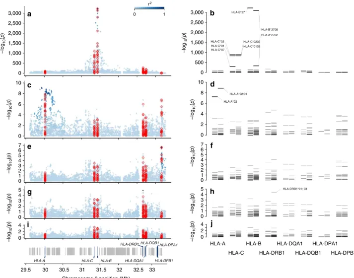
HLA-B susceptibility alleles. At the HLA-B locus, 38 classical alleles at four-digit resolution were imputed. All SNP, HLA and
amino-acid association P-values were determined by logistic
regression. As expected, the two common HLA-B*27 alleles
in the European population, B*27:02 (odds ratio (OR)¼43;
P¼1.0710122) andB*27:05(OR¼62;Po10321), were the
most significantly associated with disease risk (Fig. 1a–b; Tables 1 and 2). Controlling for the effect of the twoB*27alleles,
we identified the protective alleles HLA-B*07:02 (OR¼0.82;
P¼5.04106) andHLA-B*57:01(OR¼0.75;P¼5.13104; Table 2). Moderate association was also observed, sequentially, with the risk alleles HLA-B*51:01(OR¼1.33; P¼2.14103),
HLA-B*47:01 (OR¼2.35; P¼2.25103), HLA-B*40:02
(OR¼1.59; P¼4.65103), HLA-B*13:02 (OR¼1.43; P¼
4.29103) and HLA-B*40:01 (OR¼1.22; P¼4.93103).
No evidence of further susceptibility alleles was observed after controlling for the risk and protective alleles identified above (P40.05; Fig. 1d). TheHLA-Bassociations were similar in both
HLA-B*27-positive and HLA-B*27-negative restricted analyses (Supplementary Tables 1–6).
Non-HLA-Bsusceptibility loci in the MHC. To assess whether other MHC loci affect disease susceptibility independently from theHLA-B locus, we performed additional conditional analyses.
Adjusting for the HLA-B susceptibility alleles identified, we
observed an association signal with SNPs in the HLA-A locus
(rs2975033; OR¼1.22;P¼6.161010) and with the classical
allele HLA-A*02:01 (OR¼1.22; P¼1.41109; Fig. 1c–d).
The risk allele ‘A’ of rs2975033 was in near perfect linkage disequilibrium with the risk alleleHLA-A*02:01(r2¼0.97).
Further controlling for the effect of the susceptibility SNP
rs2975033 inHLA-A revealed an independent signal with SNPs
(rs1126513; OR¼1.21; P¼2.46107) in the class II locus
HLA-DPB1(Fig. 1e); no association of similar strength to those
seen with SNPs (P4105) were observed with classical
HLA-DPB1 alleles (Fig. 1f). After controlling for the effect of the SNP
rs1126513 in HLA-DPB1, we observed an association with the
SNP rs17885388 (OR¼1.16;P¼1.27105) in theHLA-DRB1
locus, and a similar level of significance was also observed with the class II alleleHLA-DRB1*01:03(P¼3.78105). No further associations were observed after controlling for all identified effects (P45105; Fig. 1i–j).
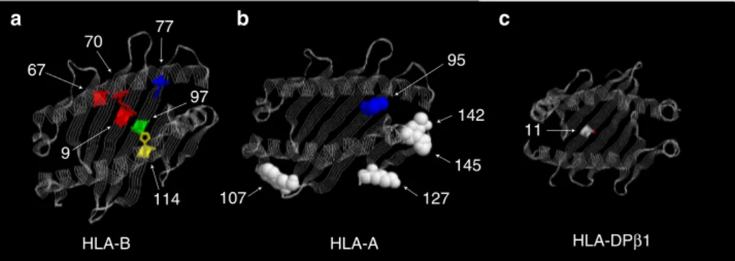
specificity of the peptides presented to CD8þ and CD4þ T lymphocytes. We, therefore, analysed the polymorphic amino-acid residues at these proteins to assess their effect in disease susceptibility. In this analysis, the strongest association was observed for amino-acid position 97 in HLA-B (omnibus
Po103221; Table 1; Fig. 1a–b). In addition, through conditional
analysis, we found that the association at the HLA-B*27 allele,
and other HLA-B*27-associated polymorphisms, was explained
by position 97 while the reverse was not true (Supplementary Table 7). This polymorphic position carries as many as six different amino-acid residues in the population (Fig. 2), each conferring a different degree of risk (or protection) to disease, consistent with the analysis ofHLA-B alleles mentioned above (Table 2). Position 97 lies in the floor of the HLA-B peptide-binding groove (Fig. 3), located in the C/F pocket, also referred as the C-terminal pocket, which anchors the side chain of the C-terminal peptide residue29. Asparagine at position
97 is uniquely observed in HLA-B*27 alleles. Threonine at
position 97 (predominantly found inHLA-B*51alleles) was also
found to increase disease risk (OR¼1.12; P¼4.50103);
serine (found in HLA-B*07 and *08 alleles) decreased risk of
disease (OR¼0.86; P¼5.2108); and valine (found in
HLA-B*57 alleles) was also protective (OR¼0.68;
P¼1.4108; Table 3).
Strong associations were also observed with the amino-acid positions 70, 114, 77 and 67 of HLA-B but these signals were strongly attenuated after conditioning on amino-acid position 97. In contrast, none of these positions could explain the association at position 97. In particular, there was little evidence of association at position 67 (that is, the position where disulphide bonding of unlinked cysteine residues might occur) after
conditioning on position 97 (P-value¼0.04; Supplementary
Table 8).
The most strongly associated amino acid of the HLA-A molecule, after conditioning on associated HLA-B alleles,
was amino acid valine at position 95 (P¼3.70109).
HLA-A HLA-C HLA-B
HLA-DRB1
Chromosome 6 posiition (Mb) HLA-DQA1
HLA-DQB1
HLA-DPB1
HLA-DPA1 HLA-A HLA-C
HLA-B
HLA-DRB1 HLA-DQA1
HLA-DQB1 HLA-DPA1
HLA-DPB1 3,000
2,500
2,000
1,500
1,000
500
0
10
8
6
6 4
4 2
2 0
0 1 3 5
4
4 2
2 0
0
29.5 30 30.5 31 31.5 32 32.5 33 1
3 5 7
3,000
–log
10
(
p
)
–log
10
(
p
)
–log
10
(p)
–log
10
(p)
–log
10
(p)
2,500
2,000
1,500
1,000
500
0
10
8
6
6 4
4 2
2 0
0
HLA-DRB1*01: 03 HLA-A*02:01
HLA-C*02 HLA-C*01 HLA-C*07
HLA-C*0202 HLA-C*0102
HLA-B*2705 HLA-A*2702 HLA-B*27
0 r2
1
HLA-A*02
1 3 5
4
4 2
2 0
0 1 3 5 7
–log
10
(
p
)
–log
10
(p)
–log
10
(p)
–log
10
(p)
–log
10
(
p
)
Figure 1 | Association results with ankylosing spondylitis susceptibility in the MHC.Omnibus SNP and amino-acid association tests are shown ina,c,e,
gandi, and classical allele association tests with two- and four-digit resolution inb,d,f,handj. The strongest association was found with positions in the polymorphic nucleotide rs41558317 and in the polymorphic amino acid 97 of HLA-B (a), and with theHLA-B*27allele (b). Controlling for the effect of
HLA-Bsusceptibility alleles, an independent association was observed with SNPs and amino-acid position in theHLA-Alocus (c) corresponding to the
HLA-A*02:01allele (d). Further conditioning onHLA-AandHLA-Bloci an independent association with SNPs and amino-acid positions in theHLA-DPB1
The association with this amino acid was statistically equivalent with that observed with the SNP rs2975033 and with the classical
allele HLA-A*02:01. This amino acid is positioned within the
binding site of HLA-A (Fig. 3).
Independent associations were observed at the two class II loci
HLA-DPB1 and HLA-DRB1, and these were highly correlated with polymorphic amino acids in the peptide-binding site
of these molecules (Fig. 3). At the HLA-DPB1locus, rs1126513
showed the strongest association and the risk allele for rs1126513 was perfectly correlated with the presence of leucine at position
11 of the HLA-DPB1 molecule (position 11; OR¼1.21;
P-value¼2.46107). At the HLA-DRB1 locus, the strongest
association with an amino acid was observed with aspartic acid at position 70 (OR¼1.16; P-value¼3.44105); due to linkage disequilibrium this association was statistical equivalent to the one observed with the SNP rs17885388.
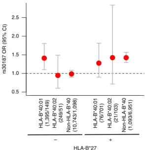
Gene–gene interactions and susceptibility loci. We have previously observed that the association with the variant rs30187 in the ERAP1 locus is restricted to HLA-B*27-positive subjects, consistent with an epistatic interaction between these two loci7. Here we investigated the possibility of interaction between the otherHLA-Bsusceptibility alleles and the variant rs30187. When testing for interaction with theHLA-B*40 alleles, we found that rs30187-T increased the risk of disease in the strata where
HLA-B*27 was present, as previously shown, or when
HLA-B*40:01 was present in the absence of HLA-B*27 (OR¼1.41;P¼5.81103); rs30187 had no effect on disease
susceptibility when both HLA-B*27 and HLA-B*40 alleles were
absent or in thenon-HLA-B*27/HLA-B*40:02stratum (Fig. 4). No evidence of interaction was observed between rs30187 and the otherHLA-Bsusceptibility alleles. This suggests that the rs30187
variant interacts with the HLA-B*40:01 allele; although no
evidence to support an interaction was observed with
HLA-B*40:02, the study had low power to detect such an effect. There was no evidence of interaction between either of the
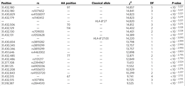
HLA-B*40 alleles and any of the other independently associated Table 1 | Most significant polymorphic positions (omnibus test) and imputed classical alleles associated with ankylosing
spondylitis susceptibility (P-valueo1102000).
Position rs AA position Classical allele v2 DF P-value
31,432,180 — 97 — 14,857 5 o103,221
31,432,180 rs1071652 — — 14,841 3 o103,221
31,430,829 rs41558317 — — 14,823 1 o103,221
31,432,179 rs1140412 — — 14,823 2 o103,219
— — — HLA-B*27 14,820 1 o103,221
31,432,506 — 70 — 14,812 3 o103,215
31,432,129 — 114 — 14,402 2 o103,128
31,432,130 rs709055 — — 14,401 2 o103,128
31,432,131 rs1050628 — — 14,389 1 o103,127
— — — HLA-B*27:05 14,220 1 o103,090
31,430,834 rs3819282 — — 13,798 1 o102,999
31,430,345 rs3819299 — — 13,757 1 o102,990
31,430,346 rs3819299 — — 13,757 1 o102,990
31,451,646 rs4463302 — — 12,898 1 o102,803
31,432,485 — 77 — 12,871 2 o102,795
31,432,486 rs1131217 — — 12,849 1 o102,793
31,377,108 rs2394967 — — 11,613 1 o102,524
31,381,125 rs6905036 — — 11,552 1 o102,511
31,432,208 rs41556113 — — 10,929 1 o102,376
31,432,843 rs41553720 — — 10,299 2 o102,237
31,432,515 — 67 — 9,741 4 o102,112
31,432,515 rs1071816 — — 9,725 3 o102,110
31,518,387 rs2844510 — — 9,525 1 o102,071
AA, amino acid; DF, degrees of freedom.
Table 2 | Evidence for association ofHLA-Balleles with
susceptibility to ankylosing spondylitis. Effect sizes and levels of significance were estimated in stepwise conditional procedure, where for rounds 2 and onwards the test conditioned on the previous alleles.
Round HLA-Ballele OR (95% CI) P-value 1 27:05 62.41 (56.90–68.45) o10321 2 27:02 43.41 (29.80–63.23) 1.0710122 3 07:02 0.82 (0.74–0.91) 5.04106 4 57:01 0.75 (0.61–0.92) 5.13104 5 51:01 1.33 (1.14–1.56) 2.14103 6 47:01 2.35 (1.43–3.86) 2.25103 7 40:02 1.59 (1.19–2.14) 4.65103 8 13:02 1.43 (1.14–1.80) 4.29103 9 40:01 1.22 (1.06–1.40) 4.93103
All other alleles 40.05
CI, confidence interval; OR, odds ratio;
HLA-B
97 11
HLA-A 0.8
Control Case
0.6
0.4
0.2
0.0
Ser Arg Thr Trp Asn Val Ile Val Leu Gly Leu
95
HLA-DPβ1
Table 3 | Haplotype analysis of SNPs encoding the amino acid 97 of HLA-B.
HLA-Bcodon 97 position
1 2 3
SNP rs41558317 rs1140412 rs1071652 rs41556417 Amino-acid residue Position (HG18) 31,430,829 31,432,179 31,432,180 31,432,181
Reference allele or amino acid in HG18
A G C T Serine (S)
Alternate allele(s) G C/A G/A/T A/C Allele frequency in
controls (ref/alt(s))
0.95/0.05 0.29 0.67/0.05
0.82 0.10/0.04/
0.05
0.92 0.04/0.04
Single locus univariateP-value
o103221 o103219 o103221 2.101065 Multivariate OR
(95% CI)
Unadjusted haplotype frequency
P HLA-BAllele
Risk allele univariate OR (95% CI)
60.36 (55.47–
65.74)
59.99 (55.13– 65.33)
59.99 (55.14– 65.34)
2.03 (1.86– 2.21)
Controls Cases
Haplotype G/A A T T Asparagine
(N)
16.51 (15.43–17.69) 0.045 0.449 o10300 *27:02 *27:04 *27:05
G/A C G T Threonine
(T)
1.12 (1.03–1.21) 0.097 0.065 4.50103 *13:02 *39:06 *40:06 *51:01 *51:08 *52:01 *55:01
*56:01
G/A C C T Arginine
(R)
1.00 (Reference) 0.493 0.297 1 *15:01 *15:03 *15:10 *15:16 *15:17
*15:18 *18:01 *35:01
*35:02 *35:03 *35:08 *35:12
*37:01 *38:01 *38:02
*39:01 *39:10 *40:01
*41:01 *44:02 *44:03 *44:04 *44:05 *45:01 *47:01 *49:01 *50:01 *53:01 *58:01
G/A C C A Tryptophan
(W)
1.00 (0.89–1.12) 0.042 0.025 0.95 *14:01 *14:02
G/A G C T Serine (S) 0.86 (0.81–0.91) 0.286 0.148 4.81108 *07:02 *07:05 *08:01 *15:07
*27:07 *40:02 *41:02 *48:01
A C A C Valine (V) 0.68 (0.59–0.78) 0.038 0.016 1.41108 *57:01 *57:03
CI, confidence interval; OR, odds ratio.
67
70 77
97
9
114
142
145 95
107 127
11
HLA-B HLA-A HLA-DPβ1
susceptibility SNPs in the loci encoding the aminopeptidases
ERAP1,ERAP2andNPEPPS(P40.1).
We then examined whether our data supported a model where the HLA-B*27 and HLA-B*40 alleles increased disease suscept-ibility beyond their inferred independent effects, as previously reported30. No support for an interaction between these alleles was observed in this data set (Supplementary Table 1).
Discussion
Independently of the expected HLA-B associations, this study
demonstrates that both HLA-B*40:01 and -B*40:02 are disease
associated alleles, and identified three furtherHLA-Brisk alleles,
HLA-B*51:01, B*47:01 and B*13:02. The allele HLA-B*51:01 is
also the major genetic risk factor for Behc¸et’s disease
31, a seronegative disease complicated by sacroiliitis resembling
AS in up to 10% of cases32. In addition to the seven HLA-B
risk alleles, we identified two protective alleles at this locus,
HLA-B*07:02 andHLA-B*57:01. Interestingly, in theHLA-B*27
-transgenic rat model of AS, the HLA-B*27-negative control
carries the HLA-B*07 allele, and does not develop disease,
consistent with the protective effect of this allele in humans33.
It has recently been shown in HLA-B7/B27 co-expressing
mice that there is partial negative selection of HLA-B27þ
T cells in the course of defining the immunodominant response to influenza infection34. Further, in Erap-deficient, influenza-infectedHLA-B27-positive mice, there was a marked reduction in presentation of the HLA-B27 immunodominant epitope, and T-cell immunity to that epitope, presumed to be because the HLA-B27-related immunodominant flu epitope requires cleavage
by Erap to be presented by HLA-B27. In contrast, in HLA-B7
-transgenic mice,Erapdeficiency had no effect on presentation of the HLA-B7 immunodominant epitope or the corresponding T-cell response to it, suggesting that it does not require Erap cleavage for presentation35. This provides a potential mechanism to explain the genetic effects observed in humans with AS, with ERAP1 loss of function protecting against HLA-B27-associated AS, but having no effect in HLA-B7 carriers, where an HLA-B7 protective association is observed.
Outside the HLA-B locus, we identified three independent
significant signals associated with AS; one was in the HLA class I
locusHLA-A, and one each in the HLA class II lociHLA-DPB1
and HLA-DRB1. The association in the HLA-A locus corre-sponded to the classical alleleHLA-A*02:01, which has also been implicated in multiple sclerosis36; however, while this allele is protective in multiple sclerosis, it increases the risk of AS.
Previous studies have hinted atHLA-DPB1associations with AS,
which we have confirmed here. HLA-DPB1, in conjunction with HLA-DPA1, forms the HLA-DP heterodimer, which typically plays a role in the presentation of exogenously derived peptides,
such as microbial peptides, to CD4þ T lymphocytes. The
strongest association was found with an amino-acid position located in the base of the peptide-binding groove of HLA-DP, suggesting that this polymorphism might impact on the peptide repertoire presented by HLA-DP.
Previous findings thatERAP1variants influence risk of disease in HLA-B*27 positive, but not negative individuals, strongly support the notion that both these molecules act in the same biological pathway to affect disease susceptibility7. We have now
shown that HLA-B*40:01 interacts with ERAP1 variants in the
same manner. Similar genetic interactions involvingERAP1have
been observed in two other immune-mediated disorders—
psoriasis with HLA-Cw6 (ref. 37) and Behc¸et’s syndrome with
HLA-B*51(ref. 38), two disorders that are already known to share genetic susceptibility factors with AS. It is likely that the similar molecular mechanisms are involved in these disorders, and that these include the pathways of MHC class I antigen presentation. To our knowledge, there is no evidence that HLA-B40, HLA-B51 or HLA-Cw6 have non-canonical disease-related properties such as those by which HLA-B27 is proposed to function in the pathogenesis of AS.
Analysis of polymorphic amino-acid positions in these AS-associated HLA molecules showed that the SNPs with the strongest evidence of association at each of these three loci were highly associated with amino-acid positions located in the peptide-binding groove of these proteins. From these results, we
infer that antigen presentation to both CD4þ and CD8þ T
lymphocytes is likely to be important in the pathogenesis of AS and/or its tissue specificity, although other mechanisms under-lying the associations cannot formally be excluded.
MHC class I molecules contain six specificity pockets in the peptide-binding groove, alphabetically named A to F, which serve to anchor particular side chains of the bound peptide39. Position 97 of HLA-B is located in the C/F pocket, also referred to as the C-terminal pocket, which anchors the side chain of the
C-terminal peptide residue29. Experimental evidence suggests
that this position is important for protein function and shaping the peptide repertoire presented by HLA-B. Mutagenesis
experiments have shown that Asn97 is important for
HLA-B*27:04 surface expression; mutating this residue from Asn97 to Asp97 results in reduced surface expression and increased accumulation of unfolded protein in the ER, as well as reduced homodimers formation40; thus, Asn97 relative to Asp97 reduces ER stress and B27 homodimer formation, yet is associated with AS risk. Moreover, work in the mouse homologue has shown that changing residue 97 (W97R) results in altered peptide specificity and affinity forb2-microglobulin41, and previous crystallographic
studies of viral peptides bound to HLA-B27 have shown that this position influences the location of the peptide in the binding groove of the molecule42. Last, this position was also found to be associated with HIV-1 viral control, where Val97 was found to provide the strongest protective effect to progression to AIDS,
hypothesized to be through a mechanism of peptide
presentation43. Asp97 is not shared by the AS-associated
subtype HLA-B*27:07, where it is substituted by serine. Serine is also a polar amino acid and the substitution would be expected to have only minor effects on the protein structure. While AS is
HLA-B*27 2.5
2.0
1.5
1.0
0.5
+ –
rs30187 OR (95% CI)
HLA-B*40:01 (1,395/149) HLA-B*40:02
(249/51)
Non-HLA-B*40 (10,743/1,098) HLA-B*40:01
(76/701)
HLA-B*40:02
(21/103)
Non-HLA-B*40 (1,093/6,951)
known to occur in individuals carryingHLA-B*27:07, its relative
strength of association compared with other AS-associated
HLA-B*27subtypes is unknown.
In summary, with high-density genotyping of the MHC, we have demonstrated independent association signals located in HLA class I and II loci. Imputation of amino-acid residues in the classical HLA class I and II proteins resolved the peak of association at each of these loci to an amino-acid residue located in the peptide-binding groove of these proteins. Refining this analysis by imputation of classical HLA alleles showed that there are multiple risk and protective haplotypes in the HLA-Blocus.
Further, epistatic interaction was demonstrated betweenERAP1
and the HLA class I allelesHLA-B*27andHLA-B*40:01.
Methods
Sample collection.All cases met the modified New York classification criteria for AS44. Nine thousand sixty-nine cases and 13,578 controls were recruited through a multi-center study coordinated by the International Genetics of Ankylosing Spondylitis Consortium8, and all samples were unrelated and met European ancestry criteria as detailed therein. All subjects provided written informed consent and the study was approved by the Princess Alexandra Hospital Research Ethics Committee (reference HREC/05/QPAH/221) and University of Queensland Research Ethics Committee (Project Clearance No: 2006000102). All samples were genotyped with the Illumina (San Diego, CA, USA) Infinium platform
Immunochip45, and the current study was restricted to 7,264 markers in the MHC (chromosome 6, bps 29,602,876–33,268,403, NCBI Build 36 human genome coordinates).
Imputation.We imputed SNPs across the MHC, and classical HLA class I and II alleles (HLA-A,HLA-C,HLA-B,HLA-DRBI,HLA-DQA1,HLA-DQB1,HLA-DPA1 andHLA-DPB1) and their corresponding amino acids determinants with SNP2HLA28. Samples with cumulative dosage above 2.5, across all four-digit alleles for any one of the HLA loci, were removed from the analysis. SNPs, alleles or amino-acid residues were excluded from the analysis if ther2imputation quality score was below 0.2.
Classical allele imputation at theHLA-Blocus resulted in high-quality data, with a median sensitivity and specificity for imputedHLA-Balleles of 0.958 and 0.998, respectively (Supplementary Fig. 1). With our imputation strategy, similar imputation performance has previously been shown for the other HLA class I and II loci (HLA-A,HLA-C,HLA-DRB1,HLA-DQA1,HLA-DQB1,HLA-DPA1and HLA-DPB1), suggesting that imputation performance for these loci was also accurate in our study28,43,46,47.
Statistical framework for association analysis.Associations of SNPs, HLA protein amino-acid positions and non-HLA-Balleles across the MHC locus were assessed with logistic regression, assuming an additive risk effect on the log-odds scale. To account for population stratification, we included as covariates 10 prin-cipal components for each individual, computed with 16,145 unlinked autosomal, non-MHC, SNPs with the tool shellfish (http://www.stats.ox.ac.uk/Bdavison/ software/shellfish/shellfish.php). The omnibus association test compares, via like-lihood ratio test, the null modelH0, where there is no risk effect at the position
tested, against the alternative modelH1, where the risk effect at the position is
included in the model as a fixed effect:
H0:logit yð Þ ¼i yþ X10
k¼1
pkpi;k ð1Þ
H1:logit yð Þ ¼i yþ X m1
a¼1
baga;iþ X10
k¼1
pkpi;k; ð2Þ
whereyidenotes the binary phenotype code for individuali(0¼control and 1¼case). Thepkparameter is the effect associated with each of the principal components andpi,kis the value of thekth principal component for individuali. Theyparameter represents the sampling fraction (that is, the logistic regression intercept). In the alternative model,aindicates the specific allele being tested and ga,iis the dosage (imputed or genotyped) of alleleain individuali. Theba para-meter represents the effect on the log odds of disease per allele. For testing a multi-allelic locus, nucleic or amino-acid positions, withmpossible alleles we included m-1bparameters, one for each allele, where the most common allele was selected as the reference allele. The likelihood ratio test that compares modelH0withH1
results in a test statistic that isw2distributed withm-1 degrees of freedom. When testing for association with imputed classical HLA alleles, we defined a series of binary markers coding the presence or absence of the allele being tested, and each different allele was tested as a biallelic position as described above.
To identify independent effects. we performed conditional logistic regression by including the most strongly associated position/polymorphism as a fixed effect in
both the null modelH0and the alternative modelH1. We then analysed all
positions as described above. Conditional analysis was repeated in an iterative fashion by sequentially adding the most significant positions as fixed effects until no significant position or polymorphism was observed. Allelic associations were deemed significant withPo105, this statistical significance threshold accounted
for 5,000 independent tests using Bonferroni correction. Two tests were considered independent if the two SNPs had a pairwise correlation (r2)o0.90, which resulted
in 3,252 SNPs independent tests. For the special case ofHLA-Balleles where we had a higher prior probability of association, we defined significance asPo103as
only 38 alleles were tested.
References
1. Brown, M. A.et al.Susceptibility to ankylosing spondylitis in twins: the role of genes, HLA, and the environment.Arthritis Rheum.40,1823–1828 (1997). 2. Brewerton, D. A.et al.Ankylosing spondylitis and HL-A 27.Lancet1,904–907
(1973).
3. Caffrey, M. F. & James, D. C. Human lymphocyte antigen association in ankylosing spondylitis.Nature242,121 (1973).
4. Schlosstein, L., Terasaki, P. I., Bluestone, R. & Pearson, C. M. High association of an HL-A antigen, W27, with ankylosing spondylitis.N. Engl. J. Med.288, 704–706 (1973).
5. Burton, P. R.et al.Association scan of 14,500 nonsynonymous SNPs in four diseases identifies autoimmunity variants.Nat. Genet.39,1329–1337 (2007). 6. Reveille, J. D.et al.Genome-wide association study of ankylosing spondylitis
identifies non-MHC susceptibility loci.Nat. Genet.42,123–127 (2010). 7. Evans, D. M.et al.Interaction between ERAP1 and HLA-B27 in ankylosing
spondylitis implicates peptide handling in the mechanism for HLA-B27 in disease susceptibility.Nat. Genet.43,761–767 (2011).
8. International Genetics of Ankylosing Spondylitis Consortium (IGAS)et al. Identification of multiple risk variants for ankylosing spondylitis through high-density genotyping of immune-related loci.Nat. Genet.45,730–738 (2013). 9. Benjamin, R. & Parham, P. HLA-B27 and disease: a consequence of inadvertent
antigen presentation?Rheum. Dis. Clin. North Am.18,11–21 (1992). 10. Kenna, T. J. & Brown, M. A. Immunopathogenesis of ankylosing spondylitis.
Int. J. Clin. Rheumatol.8,265–274 (2013).
11. Sherlock, J. P., Buckley, C. D. & Cua, D. J. The critical role of interleukin-23 in spondyloarthropathy.Mol. Immunol.57,38–43 (2014).
12. Chang, S. C., Momburg, F., Bhutani, N. & Goldberg, A. L. The ER aminopeptidase, ERAP1, trims precursors to lengths of MHC class I peptides by a ’molecular ruler’ mechanism.Proc. Natl Acad. Sci. USA102,17107–17112 (2005).
13. Saveanu, L.et al.Concerted peptide trimming by human ERAP1 and ERAP2 aminopeptidase complexes in the endoplasmic reticulum.Nat. Immunol.6, 689–697 (2005).
14. Allen, R. L., O’Callaghan, C. A., McMichael, A. J. & Bowness, P. Cutting edge: HLA-B27 can form a novel beta 2-microglobulin-free heavy chain homodimer structure.J. Immunol.162,5045–5048 (1999).
15. Chan, A. T., Kollnberger, S. D., Wedderburn, L. R. & Bowness, P. Expansion and enhanced survival of natural killer cells expressing the killer
immunoglobulin-like receptor KIR3DL2 in spondylarthritis.Arthritis Rheum. 52,3586–3595 (2005).
16. Diaz-Pena, R.et al.Influence of HLA-B*5703 and HLA-B*1403 on susceptibility to spondyloarthropathies in the Zambian population.J. Rheumatol.35,2236–2240 (2008).
17. Lopez-Larrea, C.et al.Association of ankylosing spondylitis with HLA-B*1403 in a West African population.Arthritis Rheum.46,2968–2971 (2002). 18. D’Amato, M.et al.Relevance of residue 116 of HLA-B27 in determining
susceptibility to ankylosing spondylitis.Eur. J. Immunol.25,3199–3201 (1995). 19. Nasution, A. R.et al.HLA-B27 subtypes positively and negatively associated
with spondyloarthropathy.J. Rheumatol.24,1111–1114 (1997).
20. Turner, M. J.et al.HLA-B27 misfolding in transgenic rats is associated with activation of the unfolded protein response.J. Immunol.175,2438–2448 (2005).
21. DeLay, M. L.et al.HLA-B27 misfolding and the unfolded protein response augment interleukin-23 production and are associated with Th17 activation in transgenic rats.Arthritis Rheum.60,2633–2643 (2009).
22. Campbell, E. C., Fettke, F., Bhat, S., Morley, K. D. & Powis, S. J. Expression of MHC class I dimers and ERAP1 in an ankylosing spondylitis patient cohort. Immunology133,379–385 (2011).
23. Ciccia, F.et al.Evidence that autophagy, but not the unfolded protein response, regulates the expression of IL-23 in the gut of patients with ankylosing spondylitis and subclinical gut inflammation.Ann. Rheum. Dis.73,1566–1574 (2014).
24. Kenna, T. J.et al.Disease-associated polymorphisms in ERAP1 do not alter endoplasmic reticulum stress in patients with ankylosing spondylitis.Genes Immun.16,35–42 (2015).
26. Brown, M. A.et al.HLA class I associations of ankylosing spondylitis in the white population in the United Kingdom.Ann. Rheum. Dis.55,268–270 (1996).
27. Wei, J. C., Tsai, W. C., Lin, H. S., Tsai, C. Y. & Chou, C. T. HLA-B60 and B61 are strongly associated with ankylosing spondylitis in HLA-B27-negative Taiwan Chinese patients.Rheumatology (Oxford)43,839–842 (2004).
28. Jia, X.et al.Imputing amino Acid polymorphisms in human leukocyte antigens.PLoS One8,e64683 (2013).
29. Madden, D. R., Gorga, J. C., Strominger, J. L. & Wiley, D. C. The three-dimensional structure of HLA-B27 at 2.1 A resolution suggests a general mechanism for tight peptide binding to MHC.Cell70,1035–1048 (1992). 30. van Gaalen, F. A.et al.Epistasis between two HLA antigens defines a subset of
individuals at a very high risk for ankylosing spondylitis.Ann. Rheum. Dis.72, 974–978 (2012).
31. Takano, M., Miyajima, T., Kiuchi, M., Ohmori, K. & Amemiya, H. Behcet disease and the HLA system.Tissue Antigens8,95–99 (1976).
32. Ait Badi, M. A.et al.[Skeletal manifestations in Behcet’s disease. A report of 79 cases].Rev. Med. Interne29,277–282 (2008).
33. Taurog, J. D. & Hammer, R. E. Experimental spondyloarthropathy in HLA-B27 transgenic rats.Clin. Rheumatol.15(Suppl 1): 22–27 (1996).
34. Akram, A. & Inman, R. D. Co-expression of HLA-B7 and HLA-B27 alleles is associated with B7-restricted immunodominant responses following influenza infection.Eur. J. Immunol.43,3254–3267 (2013).
35. Akram, A., Lin, A., Gracey, E., Streutker, C. J. & Inman, R. D. HLA-B27, but not HLA-B7, immunodominance to influenza is ERAP dependent.J. Immunol. 192,5520–5528 (2014).
36. Sawcer, S.et al.Genetic risk and a primary role for cell-mediated immune mechanisms in multiple sclerosis.Nature476,214–219 (2011).
37. Strange, A.et al.A genome-wide association study identifies new psoriasis susceptibility loci and an interaction between HLA-C and ERAP1.Nat. Genet. 42,985–990 (2010).
38. Kirino, Y.et al.Genome-wide association analysis identifies new susceptibility loci for Behcet’s disease and epistasis between HLA-B*51 and ERAP1.Nat. Genet.45,202–207 (2013).
39. Saper, M. A., Bjorkman, P. J. & Wiley, D. C. Refined structure of the human histocompatibility antigen HLA-A2 at 2.6 A resolution.J. Mol. Biol. 219,277–319 (1991).
40. Blanco-Gelaz, M. A.et al.The amino acid at position 97 is involved in folding and surface expression of HLA-B27.Int. Immunol.18,211–220 (2006).
41. Smith, R. A., Myers, N. B., Robinson, M., Hansen, T. H. & Lee, D. R. Polymorphism at position 97 in MHC class I molecules affects peptide specificity, cell surface stability, and affinity for beta2-microglobulin.J. Immunol.169,3105–3111 (2002).
42. Kumar, P.et al.Structural basis for T cell alloreactivity among three HLA-B14 and HLA-B27 antigens.J. Biol. Chem.284,29784–29797 (2009).
43. Pereyra, F.et al.The major genetic determinants of HIV-1 control affect HLA class I peptide presentation.Science330,1551–1557 (2010).
44. van der Linden, S., Valkenburg, H. A. & Cats, A. Evaluation of diagnostic criteria for ankylosing spondylitis. A proposal for modification of the New York criteria.Arthritis Rheum.27,361–368 (1984).
45. Cortes, A. & Brown, M. A. Promise and pitfalls of the Immunochip.Arthritis Res. Ther.13,101 (2011).
46. McLaren, P. J.et al.Fine-mapping classical HLA variation associated with durable host control of HIV-1 infection in African Americans.Hum. Mol. Genet.21,4334–4347 (2012).
47. Raychaudhuri, S.et al.Five amino acids in three HLA proteins explain most of the association between MHC and seropositive rheumatoid arthritis.Nat. Genet.44,291–296 (2012).
Acknowledgements
We thank all participating subjects with AS and healthy individuals who provided the DNA and clinical information necessary for this study. This work was in part funded by grants from Arthritis Research UK (19536 and 18797), the NIHR Oxford comprehensive Biomedical Research Centre (immunity and inflammation theme A93081) and NIHR Thames Valley collaborative research network and National Ankylosing Spondylitis Society (UK). SPARCC was established through the support of the Arthritis Society of Canada. Support was received from National Institutes of Health/National Institute of Allergy and Infectious Diseases grant 1U01AI09090-01. This work was supported in part by grant PI12/02587 (Inst. Carlos III, Spain) and by European Union ‘Fondos FEDER’. Support was received from Agence Nationale de la Recherche (grant ANR 2010 GEMISA and Investissements d’Avenir programme ANR-11-IDEX-0005-02), the Socie´te´ Franc¸aise de Rhumatologie (SFR) and the Arthritis Foundation. M.W. is funded by the
Intramural Research Program, National Institute of Arthritis and Musculoskeletal and Skin Diseases, National Institutes of Health. M.A.B. is funded by a National Health and Medical Research Council (Australia) Senior Principal Research Fellowship. D.M.E. is funded by an Australian Research Council Future Fellowship (FT130101709). P.I.W.d.B. is funded in part by the Netherlands Organization for Scientific Research (VIDI Vernieuwingsimpuls project 016.126.354) and by the National Institutes of Health (1R01AR062886-1).
Author contributions
All authors contributed to the manuscript preparation and approved the final manu-script. Case and control recruitment was performed by P.L.C., M.H.H., M.W., L.S.G., J.H., O.F., J.T., K.L., L.A.B., D.E., R.B-V., S.S., C.F., N.H., J.M., F.J.B., M.A.G-G., C.L-L, P.B., K.G., H.G., D.D.G., P.R., W.P.M., I.V.H-B., R.V-O., C.R-S., I.M.H., F.M.P-S., R.D.I., M.B., B.P.W., J.D.R. and M.A.B. Study design was contributed by A.C., P.J.L., R.D.I., M.B., B.P.W., J.D.R., D.M.E., P.I.W.d.B. and M.A.B. DNA preparation and genotyping were performed by J.J.P., X.Z., H-J.G., G.C., B.A.L., L.A., J.L., J.B.A.C., F.M.P-S. and M.A.B.. Analysis and interpretation were performed by A.C., S.L.P., P.J.L., P.C.R., D.M.E., P.I.W.d.B. and M.A.B. The manuscript preparation was performed by A.C., P.J.L., D.M.E., P.I.W.d.B. and M.A.B.
Additional information
Supplementary Informationaccompanies this paper at http://www.nature.com/ naturecommunications
Competing financial interests: The authors declare no competing financial interests.
Reprints and permissioninformation is available online at http://npg.nature.com/ reprintsandpermissions/
How to cite this article: Cortes, A.et al.Major histocompatibility complex associations of ankylosing spondylitis are complex and involve further epistasis with ERAP1. Nat. Commun.6:7146 doi: 10.1038/ncomms8146 (2015).