online | memorias.ioc.fiocruz.br
Molecular phylogeny of the Myzorhynchella Section
of
Anopheles
(
Nyssorhynchus
) (Diptera: Culicidae):
genetic support for recently described and resurrected species
Brian Patrick Bourke1, Sandra Sayuri Nagaki1, Eduardo Sterlino Bergo2,
Jáder da Cruz Cardoso1,3, Maria Anice Mureb Sallum1/+
1Departamento de Epidemiologia, Faculdade de Saúde Pública, Universidade de São Paulo, Avenida Dr. Arnaldo 715, 01246-904,
São Paulo, SP, Brasil 2Superintendência de Controle de Endemias, Secretaria de Estado da Saúde de São Paulo, Araraquara, SP, Brasil 3Divisão de Vigilância Ambiental em Saúde, Centro Estadual de Vigilância em Saúde, Secretaria da Saúde do Estado do Rio Grande do Sul,
Porto Alegre, RS, Brasil
Phylogenetic relationships among species of the Myzorhynchella Section of Anopheles (Nyssorhynchus) were investigated using the nuclear ribosomal DNA second internal transcribed spacer (ITS2), the nuclear whitegene and mitochondrial cytochrome oxidase subunit I (COI) regions. The recently described Anopheles pristinus and resurrected Anopheles guarani were also included in the study. Bayesian phylogenetic analyses found Anopheles parvus to be the most distantly related species within the Section, a finding that is consistent with morphology. An. pristinus and An. guarani were clearly resolved from Anopheles antunesi and Anopheles lutzii, respectively. An. lut-zii collected in the same mountain range as the type locality were found within a strongly supported clade, whereas individuals from the southern state of Rio Grande do Sul, tentatively identified as An. lutzii based on adult female external morphology, were distinct from An. lutzii, An. antunesi and from each other, and may therefore represent two new sympatric species. A more detailed examination of An. lutziisensu latoalong its known geographic range is recommended to resolve these anomalous relationships.
Key words: phylogeny - Anopheles - Myzorhynchella - white - ITS2 - COI
Mosquitoes (Culicidae) are highly diverse geo-graphically widespread taxa containing approximately 3,529 species (Harbach 2011a). They are also of major medical importance as a consequence of being vectors of pathogens that cause diseases in humans such as dengue, filariasis and malaria (WHO 1989). All known vectors of malarial parasites belong to the genus Anopheles Mei-gen and are responsible for an estimated 243 million ma-laria cases annually (WHO 2009). Within the Americas there are an estimated one million cases of malaria an-nually, the majority of which are caused by vectors of the subgenus Nyssorhynchus Blanchard (Zimmerman 1992, WHO 2009). This subgenus contains 39 species (Har-bach 2011b, Nagaki et al. 2011), at least seven of which are known to harbour malarial parasites (Rosa-Freitas et al. 1998). Whereas the subgenus is a well supported monophyletic group (Sallum et al. 2000, 2002, Harbach & Kitching 2005), relationships among species within this group are less well defined.
The subgenus has traditionally been divided into three sections based on morphological characters (Pey-ton et al. 1992): the Argyritarsis, Albimanus and Myzo-rhynchella Sections. Until recently the MyzoMyzo-rhynchella Section was comprised of four nominal species, Anophe-
Financial support: FAPESP (2005/53973-0 to MAMS), CNPq (BPP 300351/2008-9 to MAMS)
+ Corresponding author: [email protected] Received 28 February 2011
Accepted 13 July 2011
les lutzii Cruz, Anopheles parvus (Chagas), Anopheles nigritarsis (Chagas) and Anopheles antunesi Galvão & Amaral (Harbach 2004). The Myzorhynchella Section is currently the only section in Nyssorhynchus without species implicated in malaria transmission and the only section with some genetic support as a natural grouping. Genetic data also indicate that species diversity within the Myzorhynchella Section is perhaps underestimated due to the existence of species complexes (Bourke et al. 2010). Nagaki et al. (2011) recently resurrected Anophe- les guarani from An. lutzii and described a new species, Anopheles pristinus Nagaki & Sallum, distinguished from An. antunesi. Members of species complexes fre-quently vary in their ability to transmit malaria and an effective assessment of mosquito susceptibility to the malaria parasite relies on the ability to accurately resolve and identify species. Recent studies have worked to-wards identifying malaria refractory genes in Anopheles (Marshall & Taylor 2009, Corby-Harris et al. 2010). If the Myzorhynchella Section is a natural grouping within Nyssorhynchus without vectors of human malarial pro-tozoa, it may prove to be a useful group for the study and identification of such genes.
MATERIALS AND METHODS
Mosquito collection - A description of the specimens used in this study can be found in Table I. These spec-imens included the offspring of females caught in the field using a Shannon trap (Shannon 1939) and larvae and pupae collected from immature habitats, which were then raised to adulthood. Species identification of all but two specimens was based on either adult male genitalia or fourth-instars larval characteristics. Specimens An. lutzii A325 and An. lutzii B369 were identified only by female morphology.
DNA extraction - DNA was extracted from each specimen according to the animal tissue DNA extraction protocol provided by the QIAgen DNeasy® Blood and
Tissue Kit (QIAGEN Ltd, Crawley, UK). All extractions
were diluted to 200 μL with the buffer provided and ex -traction solutions were retained for storage at -80ºC in the entomological frozen collection of the School of Pub-lic Health, University of São Paulo, Brazil.
ITS2 region - This region was amplified using the 5.8SF (5’-ATCACTCGGCTCGTGGATCG-3’) and 28SR (5’-ATGCTTAAATTTAGGGGGTAGTC-3’) primers using the same protocol adopted by Sallum et al. (2008). The polymerase chain reaction (PCR) was conducted
in a total volume of 25 μL containing 1 μL of DNA
extraction solution, 1 × PCR buffer (Invitrogen), 1.5 mM MgCl2 (Invitrogen), 1.25 μL dimethly sulfoxide
(Sigma), 0.1 μM of each primer, 200 mM each dNTPs
(Amresco) and 1.25 U Taq Platinum polymerase (In- vitrogen). The reaction proceeded under the following temperature regime: 94ºC for 2 min, 34 cycles of 94ºC for 30 s, 57ºC for 30 s and 72ºC for 30 s and a final extension at 72ºC for 10 min.
COI mitochondrial gene - This region was amplified using LCO1490 (5’-GGTCAACAAATCATAAAGA-TATTGG-3’) and HCO2198 (5’-TAAACTTCAGGGT-GACCAAAAAATCA-3’) primers (Folmer et al. 1994). DNA working solution was first made by diluting the DNA extraction solution to 1:20 using ultra-pure auto-claved water. The PCR reaction was conducted as above for ITS2. The reaction proceeded under the following temperature profile: 95ºC for 2 min, 35 cycles of 94ºC for 1 min, 57ºC for 1 min and 72ºC for 1 min and a final extension at 72ºC for 7 min.
White nuclear gene - This gene was amplified us-ing WZ2E and WZ11 primers (Besansky & Fahey 1997). This amplification product then served as a template in a sequencing reaction using internal primers W1F (5’-GATCAARAAGATCTGYGACTCGTT-3’) and W2R (5’GCCATCGAGATGGAGGAGCTG-3’). The initial
PCR reaction contained approximately 3 μL of DNA ex
-traction solution in a total volume of 25 μL containing
1 × PCR buffer (Invitrogen), 1.5 mM of MgCl2
(Invitro-gen), 200 mM of each dNTP (Amresco), 2 μM of each
primer and 0.625 U Taq DNA Polymerase (Invitrogen). The reaction proceeded under the following temperature regime: 94ºC for 5 min, 35 cycles at 94ºC for 30 s, an an-nealing temperature of 50ºC for 1 min and then 72ºC for
2 min followed by a final extension at 72ºC for 10 min. The next step then involved taking this PCR product for sequencing using the W1F and W2R internal primers.
Cloning - ITS2 PCR amplicons obtained from An. parvus were purified using PEG precipitation (20% polyethylene glycol 8000/2.5 M NaCl) and cloned into pGem-T Easy Vector (Promega, Madison, WI, USA). Three positive clones were sequenced. For the nuclear white gene, females were cloned when the direct se-quence showed either unreadable peaks or double peaks. Individuals that had either ITS2 or white gene cloned are shown in Table I.
Sequencing and sequence alignment - Sequencing reactions were carried out in both directions using a Big Dye Terminator cycle sequencing kit v3.1 (Applied Biosystems) and Applied Biosystems 3130 DNA Ana-lyzer (Applied Biosystems). The COI and white gene se-quences were aligned first by nucleotides using ClustalX (Thompson et al. 1997) and then by amino acid using TranslatorX (Abascal et al. 2010). The intron in the white gene could not be reliably aligned and so was excluded from further analyses. In addition, only a single ran-domly selected sequence was used to represent cloned individuals in the combined gene analysis.
In the case of ITS2, sequences were first annotated for the 5.8S and 28S ends. Template ITS2 secondary structure was then predicted for An. antunesi RJ036 with Predict ITS2 Structure in the ITS2 Database (Koet-schan et al. 2010). The remaining secondary structures were modelled at this source with Custom Modelling using the predicted An. antunesi RJ036 ITS2 secondary structure (above) as a template. Sequences with second-ary structures were then aligned and edited in 4Sale (Seibel et al. 2006, 2008). It was not possible to align the complete ITS2 sequences and so those regions that could not be aligned were excluded from further analysis. Ad-ditionally, we were unable to align ITS2 sequences with an appropriate outgroup and so trees constructed using ITS2 sequence data (including the combined gene trees) are unrooted.
Phylogenetic analyses - Bayesian analyses were ap-plied to ITS2, COI and white sequence data using a parti-tioning strategy to allow different partitions to have their own model characteristics (composition, rate matrix and among-site variation) and to allow for among-partition rate variation. Datasets could be left unpartitioned, partitioned by gene, partitioned by codon position or partitioned by both gene and codon position. Optimal evolutionary models were determined for isolated parti-tions using the Akaike Information Criterion (AIC) in Modeltest (Posada & Crandall 1998).
TABLE I
Specimen information, including specimen and voucher numbers, localities, geographical coordinates, species included in the study, and GenBank acessions of the second internal transcribed spacer (ITS2) ribosomal DNA,
cytochrome oxidase subunit I (COI) mitochondrial and white nuclear genes
Sample Specimen. Locality (state) Coordinates Species ITS2 COI white
RJ0311 RJ0312 RJ0313 RJ036 RJ03(11) RJ03(12) RJ03(13) RJ03(6)
Itatiaia (RJ) -44.622139, -22.416306
Anopheles antunesi GU989325 GU989326 GU989327 GU989324 GU989343 GU989344 GU989345 GU989342 JN392474 JN392475 JN392476 JN392473 VP0917 VP11b VP09-17 VP11b
Pindamonhangaba (SP) -45.515500, -22.758806
An. antunesi GU989328 GU989329
GU989346 GU989347
JN392477 FJ147290 PR29 PR29 Foz do Iguaçu (PR) -54.586667,
-25.480556
Anopheles guarani JN023041 JF923659 JN392478
SP0210 SP0211 SP0212 SP1213 SP0214 SP0215 SP029 SP02(10)-5 SP02(11)-9 SP02(12)-1 SP02(13)-3 SP02(14)-6 SP02(15)-5 SP02(9)-2
Pariquera-Açu (SP) -47.949056, -24.749583
Anopheles lutzii JN023045 JN023046 JN023047 JN023048 JN023049 JN023050 JN023044 JF923665 JF923666 JF923667 JF923668 JF923669 JF923670 JF923664 FJ147284 JN392483 JN392484 JN392485 JN392486 JN392487 FJ147283 A325 B369 Clone 3 Clone 4 Clone 5 A325 B369
Maquiné (RS) -50.213611, -29.661111
An. lutzii JN023042 JN023043 -JF923662 JF923663 -JN392479 -JN392480 JN392481 JN392482 PR2818 PR2851 Clone 1 Clone 2 PR2865 PR28(18)-1 PR28(5)-1 PR28(65)-6
Guaíra (PR) -54.290556, -24.271500
Anopheles parvus JN023064 -JN023062 JN023063 JN023065 JF923678 JF923677 -JF923679 FJ147288 JN392498 -JN392499 AS51 AS52 Clone 1 Clone 2 Clone 3 AS53 Clone 1 Clone 3 AS5-1 AS5-2 AS5-3
Pilar de Goiás (GO) -49.539834, -14.809194
An. parvus JN023051 -JN023052 JN023053 JN023054 JN023055 -JF923671 JF923672 -JF923673 -JN392488 -JN392489 JN392490 JN392491 -JN392492 JN392493 AS54 Clone 1 Clone 2 Clone 3 AS5-4 -JN023056 JN023057 JN023058 JF923674 -JN392494 JN392495 -MG562 Clone 2 Clone 3
MG56-2 Campo Belo (MG) -45.121778, -20.795111
An. parvus JN02306 -JF923676 -JN392496 JN392497 MG07920 Clone 1 Clone 2
MG07(9)-20 Frutal (MG) -49.076500, -20.025278
An. parvus
-JN023059 JN023060 JF923675 -FJ147287 -SP50a SP50b SP51100 SP53100 SP53101 SP534 SP535 SP552 SP554 SP50a SP50b SP51-100 SP53-100 SP53-101 SP53-4 SP53-5 SP55(2) SP55(4)
Pindamonhangaba (SP) -45.515278, -22.758472
Anopheles pristinus GU989333 GU989334 GU989335 GU989338 GU989339 GU989336 GU989337 GU989340 GU989341 GU989351 GU989352 GU989353 GU989356 GU989357 GU989354 GU989355 GU989358 GU989359 JN392500 JN392501 JN392502 JN392505 JN392506 JN392503 JN392504 JN392507 JN392508 VP11a VP11a Pindamonhangaba (SP) -45.515500,
-22.758806
An. pristinus GU989331 GU989348 FJ147289
significant support for partition two, values between 10 and -10 indicate ambiguity and values less than -10 in-dicate significant support for partition one. All Bayesian analyses were performed using MrBayes (Ronquist & Huelsenbeck 2003) and each analysis consisted of two runs to provide confirmation of convergence of posterior probability distribution.
For COI, white, ITS2 and all but one of the combined analyses of gene partition strategies, each run was four million generations long and the first two million were discarded as burn-in. The Metropolis-coupled Markov chain Monte Carlo strategy was used with four heated chains; adequate mixing was achieved by setting the chain temperature to 0.1 for isolated genes and com-bined genes. Convergence of topology between the two runs was monitored using the average standard devia-tion of split frequencies - this index consistently fell to below 0.015 in the post-burn-in samples. Convergence was also monitored by noting that the potential scale reduction factor values were all approximately 1.0 in the post-burn-in samples. The one exceptional partition strategy (partitioning by gene and codon position with among partition rate variation) did not converge (runs were varied from two-40 million generations, with four-eight heated chains and chain temperatures from 0.05-0.25) and so was excluded from further analysis. Post burn-in samples were then used to construct a consensus tree containing nodes with greater that 70% posterior probability support. Trees were drawn using the R pack-age APE (Paradis et al. 2004).
Pairwise genetic distances for COI were calculated in MEGA4 (Tamura et al. 2007) using Kimura’s (1980) two-parameter (K2P) model. Uncorrected pairwise p-distances were calculated in MEGA4 from a separate An. parvus ITS2 alignment using ClustalX.
RESULTS
The alignment included sequences from 35 individu-als. Difficulties with aligning ITS2 (at positions 143-199, 223-285, 386-635 and 714-797) and the intron in the white gene resulted in these positions being excluded from the phylogenetic analyses. Non-overlapping sites from the white gene were also excluded. In total, 1,539 sites were included in the analyses, consisting of 363 sites from ITS2, 657 sites from COI and 519 sites from the white gene. GenBank accessions of the nuclear white, mito-chondrial COI and the ITS2 are in Table I. Anopheles gambiae Giles and Anopheles strodei Root were used as outgroup taxa in the analyses of COI and white gene. It was not possible to find an appropriate outgroup for the ITS2 sequences and so ITS2 and combined COI-white -ITS gene trees were unrooted.
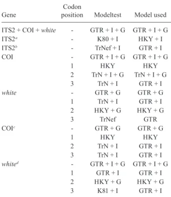
The optimal models of evolution determined for each sequence partition are displayed in Table II. Where the selected rate matrix was unavailable in MrBayes (i.e. TrN and TIM), the most similar rate matrix available was selected (i.e. GTR). ITS2 is a non-coding region and so was left unpartitioned in the analysis. The best evo-lutionary model for this region was the TrNef+I model (substituted with the GTR+I model in Bayesian analy-ses). BF (Tables III, IV, V) found the optimal partitioning
TABLE II
Models used for gene and codon positions
Gene
Codon
position Modeltest Model used
ITS2 + COI + white - GTR + I + G GTR + I + G
ITS2a - K80 + I HKY + I
ITS2b - TrNef + I GTR + I
COI - GTR + I + G GTR + I + G
1 HKY HKY
2 TrN + I + G TrN + I + G
3 TrN + I GTR + I
white - GTR + G GTR + G
1 TrN + I GTR + I
2 HKY + G HKY + G
3 TrNef GTR
COIc - GTR + G GTR + G
1 HKY HKY
2 TrN + I GTR + I
3 TrN + I GTR + I
whited - GTR + I + G GTR + I + G
1 GTR + I GTR + I
2 HKY + G HKY + G
3 K81 + I GTR + I
a: including clones for isolated gene tree: b: excluding clones for combined gene tree; c: with outgroup; d: with outgroup and clones; COI: cytochrome oxidase subunit I; ITS2: second inter-nal transcribed spacer.
TABLE III
Bayes factors calculated from the harmonic mean of the likelihoods for all partitions at cytochrome oxidase subunit I
None Codon (-) Codon (+)
None - 126.35 563.01
Codon (-) -126.35 - 436.66
Codon (+) -563.01 -436.66
-(+), (-): inclusion and exclusion of among partition rate varia-tion, respectively.
TABLE IV
Bayes factors calculated from the harmonic mean of the likelihoods for all partitions at the white gene
None Codon (-) Codon (+)
None - 163.59 190.35
Codon (-) -163.59 - 26.76
Codon (+) -190.35 -26.76
strategies for COI, white and combined genes. The best model for the COI gene (with an outgroup taxa) was one that partitioned the data by codon position and included among-partition rate variation (APRV) (Table III). The model chosen for the white gene (with an outgroup taxa) was one that partitioned the data by codon position and included APRV (Table IV). The model chosen for com-bined genes was one that partitioned the data by gene and codon position (Table V).
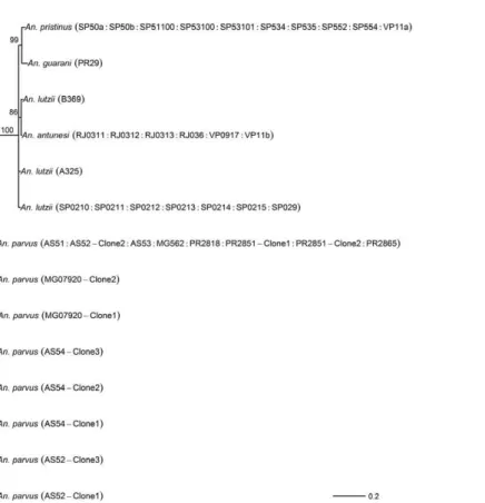
The results of Bayesian analyses show a high degree of congruence among the ITS2, COI, white and combined gene trees (Figs 1-4, respectively). An. parvus, An. pristi-nus and An. guarani are strongly supported as species across all trees. In addition, the majority of the An. lutzii individuals (7 of 9) form a strongly supported clade across all trees with support of 100% Bayesian posterior proba-bility (BPP). Some incongruence also exists between gene trees. The remaining two An. lutzii individuals (A325 and
TABLE V
Bayes factors calculated from the harmonic mean of the likelihoods for all partitions at combined genes
None Gene (-) Gene (+) Codon (-) Codon (+) Gene/Codon (-) Gene/Codon (+)
None - 203.64 174.7 355.22 451.35 469.2 Na
Gene (-) -203.64 - -28.94 151.58 247.71 265.56 Na
Gene (+) -174.7 28.94 - 180.52 276.65 294.5 Na
Codon (-) -355.22 -151.58 -180.52 - 96.13 113.98 Na
Codon (+) -451.35 -247.71 -276.65 -96.13 - 17.85 Na
Gene/Codon (-) -469.2 -265.56 -294.5 -113.98 -17.85 - Na
Gene/Codon (+) Na Na Na Na Na Na
-Na: not applicable; (+), (-): inclusion and exclusion of among partition rate variation, respectively.
Fig. 1: Bayesian tree of the second internal transcribed spacer of species of the Myzorhynchella Section. Numbers at branches indicate Bayesian
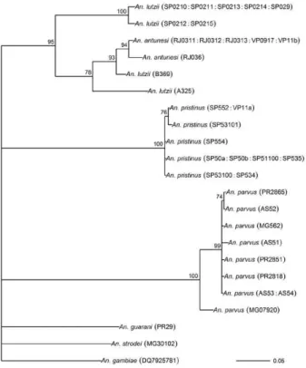
Fig. 2: Bayesian tree of the mitochondrial cytochrome oxidase subunit I gene of species of the Myzorhynchella Section. The data were
par-titioned by codon position with among partition variation. Numbers at branches indicate Bayesian posterior probability (≥ 70%). Anopheles
gambiae and Anopheles strodei were included as outgroup taxa.
Fig. 3: Bayesian tree of the nuclear white gene of species of the Myzorhynchella Section. The data were partitioned by codon position with
among partition variation. Numbers at branches indicate Bayesian posterior probability (≥70%). Anopheles gambiae and Anopheles strodei
B369) cluster differently across trees. In the white and combined gene trees (Figs 2, 4), individual A325 forms a clade with the An. lutziisensu stricto group described above (> 85% BPP), whereas the other (represented by the 3 clones from B369) forms a clade with An. antunesi (100% BPP). Both individuals form a clade with An. an-tunesi at the COI gene (78% BPP) (Fig. 2), whereas A325 was unclustered with An. lutzii at ITS2 (Fig. 1).
The COI and white gene trees (Figs 2, 3) were rooted with an outgroup. In the white gene tree (Fig. 3), An. stro-dei and An. parvus were sisters to a clade containing the remaining species in the Myzorhynchella Section (96% BPP). In the COI tree (Fig. 2), An. strodei and An. par-vus were sisters to An. pristinus, An. guarani and the An. lutzii-An. antunesi clade. In the combined gene tree (Fig. 4), An. pristinus was sister to a clade containing An. gua-rani, An. antunesi and An. lutzii (98% BPP) and within this clade An. guarani was sister to a clade containing all individuals of An. antunesi and An. lutzii (100% BPP).
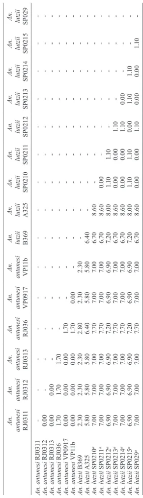
COI pairwise genetic distances among An. lutzii and An. antunesi were calculated under the K2P model (Table VI). Pairwise distances among individuals within An. antunesi and An. lutzii s.s. were found to be less than 2%, whereas differences between individuals from each group were in the range 6.90-7.70%. Individuals An. lut-zii A325 and An. lutzii B369 were most distant to those from the An. lutzii s.s. group (8.00-8.60 and 6.40-7.20%, respectively). The distances between A325 and An. an-tunesi individuals ranged from 5.80-6.40, whereas the
distances between B369 and An. antunesi ranged from 2.30-2.80%. An. lutzii A325 and An. lutzii B369 differed from each other by 6.4%.
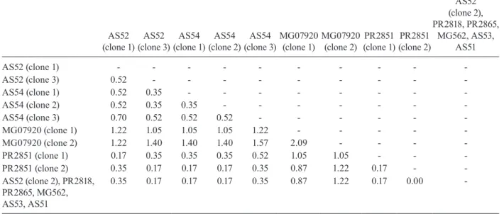
Difficulty in aligning ITS2 sequences resulted in large numbers of polymorphic sites being excluded from the phylogenetic analyses. It is likely the exclusion of such sites contributed to the existence of the large poly-tomy observed among An. parvus individuals and the lack of resolution for An. lutzii s.s. and An. lutzii A325. A separate alignment of An. parvus individuals was con-structed to include all sites and pairwise p-distances were calculated to describe variation among these individuals (p-distances) (Table VII). An. parvus was the only spe-cies in the study to have intragenomic variation at ITS2. An. parvus showed higher intragenomic (0.17-2.09%) than intraspecific (0-1.57%) variation. Two clones from a single individual from the state of Minas Gerais (MG) differed from each other by 2.09% and from other con-specifics by 0.87-1.57%. This same individual was also clearly resolved from the remaining An. parvus individ-uals at the COI gene. A separate alignment of An. lutzii and An. antunesi could not be constructed because of the frequency of ambiguous sites and so p-distances could not be calculated as in the case of An. parvus. Instead, we calculated the proportion of unambiguously aligned sites for these ITS2 sequences on a pairwise basis us-ing secondary structure (Table VIII). An. lutzii and An. antunesi consisted of four unique ITS2 sequences, one each for An. lutzii A325, An. lutzii B369, An. antunesi
Fig. 4: Bayesian tree of the combined second internal transcribed spacer, single-copy nuclear white gene and mitochondrial cytochrome oxidase
T A B L E V I C y to ch ro m e o x id a se s u b u n
it I (
C O I) p a ir w is e d is ta n ce K im u ra ’s t w o -p a ra m et e r ( K 2 P ) v alu e s ( % ) a m o n g i n d iv id u al s o f A n o p h el e s a n tu n e si a n d A n o p h el e s l u tz ii An.
antunesi RJ031
1
An.
antunesi RJ0312
An.
antunesi RJ0313
An.
antunesi RJ036
An.
antunesi VP0917
An.
antunesi VP1
1b
An. lutzii B369 An. lutzii A325 An. lutzii
SP0210
An. lutzii
SP021
1
An. lutzii
SP0212
An. lutzii
SP0213
An. lutzii
SP0214
An. lutzii
SP0215
An. lutzii
SP029 An. antunesi RJ031 1 -An. antunesi RJ0312 0 .0 0 -An. antunesi RJ0313 0 .0 0 0 .0 0 -An. antunesi RJ036 1 .7 0 1 .7 0 1 .7 0 -An. antunesi VP0917 0 .0 0 0 .0 0 0 .0 0 1 .7 0 -An. antunesi VP1 1b 0 .0 0 0 .0 0 0 .0 0 1 .7 0 0 .0 0 -An. lutzii B369 2 .30 2 .30 2 .30 2 .8 0 2 .30 2 .30 -An. lutzii A325 5. 8 0 5. 8 0 5. 8 0 6 .4 0 5. 8 0 5. 8 0 6 .4 0 -An. lutzii SP0210 a 7. 0 0 7. 0 0 7. 0 0 7. 7 0 7. 0 0 7. 0 0 6 .7 0 8 .6 0 -An. lutzii SP021 1 a 7. 0 0 7. 0 0 7. 0 0 7. 7 0 7. 0 0 7. 0 0 6 .7 0 8 .6 0 0 .0 0 -An. lutzii SP0212 a 6 .9 0 6 .9 0 6 .9 0 7. 2 0 6 .9 0 6 .9 0 7. 2 0 8 .0 0 1 .1 0 1 .1 0 -An. lutzii SP0213 a 7. 0 0 7. 0 0 7. 0 0 7. 7 0 7. 0 0 7. 0 0 6 .7 0 8 .6 0 0 .0 0 0 .0 0 1 .1 0 -An. lutzii SP0214 a 7. 0 0 7. 0 0 7. 0 0 7. 7 0 7. 0 0 7. 0 0 6 .7 0 8 .6 0 0 .0 0 0 .0 0 1 .1 0 0 .0 0 -An. lutzii SP0215 a 6 .9 0 6 .9 0 6 .9 0 7. 2 0 6 .9 0 6 .9 0 7. 2 0 8 .0 0 1 .1 0 1 .1 0 0 .0 0 1 .1 0 1 .1 0 -An. lutzii SP029 a 7. 0 0 7. 0 0 7. 0 0 7. 7 0 7. 0 0 7. 0 0 6 .7 0 8 .6 0 0 .0 0 0 .0 0 1 .1 0 0 .0 0 0 .0 0 1 .1 0 -a : A n . l u tz ii s .s . M e a n g ro u p d is ta n ce s w e re 0 .6 % a n d 0 .5 % f o r A n . a n tu n e si a n d A n . l u tz ii s .s ., r e sp e ct iv el y.
and An. lutzii s.s., giving a total of six pairwise combina-tions. The results indicated that the An. lutzii A325 and An. lutzii B369 sequence pair was the most difficult to align with only 77% of sites aligned, compared to 0.91% for An. lutzii s.s. and An. antunesi.
DISCUSSION
TABLE VII
Pairwise p-distance values (%) among complete internal transcribed spacer sequences of Anopheles parvus
AS52 (clone 1)
AS52 (clone 3)
AS54 (clone 1)
AS54 (clone 2)
AS54 (clone 3)
MG07920 (clone 1)
MG07920 (clone 2)
PR2851 (clone 1)
PR2851 (clone 2)
AS52 (clone 2), PR2818, PR2865,
MG562, AS53, AS51
AS52 (clone 1) - - -
-AS52 (clone 3) 0.52 - - -
-AS54 (clone 1) 0.52 0.35 - - -
-AS54 (clone 2) 0.52 0.35 0.35 - - -
-AS54 (clone 3) 0.70 0.52 0.52 0.52 - - -
-MG07920 (clone 1) 1.22 1.05 1.05 1.05 1.22 - - - -
-MG07920 (clone 2) 1.22 1.40 1.40 1.40 1.57 2.09 - - -
-PR2851 (clone 1) 0.17 0.35 0.35 0.35 0.52 1.05 1.05 - -
-PR2851 (clone 2) 0.35 0.17 0.17 0.17 0.35 0.87 1.22 0.17 -
-AS52 (clone 2), PR2818, PR2865, MG562, AS53, AS51
0.35 0.17 0.17 0.17 0.35 0.87 1.22 0.17 0.00
-TABLE VIII
Proportion of unambiguously aligned sites at internal transcribed spacer (excluding the 5.8S and 28S regions) based on pairwise secondary structure alignments
An. lutzii s.s An. antunesi An. lutzii A325 An. lutzii B369
Anopheles lutzii s.s. - - -
-Anopheles antunesi 0.91 - -
-An. lutzii A325 0.79 0.90 -
-An. lutzii B369 0.93 0.97 0.77
-There is general consistency in the remaining species relationships found at all genes, i.e. the existence of An. lutzii and An. antunesi species complexes and support for the recently described An. guarani and resurrected An. pristinus. An. lutzii was first described from individuals collected from the state of Rio de Janeiro (Cruz 1901). A later study synonymised An. niger Theobald and An. guarani with An. lutzii (Lane 1953). Consequently, the geographic distribution for this species became quite extensive, with records from Argentina, Brazil, Mexico and Paraguay. Recently, Nagaki et al. (2011) found mor-phological support for the resurrection of An. guarani from An. lutzii and for An. niger to be synonymised with An. guarani. Indications are that, contrary to hav-ing a continental-scale distribution, An. lutzii may be restricted to the Atlantic Forest of southeastern Brazil (Nagaki et al. 2011). Our analysis again found support for the distinction of An. guarani. An. lutzii on the other hand is found to be paraphyletic with respect to other species in the group. Whereas most An. lutzii individu-als [from the state of São Paulo (SP)] consisted of one strongly supported group likely to be An. lutzii s.s. (with the type specimen originating from the same Serra do
Fixed interspecific differences and intraspecific ho-mogeneity generally found at ITS2 have proved effective at resolving many closely related Anopheles species and our results found that An. antunesi and An. lutzii s.s. are each represented by a single ITS2 sequence. Differences observed between An. lutzii A325 and An. lutzii B369 (based on the proportion of sites successfully aligned) were greater than interspecific differences. However, the exclusion of a large numbers of potentially important sites at ITS2 from phylogenetic analysis may have ac-counted for the poor resolution between An. lutzii A325 and An. lutzii in the ITS2 tree. It is notable that An. lutzii A325 and An. lutzii B369 were the only individuals in this study to be identified solely by adult female morphology. An. lutzii can be differentiated from other members of the Myzorhynchella Section by having small patches of pale scales at the proximal and distal ends of two white spots on the vein R4+5 of the wing (versus other combina-tions of dark and white spots) (Nagaki et al. 2011). How-ever, important differences in the egg, larval and male genitalic morphology may exist that differentiate them from An. lutzii and An. antunesi.
From its original description by Galvão and Amaral (1940) from SP, An. antunesi has been recorded across a large geographical range from northeastern Brazil (Re-bêlo et al. 2007) to Argentina (Gorham et al. 1967, Darsie 1985) and Uruguay (Rodriguez & Varela 1962, Gorham et al. 1967). However, recent examination of An. antunesi from the type locality has shown that individuals for-merly described as An. antunesi can be resolved into two sympatric species, An. antunesi and An. pristinus,based on the pattern of pale and dark wing spots, male genitalia and fourth-instar larva (Nagaki et al. 2010). Our analyses support this finding by resolving An. antunesi and An. pristinus and providing strong support for the monophyly of An. pristinus. However, we also found An. antunesi clusters with An. lutzii, as mentioned earlier, which is a relationship that has been recovered in a previous study of Nyssorhynchus phylogeny (Bourke et al. 2010). As a result of these findings, we find it necessary to question the status of An. antunesi in much of its reported range and suggest that the reports of the species in these varied localities may be the result of misidentifications.
The main findings of the current study confirm the species status of An. pristinus and An. guarani and iden-tify a strongly supported An. lutzii s.s clade and two spe-cies complexes (An. antunesi and An. lutzii complexes). To further clarify phylogenetic relationships among species within the Myzorhynchella Section, we propose additional sampling and morphological analyses (egg, larval, pupal, male genitalic and adult female morphol-ogy) of An. lutzii s.l. from various localities in southern and southeastern Brazil. These individuals may then be more accurately identified, as a particular form or spe-cies, prior to additional phylogenetic analysis. In addi-tion, the findings of Nagaki et al. (2010, 2011) and the current study underlines the need for a reevaluation of the geographic distribution of the species of the Myzo-rhynchella Section in general. The principal questions raised from this study, therefore, are whether published records of An. antunesi from outside the type local-ity, such as Argentina and Uruguay, refer to the
nomi-nate species or to An. pristinus and, similarly, whether reports of An. lutzii to date refer to An. lutzii s.s., to a distinct species in an An. antunesi complex, or should be classified as An. guarani. The reports of An. lutzii from Mexico are a good example of potential confusion associated with this species. The individuals of An. ni-ger, originally described as Myzorhynchella nigra by Theobald (1907) and synonymised with An. lutzii (Cha-gas 1907, Belkin 1968), are now potentially An. guarani, as noted by Nagaki et al. (2011). Consequently, resolving the apparent complexes and undertaking a morphologi-cal re-examination of individuals identified in collec-tions as An. antunesi and An. lutzii will be the basis for providing more accurate distributions of species in the Myzorhynchella Section.
ACKNOWLEDGEMENTS
To the three anonymous reviewers, for their great contri-bution to the improvement of the text.
REFERENCES
Abascal F, Zardoya R, Telford MJ 2010. TranslatorX: multiple align-ments of nucleotide sequences guided by amino acid translations.
Nucleic Acids Res 38: W7-W13.
Arnheim N 1983. Concerted evolution of multigene families. In M Nei, R Koehn, Evolution of genes and proteins, Sinauer Associ-ates, New York, p. 38-61.
Beebe NW, Maung J, van den Hurk AF, Ellis JT, Cooper RD 2001. Ribosomal DNA spacer genotypes of the Anopheles bancroftii
group (Diptera: Culicidae) from Australia and Papua New
Guin-ea. Insect Mol Biol 10: 407-413.
Belkin JN 1968. Mosquito studies (Diptera, Culicidae) IX. The type specimens of New World mosquitoes in European museums.
Contrib Amer Entomol Inst 3: 1-69.
Besansky NJ, Fahey GT 1997. Utility of the white gene in estimating phylogenetic relationships among mosquitoes (Diptera: Culici-dae). Mol Biol Evol 14: 442-454.
Bourke BP, Foster PG, Bergo ES, Calado DC, Sallum MA 2010. Phy-logenetic relationships among species of Anopheles ( Nyssorhyn-chus) (Diptera, Culicidae) based on nuclear and mitochondrial gene sequences. Acta Trop 114: 88-96.
Brown JM, Lemmon AR 2007. The importance of data partitioning and the utility of Bayes factors in Bayesian phylogenetics. Syst
Biol 56: 643-655.
Chagas C 1907. O novo gênero Myzorhynchella de Theobald: duas novas anophelinas brasileiras pertencentes a este gênero -
Myzo-rhynchella parva (nov. sp.). Brazil Medico 21: 291-293.
Collins FH, Paskewitz SM 1996. A review of the use of ribosomal DNA (rDNA) to differentiate among cryptic Anopheles species.
Insect Mol Biol 5: 1-9.
Corby-Harris V, Drexler A, Watkins de Jong L, Antonova Y, Pakpour N, Ziegler R, Ramberg F, Lewis EE, Brown JM, Luckhart S, Riehle MA 2010. Activation of Akt signalling reduces the preva-lence and intensity of malaria parasite infection and lifespan in
Anopheles stephensi mosquitoes. PLoS Pathog 6: e1001003.
Cruz OG 1901. Contribuição para o estudo dos culicídios do Rio de Janeiro. Brazil Medico 15: 423-426.
Darsie RF 1985. Mosquitoes of Argentina. Part I. Keys for identifica-tion of adult females and fourth stage larvae in English and Span-ish (Diptera: Culicidae). Mosq Syst 17: 153-253.
subunit I from diverse metazoan invertebrates. Mol Mar Biol
Biotechnol 3: 294-297.
Forattini OP, Sallum MAM, Bergo ES, Flores DC 1998. Ultrastruc-ture of eggs of Anopheles rondoni, Anopheles lutzii and Anophe-
les parvus, three species of the subgenus Nyssorhynchus. J Am
Mosq Control Assoc 14: 256-265.
Galvão ALA 1941. Contribuição ao conhecimento das espécies de
Myzorhynchella (Diptera: Culicidae). Arch Zool Sao Paulo 2:
505-576.
Galvão ALA, Amaral ADF 1940. Estudos sobre os anofelinos do grupo Myzorhynchella com a descrição de uma espécie nova,
Anopheles (Nyssorhynchus) antunesi n. sp. (Diptera: Culicidae).
Folia Clin Biol 12: 150-160.
Gorham JR, Stojanovich CJ, Scott HG 1967. Clave ilustrada para los
mosquitos anofelinos de Sudamerica Oriental, Communicable
Disease Centre/Public and Human Health Services, Atlanta, 62 pp.
Harbach RE 2004. The classification of genus Anopheles (Diptera: Culicidae): a working hypothesis of phylogenetic relationships.
Bull Entomol Res 95: 537-553.
Harbach RE 2011a. [accessed 29 April 2011]. Family Culicidae Mei-gen, 1818. Mosquito taxonomic inventory. Available from: mos-quito-taxonomic-inventory.info/family-culicidae-meigen-1818.
Harbach RE 2011b. [accessed 29 April 2011]. Subgenus Nyssorhyn-chus Blanchard, 1902. Mosquito taxonomic inventory. Available from: mosquito-taxonomic-inventory.info/subgenus-nyssorhyn-chus-blanchard-1902.
Harbach RE, Kitching LJ 2005. Reconsideration of anopheline mos-quito phylogeny (Diptera: Culicidae: Anophelinae) based on morphological data. Syst Biodiv 3: 345-374.
Hebert PDN, Cywinska A, Ball SL, DeWaard JR 2003. Biological identifications through DNA barcodes. Proc R Soc Lond B 270: 313-321.
Hebert PDN, Stoeckle MA, Zemlak TS, Francis CM 2004. Identifica-tion of birds through DNA barcodes. PLoS Biol 2: 1657-1663.
Kimura M 1980. A simple method for estimating evolutionary rates of base substitutions through comparative studies of nucleotide sequences. J Mol Evol 15: 111-120.
Koetschan C, Forster F, Keller A, Schleicher T, Ruderisch B, Schwarz R, Muller T, Wolf M, Schultz J 2010. The ITS2 Database III - sequences and structures for phylogeny. Nucleic Acids Res 38: D275-D279.
Lane J 1953. Neotropical Culicidae. Dixinae, Chaoborinae and
Culi-cinae, tribes Anophelini, Toxorhynchitini and Culicini, vol. I,
Universidade de São Paulo, São Paulo, 548 pp.
Li C, Wilkerson RC 2005. Identification of Anopheles (
Nyssorhyn-chus) albitarsis complex species (Diptera: Culicidae) using rDNA
internal transcribed spacer 2-based polymerase chain reaction primers. Mem Inst Oswaldo Cruz 100: 495-500.
Li C, Wilkerson RC 2007. Intragenomic rDNA ITS2 variation in the neotropical Anopheles (Nyssorhynchus) albitarsis complex (Dip-tera: Culicidae). J Hered 98: 51-59.
Marshall JM, Taylor CE 2009. Malaria control with transgenic mos-quitoes. PLoS Med 6: e1000020.
Nagaki SS, da Silva AM, Sallum MAM 2011. Redescription of
Anopheles (Nyssorhynchus) lutzii and resurrection of Anopheles
guarani from synonymy with An. lutzii (Diptera: Culicidae). Ann
Entomol Soc Am104: 374-388.
Nagaki SS, Motta MA, Sallum MAM 2010. Redescription of Ano-
pheles (Nyssorhynchus) antunesi Galvão & Amaral and
de-scription of a new species of the Myzorhynchella Section (Dip-tera: Culicidae) from Serra da Mantiqueira, Brazil. Mem Inst
Oswaldo Cruz 105: 278-285.
Paradis E, Claude J, Strimmer K 2004. APE: analyses of phylogenet-ics and evolution in R language. Bioinformatics 20: 289-290.
Peyton EL, Wilkerson RC, Harbach RE 1992. Comparative analy-sis of the subgenera Kerteszia and Nyssorhynchus of Anopheles
(Diptera: Culicidae). Mosq Syst 24: 51-69.
Posada D, Crandall KA 1998. MODELTEST: testing the model of DNA substitution. Bioinformatics 14: 817-818.
Rebêlo JMM, Moraes JLP, Alves GA, Leonardo FS, da Rocha RV, Mendes WA, Costa E, Câmara LEMB, Silva MJA, Pereira YNO, Mendonça JAC 2007. Distribution of species from genus Ano-
pheles (Diptera: Culicidae) in the state of Maranhão, Brazil. Cad
Saude Publica 23: 2959-2971.
Rodriguez MF, Varela JC 1962. Anopheles (Myzorhynchella)
an-tunesi, especie nueva para el Uruguay. An Fac Med Montev 47:
246-249.
Ronquist F, Huelsenbeck JP 2003. MRBAYES 3 Bayesian phylogenetic inference under mixed models. Bioinformatics 19: 1572-1574.
Root FM 1927. Studies on Brazilian mosquitoes. IV. Notes on some Brazilian species of Anopheles. Am J Hyg 7: 599-605.
Rosa-Freitas MG, Lourenço-de-Oliveira R, Carvalho-Pinto CJ, Flores-Mendoza C, Silva-do-Nascimento TF 1998. Anopheline species complexes in Brazil. Current knowledge of those related to malaria transmission. Mem Inst Oswaldo Cruz 93: 651-655.
Sallum MA, Marrelli MT, Nagaki SS, Laporta GZ, Dos Santos CL 2008. Insight into Anopheles (Nyssorhynchus) (Diptera: Culici-dae) species from Brazil. J Med Entomol 45: 970-981.
Sallum MAM, Schultz TR, Foster PG, Aronstein K, Wirtz RA, Wil-kerson RC 2002. Phylogeny of Anophelinae (Diptera: Culicidae) based on nuclear ribosomal and mitochondrial DNA sequences.
Syst Entomol27: 361-382.
Sallum MAM, Schultz TR, Wilkerson RC 2000. Phylogeny of Anophelinae based on morphological characters. Ann Entomol
Soc Am 93: 745-775.
Seibel PN, Muller T, Dandekar T, Schultz J, Wolf M 2006. 4SALE - A tool for synchronous RNA sequence and secondary structure alignment and editing. BMC Bioinformatics 7: 498.
Seibel PN, Muller T, Dandekar T, Wolf M 2008. Synchronous visual analysis and editing of RNA sequence and secondary structure alignments using 4SALE. BMC Res Notes 1: 91.
Shannon RC 1939. Methods for collecting and feeding mosquitoes in jungle yellow fever studies. Am J Trop Med 19: 131-138.
Tamura K, Dudley J, Nei M, Kumar S 2007. MEGA4: Molecular Evo-lutionary Genetics Analysis (MEGA) software version 4.0. Mol
Biol Evol 24: 1596-1599.
Theobald FV 1907. A monograph of the Culicidae or mosquitoes,
vol. IV, British Museum of Natural History, London, 639 pp.
Thompson JD, Gibson TJ, Plewniak F, Jeanmougin F, Higgins DG 1997. The ClustalX windows interface: flexible strategies for multiple sequence alignment aided by quality analysis tools.
Nu-cleic Acids Res 25: 4876-4882.
WHO - World Health Organization 2009. World malaria re-port 2009. Available from: who.int/malaria/publications/ atoz/9789241563901/en/index.html.
WHO - World Health Organization 1989. Geographical distribution
of arthropod-borne diseases and their principal vectors,
docu-ment WHO/VBC/89.967, WHO, Geneva, 134 pp.
Wilkerson RC, Reinert JF, Li C 2004. Ribosomal DNA ITS2 sequenc-es differentiate six specisequenc-es in the Anopheles crucians complex (Diptera: Culicidae). J Med Entomol 41: 392-401.