0085-5626/© 2015 Sociedade Brasileira de Entomologia. Published by Elsevier Editora Ltda. All rights reserved. http://dx.doi.org/10.1016/j.rbe.2015.02.010
ISSN 0085-5626
A journal on insect diversity and evolution
VOLUME 59, NÚMERO 1, JANEIRO-MARÇO 2015 VOLUME 59, NUMBER 1, JANUARY-MARCH 2015
REVISTA BRASILEIRA DE
www.sbe.ufpr.br/
A Journal on Insect Diversity and Evolution
E
ntomologia
REVISTA BRASILEIRA DE
Introduction
Anopheles (Nyssorhynchus) goeldii Rozeboom & Gabaldón, 1940
was described based on morphological characteristics of the male, female, larva, pupa, and eggs. The type specimens utilized for species designation and description were captured in a locality in the vicinity of Rio Tapajós designated as Boa Vista district (= Fordlândia). Townsend captured the type specimens from June 1932 to April 1933 (Townsend, 1934). Currently, the district of Boa Vista (= Fordlândia) is situated in the municipality of Aveiro, state of Pará, Brazil. Later, Floch
and Abonnenc (1946) synonymized An. goeldii with Anopheles (
Nysso-rhynchus) nuneztovari Gabaldón, 1940 based on specimens captured
in French Guiana. Lane (1953) adopted Floch and Abonnenc’s (1946)
hypothesis and maintained An. goeldii in the synonymy of An.
nunez-tovari. In 1980, Faran also accepted the synonymy of the two species,
however, he also argued that An. goeldii was likely a valid species.
Subsequently, Gabaldón (1981) ressurrected An. goeldii from the
syn-onymy and listed several morphological characteristics of the fourth instar larva and male genitalia to distinguish both species.
Results of several taxonomic studies carried out for An.
nunezto-vari showed that morphological features, including characteristics of
the polytene chromosomes, could differentiate populations from
distinct localities. Conn (1990) named two allopatric populations of
An. nuneztovari as chromosome type A and chromosome type B. The
former population was determined based on individuals captured in the Amazon River basin, whereas the latter population was from ar-eas in western Venezuela and ar-east of the Andes. Later, Conn et al. (1993) described a third population that could be differentiated from type A and type B based on the polytene chromosome-banding pat-tern. This population was designated as chromosome type C and was found in Colombia, western Venezuela and eastern Andes. Subse-quently, Fritz et al. (1994) employed DNA sequences using the inter-nal transcribed spacer 2 (ITS2) of the ribosomal DNA of individuals
of An. nuneztovari representing three populations from Brazil,
Boliv-ia, Venezuela, ColombBoliv-ia, and Suriname. As a result, Fritz et al. demonstrated the presence of two distinct groups, one group en-compassing individuals from Suriname and northern Brazil and a second group formed by populations from eastern and central Brazil. Conn et al. (1998) addressed genetic variation among 12 populations
of An. nuneztovari from Brazil, Bolivia, Colombia, Suriname, and
Ven-ezuela employing restriction fragment length polymorphism (RFLP) of the mitochondrial DNA. Consequently, three genetic groups were defined, one from Venezuela and Colombia, and two from the Ama-zon River basin. More recently, Bergo et al. (2007), based on morpho-logical characteristics of the male genitalia of specimens from the
Amapá state, confirmed An. goeldii as a valid species. In addition,
Calado et al. (2008), employing DNA sequences of the cytochrome oxidase subunit I (COI) mitochondrial gene, the single copy nuclear
*Corresponding author.
E-mail: masallum@usp.br (M.A.M. Sallum).
Systematics, Morphology and Biogeography
Anopheles goeldii
Rozeboom & Gabaldón (Diptera, Culicidae):
a species of the Nuneztovari Complex of
Anopheles
Meigen
Denise Cristina Sant’Ana
a, Eduardo Sterlino Bergo
b, Maria Anice Mureb Sallum
a,*
a Departamento de Epidemiologia, Faculdade de Saúde Pública, Universidade de São Paulo, São Paulo, SP, Brazil b Superintendência de Controle de Endemias, Secretaria de Estado da Saúde de São Paulo, Araraquara, SP, Brazil
A B S T R A C T
Anopheles (Nyssorhynchus) goeldii Rozeboom & Gabaldón, 1941, a species of the Nuneztovari Complex, was described based on morphological characteristics of the male, female, larva, pupa, and eggs. The type locality is Boa Vista (= Fordlândia), a district in the vicinity of Rio Tapajós, in the municipality of Aveiro, in the state of Pará, Brazil. Anopheles goeldii is redescribed based on morphological traits of the fourth instar larva, pupa, egg, and male and female. DNA sequences from the cytochrome oxidase subunit I (COI barcode region) of the mitochondrial genome were utilized for species characterization. Specimens of An. goeldii
from the Pará, Amapá, and Amazonas states were employed to redescribe the species and to compare with morphologically similar taxa.
© 2015 Sociedade Brasileira de Entomologia. Published by Elsevier Editora Ltda. All rights reserved.
Keywords: Adult Pupa
Fourth instar larva
Scanning electron microscopy eggs Cytochrome oxidase subunit I barcode Anopheles nuneztovari
A R T I C L E I N F O
Article history: Received 7 October 2014 Accepted 10 December 2014
white gene and the second internal transcribed spacer (ITS2) of the
ribosomal DNA, formally resurrected An. goeldii from the synonymy
with An. nuneztovari. Gabaldón (1981) had proposed that the species
was a valid taxon, without formally resurrecting it from the
synony-my. In the present study, An. goeldii is addressed employing both
morphological characters and COI barcode sequences obtained from specimens captured in the Amapá, Amazonas, and Pará states. Spec-imens from the state of Pará were from a locality situated in the
same ecoregion of the type locality of An. goeldii.
Material and methods
Twenty-five field-collected females were captured resting in cor-rals and cattle sheds in Urumanduba, in the Santarém municipality
(228’56.2” S, 5439’39.3” W), and in São Domingos (245’07.2”S,
5501’04.9”W), in the Belterra municipality, state of Pará, Brazil.
Blood-fed females were kept in laboratory for 48 hours. One wing was removed to induce oviposition (Foratti et al., 1997). Thirty-six hours after oviposition, 20 eggs of each female were taken from the water and fixed in alcoholic Bouin’s solution. The remaining eggs were kept in separate vials to obtain adults associated with pupa and
fourth instar larva. Males and females were identified as near An.
nuneztovari using the key proposed by Forattini (2002).
Eggs were prepared for scanning electron microscopy (SEM)
fol-lowing the protocol described by Almeida et al. (2014). External mor-phology of the eggs was examined in a scanning electron microscope (JEOL 6460LV, Japan) in the Laboratory of Thin Films, Department of Physics, Institute of Experimental Physics, Universidade de São Pau-lo, Brazil. Morphological characters of the female, male, fourth instar larva, pupa, and male genitalia were examined. Abbreviations adopt-ed for the life stages are: F, adult female; M, adult male; G, male genitalia; L, larva; P, pupa; Le, larval exuviae; Pe, pupal exuviae; E, eggs. Terminology adopted for morphological descriptions followed Sallum et al. (2005). Voucher specimens are deposited in the Coleção Entomológica de Referência (FSP-USP), Faculdade de Saúde Pública, Universidade de São Paulo.
Results
Anopheles Nyssorhynchus goeldii Rozeboom & Gabaldón, 1941
Anophelesgoeldii Rozeboom & Gabaldón (1941): 88-100.
Descrip-tion based on six males and nine females. The holotype male and the paratype males and females are deposited in the National Museum of Natural History (NMNH), USA. The type locality is near Rio
Tapa-jós, Boa Vista district (= Fordlândia), situated in the municipality of Aveiro, state of Pará, Brazil.
Description.Female. Integument dark brown, with contrasting
light and dark areas; integument pruinose. Head: Integument brown
to dark brown. Proboscis with dark, decumbent scales and short se-tae, length 1.89-2.25 mm (mean = 2.08 ± 0.1) (n = 10); maxillary pus length 1.84-2.39 mm (mean = 2.0 ± 0.17) (n = 10). Maxillary pal-pomeres 1 and 2 predominantly with semi-erect dark scales, palpomere 2 with narrow, apical white band, palpomere 3 mostly dark-scaled with white scales at apex, palpomere 4 mostly white-scaled at dorsal and outer surfaces, with dark scales at base,
pal-pomere 5 predominantly white-scaled with dark scales at base.
Tho-rax: Anterior promontory with long, white setiform scales, usually
not extending far dorsad onto acrosthical area. Legs: Fore tarsomeres
2 and 3 with pale scales at apex, tarsomere 2 length 0.10-0.17 mm (mean = 0.14 ± 0.02) (n = 9), tarsomere 3 length 0.12-0.19 mm (mean = 0.16 ± 0.02) (n = 9); tarsomere 4 variable, totally dark or with a small, pale, apical band, tarsomere 5 dark, with a yellowish, apical band. Midtarsomeres 1 and 2 with pale, apical band, segments 3 and 4 usually dark-scaled, occasionally with a few pale scales at apex, segment 5 with pale, apical scales. Hind tarsomere 2 dark-scaled in basal 0.23-0.38 (mean = 0.28 ± 0.04) (n = 10), tarsomere 5
dark-scaled in about basal 0.5, pale-dark-scaled at apex. Wing: Length
2.82-3.35 mm (mean = 3.12 ± 0.2) (n = 10), wing spot measurements in Table 1; veins with dark and pale, yellowish spots; vein costa always with basal plus prehumeral pale, prehumeral dark, humeral pale, hu-meral dark, presector pale, presector dark, distal sector dark, subcos-tal pale, preapical dark, preapical pale, and apical dark spots; sector pale, sector dark, and accessory sector pale spots present in 60% of specimens examined; ratio of length of humeral pale spot/length of prehumeral dark spot: 0.77-1.16 (mean = 0.96 ± 0.12) (n = 10); ratio of length of subcostal pale spot/ length of distal sector dark spot
0.21-0.41 (mean = 0.33 ± 0.08) (n = 10). Abdomen: integument dark
brown; terga II-V with pale scales in sub-triangular pattern, pale scales evenly distributed on terga VI-VIII; dark posterolateral scale tufts present on segments II-IV; sternum I with a few, moderately long to long setae.
Male. Essentially the same as in female except for secondary
sex-ual characters. Wing generally paler, with reduced scaling, pale spots
usually longer than in female. Head: proboscis length 2.33-2.70 mm
(mean = 2.53 ± 0.13) (n = 10); maxillary palpi length 2.16-2.70 mm (mean = 2.42 ± 0.22) (n = 6), mostly dark-scaled, with pale spots; palpomere 2 with erect, dark scales and a few pale scales; palpomere 3 with erect, dark scales basally, with pale, apical band; palpomere 4
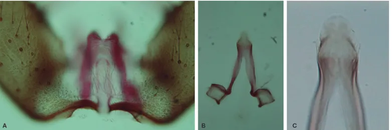
dark-scaled, with pale scales at base and apex. Male genitalia (Figs. 1
and 2) – Segment VIII: tergum and sternum moderately narrow, with spatulate scales and long setae. Segment IX: sternum moderately long, subtrapezoidal. Dorsal claspette: pedicel long, moderately broad to moderately narrow, base rounded; leaflets broad, length 0.05-0.09 mm (mean = 0.06 ± 0.03) (n = 10); dorsal leaflet with prominent, large basomesal projection. Ventral claspette: moderate-ly short, lateral margins not strongmoderate-ly tapering toward apex; apex broad, width at apex about 0.44-0.53 (mean = 0.49 ± 0.03) (n = 7) length of claspette; basal lobule moderately expanded laterally, rounded distally; spicules along basal margin short, evenly distribut-ed over basal surface; spicules long, more abundant on basomesal margin; ventral and lateral surfaces, including basal lobule, with short spicules, extending to or nearly to apex; apex with abruptly angled, rounded, sclerotized, lateral margins; preapical plate moder-ately small, circular, weakly sclerotized. Phallosome: Aedeagus length 0.13-0.19 mm (mean = 0.18 ± 0.02) (n = 8); apex moderately rounded, wider than long; subapical leaflets variable, present or ab-sent (when preab-sent, small, membranous, weakly sclerotized, non-serrated).
Pupa (Fig. 2). Position and development of setae as figured; range
and modal number of branches in Table 2. All measurements are from 20 specimens unless otherwise indicated. Integument without distinctive pattern of dark spots, mostly yellowish; sternum II with central pentagonal dark area, with minute spicules anteriorly on
ventral surface. Cephalothorax: setae 1,2-CT usually double, 2-CT
shorter than 1,3-CT, 4-5-CT usually triple, 6,8-CT single or double, 7,9-CT usually double, 10,12-CT single to triple. Trumpet: length 0.35-0.43 mm (mean = 0.38 ± 0.02), width 0.08-0.10 mm (mean = 0.09 ± 0.02) (n = 20); pinna moderately pigmented, light brown,
about 2.0-4.0 (mean = 2.98 ± 0.52) (n = 15) length of meatus.
Abdo-men: length 2.26-2.79 mm (mean = 2.54 ± 0.13). Seta 1-I dendritic,
number of branches not counted, 5-I single to triple, long, 9-I single, rarely double, long; 1-II, III moderately long, developed; 1-IV-VII sin-gle, long, strongly developed, pigmented; 9-II short, unpigmented, 11-II single, present or absent; 5-III long, developed, 9-III short, light-ly pigmented; 5-IV frequentlight-ly triple , 9-IV short, thick, heavilight-ly pig-mented, 1.72-3.25 (mean = 2.3 ± 0.44) length of seta 9-III; 5-V, VI single to triple, usually single, long, developed, 9-V thick, pigmented, acuminate, lightly curved, 1.44-2.22 (mean = 1.78 ± 0.22) length of seta 9-IV, 9-VI strong, pigmented, acuminate, curved, 0.85-1.40
Table 2.
Number and range (mode) of setal branches of the pupa of Anopheles goeldii collected in Santarém and Belterra, state of Pará, Brazil (n = 40).
Seta Cephalothorax Abdominal segments Paddle
No. CT I II III IV V VI VII VIII IX P
0 – – 2-6 (4) 4-8 (5) 4-7 (5) 3-6 (4) 2-5 (4) 3-7 (4) 1, 2 (1) – –
1 2, 3 (2) n.c.a 7-12 (9) 4-8 (5) 1 1 1, 2 (1) 1 – n.c. 1
2 2, 3 (2) 4-7 (5) 4-8 (6) 2-6 (4) 1-3 (2) 1-3 (2) 1-3 (2) 1-4 (2) – – 1, 2 (2)
3 2-4 (3) 1, 2 (1) 1, 2 (1) 1-3 (1) 3-6 (5) 1-4 (3) 1-3 (2) 2-5 (4) – – –
4 2-5 (3) 2-5 (4) 2-5 (4) 2-7 (3) 2-5 (4) 1-5 (3) 1-4 (2) 1-3 (1) 2, 3 (3) – –
5 2-5 (3) 1-3 (1) 2-6 (4) 4-9 (6) 1-6 (3) 1-3 (1) 1-3 (1) 1, 2 (1) – – –
6 1, 2 (2) 1, 2 (1) 1-3 (1) 1-3 (1) 1, 2 (1) 1, 2 (1) 1, 2 (1) 1, 2 (1) – – –
7 1-3 (2) 1-4 (3) 1-6 (3) 2-6 (4) 2-5 (4) 1-4 (3) 1 1, 2 (1) – – –
8 1, 2 (1) – – 2-5 (3) 1-4 (3) 1-4 (2) 1-4 (3) 3-6 (3) – – –
9 1-3 (2) 1, 2 (1) 1, 2 (1) 1 1 1 1 1 1 – –
10 1-3 (1) – – 1-4 (3) 1 1 – 1, 2 (1) – – –
11 4-8 (5) – 1 1, 2 (1) 1, 2 (1) 1, 2 (1) 1, 2 (1) 1-3 (2) – – –
12 1-3 (2) – – – – – – – – – –
13 – – – – – – – – – – –
14 – – – – 1 1 1 1 1 – –
a n.c. = not counted. Table 1.
Wing spot measurements (in mm) for male and female of Anopheles goeldii collected in Santarém and Belterra, state of Pará, Brazil.
Wing spot Range Mean SD (±) n =
Female
Basal pale + prehumeral pale 0.155-0.266 0.192 0.022 10
Prehumeral dark 0.102-0.225 0.159 0.034 10
Humeral pale 0.11-0.184 0.145 0.019 10
Humeral dark 0.094-0.18 0.126 0.026 10
Presector pale 0.04-0.118 0.089 0.02 10
Presector dark 0.278-0.397 0.343 0.035 10
Sector pale 0.081-0.11 0.098 0.008 6
Sector dark 0.036-0.102 0.059 0.02 6
Accessory sector pale 0.073-0.135 0.101 0.018 6
Distal sector dark 0.532-0.688 0.603 0.042 10
Subcostal pale 0.127-0.27 0.2 0.042 10
Preapical dark 0.52-0.651 0.588 0.036 10
Preapical pale 0.155-0.327 0.241 0.037 10
Apical dark 0.065-0.127 0.093 0.017 10
Male
Basal pale + prehumeral pale 0.151-0.196 0.167 0.014 10
Prehumeral dark 0.102-0.225 0.162 0.031 10
Humeral pale 0.094-0.192 0.147 0.028 10
Humeral dark 0.065-0.204 0.134 0.039 10
Presector pale 0.024-0.143 0.086 0.033 10
Presector dark 0.336-0.442 0.396 0.027 10
Sector pale 0.028-0.122 0.081 0.022 10
Sector dark 0.04-0.163 0.107 0.032 10
Accessory sector pale 0.09-0.282 0.158 0.047 10
Distal sector dark 0.34-0.594 0.508 0.07 9
Subcostal pale 0.204-0.311 0.249 0.028 9
Preapical dark 0.389-0.561 0.485 0.053 9
Preapical pale 0.188-0.274 0.231 0.03 10
Figure 2. Pupa and male genitalia of Anopheles goeldii. Pupa, CT: cephalothorax; Pa: paddle; I-IX: abdominal segments. Male genitalia. Scales in mm.
1.0 mm
0.1 mm
CT
I
II
III
IV
V
VI
VII
VIII
GL
(mean = 1.13 ± 0.15) length of 9-V; 5-VII frequently single, long, 9-VII thick, pigmented, acuminate, curved, about 1.08-1.68 (mean = 1.30 ± 0.18) length of 9-VI; 9-VIII strong, pigmented, about 0.95-1.39 (mean = 1.16 ± 0.12) length of 9-VII. Paddle: length 0.67-0.78 mm (mean = 0.72 ± 0.03), width 0.49-0.55 mm (mean = 0.52 ± 0.02), obovate, with round apex, weakly emarginated at insertion of 1-Pa, lightly pig-mented, buttress slightly darker, midrib faint, outer basolateral ser-ration prominent, filamentous spicules on outer apical margin and most of inner margin prominent, setae 1-Pa strong, dark-pigmented, 2-Pa single or double.
Fourth instar larva (Fig. 3). Position and development of setae as
figured; range and modal number of branches in Table 3. All
mea-surements from 20 specimens, unless otherwise indicated. Head:
integument light brown to yellowish, with dark spots, not forming distinct pattern; length 0.42-0.54 mm (mean = 0.49 ± 0.04) (n = 11), width 0.54-0.58mm (mean = 0.56 ± 0.01) (n = 11). Setae 2,3-C single, barbed, 2-C length 0.14-0.18mm (mean = 0.14 ± 0.05) (n = 10), 3-C length 0.11-0.14 mm (mean = 0.13 ± 0.01) (n = 11); 0.04-0.06 mm (mean = 0.04 ± 0.01) (n = 10) distance between bases of 2-C; 0.06-0.08 mm (mean = 0.06 ± 0.02) (n = 10) distance between bases of 2-C and 3-C; 4-C frequently double, forked; 5-C long, plumose; 9-C with
4-8 branches. Collar dark brown, heavily pigmented. Antenna:
length 0.19-0.29 mm (mean = 0.25 ± 0.03) (n = 11), width 0.03-0.04 mm (mean = 0.04 ± 0) (n = 11), slightly pigmented; 1-A small,
insert-ed 0.05-0.07 mm (mean=0.06 ± 0.01) (n = 11) distant from base.
Tho-rax: setae 1-2-P normally on separate tubercles, 1-P palmate,
lance-olate, moderately narrow, pigmented leaflets; 3-T palmate,
moderately narrow, semitransparent leaflets. Abdomen: setae
0-II-VII moderately long; 1-I-0-II-VII palmate, 1-I moderately narrow, semi-transparent leaflets, 5-I moderately short, usually triple, 13-I short; 1-II, VII with leaflets slightly smaller than those from setae 1-III-VI; 2-IV, V usually single; 13-V larger than 13-IV. Pecten plate with 4-6 long spines alternating with 10-14 short spines, long spines length 0.09-0.1 mm (mean = 0.1 ± 0) (n = 11), short spines length 0.04-0.05 mm (mean = 0.04 ± 0) (n = 11); setae 1-X long, single, inserted at ventral border of saddle.
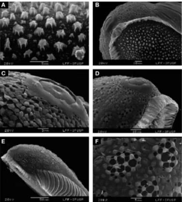
Eggs (Fig. 4). Length 361-400 µm (mean = 389 ± 10.37); width
104-110 µm (mean = 107 ± 1.7), ratio of length/width 3.37-3.74 (mean
= 3.63 ± 0.1) (n = 13).
Overall appearance. Black in color, boat-shaped in dorsal and
lat-eral views (Figs. 4A and 4C), in latlat-eral view the contour is slightly concave dorsally and curved ventrally (Fig. 4C). Floats lateral in posi-tion, long, well developed, not reaching the end of the egg (Figs. 4D and 4E), dorsal frill in the anterior and posterior poles, frill of the anterior pole is more developed than the posterior pole, connected
to the float (Fig. 4A-B, 5C). Dorsal surface. Deck with anterior end
wider than the posterior end (Fig. 4B). Deck with tubercles irregular-ly shaped with tiny tubercles intermixed with larger ones (Figs 4F and 5A); tubercles present on anterior and posterior parts of deck
larger and more spacious than those on middle part (Fig. 4B). Ventral
and lateral surfaces. Composed of chorionic cells (Figs. 4D-E and 5F)
with rounded pores of different sizes, central pores larger. Float
de-veloped, occupying a lateral position (Figs 4C, 4E, and 5E). Anterior
end. Rounded (Figs. 4B, 5B, and 5D), frill well developed involving all
dorsal anterior end of the egg (Figs. 4A and 4B). Micropylar collar separated from lower frill margin (Figs. 5B and 5D), collar surface smooth, micropylar disk with a continuous ring, with 7, 8 sectors,
and limited by short rays (Fig. 5C). Posterior end. Narrower than
an-terior end, with rounded contour, frill developed (Figs. 4A and 4B).
Molecular characterization. The inferred phylogeny was pre-sented by Foster et al. (2013) using DNA sequence data from the
white nuclear gene, the carbamoyl-phosphate synthetase 2,
aspar-tate transcarbamylase, and dihydroorotase(CAD) nuclear gene, and
the cytochrome oxidase subunit I (COI) mitochondrial gene. The
sta-tistical support values for the An. goeldii lineage varied depending on
the gene and concatenated data sets. Accordingly, the support from the three concatenated genes varied from 0.600 to 0.790 Bayesian posterior probability depending on the data partition, whereas only
the COI gene failed to support the monophyly of An. goeldii and its
closest species, An. nuneztovari. The statistical support for the clade
composed of An. nuneztovari and An. goeldii varied depending on the
gene and concatenated data sets. The monophyly of An. nuneztovari
was strongly supported by the white gene and the three
concatenat-ed genes (see Foster et al., 2013, for further details).
Distribution. Considering that An. goeldii was resurrected from
the synonymy with An. nuneztovari by Gabaldón (1981), and the
Table 3.
Number and range (mode) of setal branches of the fourth instar larva of Anopheles goeldii collected in Santarém and Belterra, state of Pará, Brazil (n = 40).
Head Thorax Abdominal segments
No C P M T I II III IV V VI VII VIII X
0 1 1 – – – 3-8 (6) 3-7 (5) 4-8 (5) 5-8 (6) 4-8 (5) 3-8 (5) 2-5 (3) –
1 1 11-16 (13) 23-34 (29) 1, 2 (1) 8-17 (14) 19-27 (23) 21-32 (26) 21-29 (26) 20-32 (26) 19-28 (25) 18-29 (25) 1 1, 2 (1)
2 1 12-23 (17) 1-3 (1) 1 2-4 (3) 4-8 (6) 3-6 (4) 1 1-3 (1) 3-9 (5) 5-9 (6) 5-10 (8) 15-22 (19)
3 1 1 1 10-20 (14) 1, 2 (1) 1-3 (1) 1 2-4 (3) 1 1 2-4 (3) 7-16 (12) 6-12 (8)
4 1-3 (2) 14-21 (17) 1-4 (3) 2-5 (3) 3-6 (4) 4-7 (5) 2-4 (3) 2-4 (3) 2-5 (3) 1 1 1 8
5 14-23 (16) 26-41 (32) 1 29-42 (37) 2-4 (3) 5-10 (8) 5-12 (7) 3-5 (4) 4-7 (6) 5-9 (7) 5-10 (8) 5-10 (6) –
6 11-22 (15) 1 2-6 (3) 2, 3 (2) 30-40 (32) 31-43 (35) 21-36 (26) 1 1 1, 2 (1) 3-6 (5) 1-S 4-8 (6)
7 15-24 (18) 27-43 (31) 3-5 (3) 28-40 (36) 25-38 (28) 26-40 (30) 3-5 (4) 3-5 (4) 2-4 (3) 1-3 (3) 4-7 (5) 2-S 3-7 (5)
8 1-4 (3) 28-40 (35) 17-29 (22) 29-43 (35) – 2, 3 (3) 2-4 (3) 2, 3 (3) 2-4 (3) 2-4 (3) 4-8 (5) 6-S 1, 2 (1)
9 4-8 (6) 1 1 1 3-7 (5) 6-11 (7) 6-12 (10) 5-11 (8) 6-12 (9) 5-13 (9) 6-12 (8) 7-S 1-3 (2)
10 1-4 (3) 1 1 1 1 2-4 (3) 1 1 1 2-4 (3) 3-6 (5) 8-S 2-5 (4)
11 n.c.a 1-3 (2) – – 2-5 (3) 1 2, 3 (2) 2, 3 (2) 1-3 (2) 1-3 (2) 1-3 (2) 9-S 2-5 (4)
12 2-4 (3) 1 1, 2 (1) 1-3 (2) 1-3 (2) 1 2-4 (3) 2-5 (3) 1, 2 (2) 1 1, 2 (1) – –
13 2-5 (4) 3-4 (3) 4-9 (6) 2-4 (2) 4-8 (6) 6-11 (7) 6-10 (7) 4-8 (5) 4-7 (5) 6-14 (8) 4-6 (5) – –
14 n.c. 5-10 (7) 6-13 (10) – – 1 1 1 1 1 1 1 –
15 3 – – – – – – – – – – – –
Figure 3. Fourth-instar larva of Anopheles goeldii. A: antenna; C: cranium; Dm: dorsomentum; M: mesothorax; P: prothorax; T: metathorax; Vm: ventromentum; I-VIII: abdom-inal segments; X: anal lobe. Scales in mm.
0.5 mm
1.0 mm
VII
VI V IV III II T M P
VIII
X
1–IV
C
Dm
morphological similarity between these two species and Anopheles
(Nyssorhynchus) dunhami Causey, 1945, the geographical distribution
of An. goeldii is incompletely resolved. Calado et al. (2008) proposed
that the records from the published literature relative to An.
nunez-tovari cytotype A collected in localities along the Amazon River basin
in thenAmazonas, Pará, and Amapá states could also include An.
goel-dii. In addition, because Anopheles dunhami can be easily
misidenti-fied as either An. nuneztovari A or An. goeldii, the distribution of these
three species needs to be redefined. Ruiz et al. (2010) recorded the
occurrence of An. dunhami in Leticia, Colombia, at the Brazilian
bor-der, and Foster et al. (2013) confirmed the presence of An.
nunezto-vari A in the Rondônia state. Based on the specimens employed in the
present study, An. goeldii occurs in localities from the Amazonas,
Amapá, and Pará states within the Amazon River basin.
Medical importance. It is unknown; however, it is plausible to suppose that this species may be playing an important role, either as a primary or secondary vector, in the dynamics of human malaria in the Amazon River basin. In agreement with this hypothesis, Galardo et
al. (2007) demonstrated specimens identified as An. nuneztovari from
Pará and Amapá as infected with Plasmodium. In addition, Tadei and
Thatcher (2000) assessed the infectivity of mosquitoes identified as
An. nuneztovari and found that they were infected with Plasmodium
(Plasmodium) vivax Grassi & Feletti, 1890 and Plasmodium (Laverania)
falciparum Welch, 1897. Moreover, specimens of An. nuneztovari were
also found infected with Plasmodium malariae (Feletti & Grassi, 1889)
along the highway BR-174 (Tadei and Thatcher, 2000). Further studies
will be necessary to define the potential association of An. goeldii with
the dynamics of malaria transmission in the Amazon.
Bionomics. Blood fed females were collected in corrals. Larvae and pupae were found in ground pools in pastures, in flooded areas, artificial ponds, in full sun or partially shaded. The larval habitat con-sisted of temporary, stagnant, fresh, clear, or turbid water, with abundant floating and emergent vegetation, the water temperature
averaging at 28 C. The mosquito species collected in these habitats
with An. goeldii were An. (Nyssorhynchus) marajoara (Galvão &
Da-masceno), 1942; An. (Nyssorhynchus) triannulatus (Neiva & Pinto,
1922); An. (Nyssorhynchus) braziliensis, An. (Nyssorhynchus) darlingi
(Root, 1926); An. (Anopheles) intermedius (Peryassu, 1908); An.
(Anopheles) peryassui (Dyar & Knab, 1908); Aedeomyia (Aedeomyia)
squamipennis (Lynch Arribalzaga, 1878); Culex (Culex) mollis (Dyar &
Knab, 1906); Culex (Melanoconion) bastagarius (Dyar & Knab), 1906;
Culex (Melanoconion) batesi (Rozeboom & Komp, 1948); Culex (
Mela-noconion) evansae (Root, 1927); Culex (Melanoconion) theobaldi (Lutz,
1904); Culex (Melanoconion) intrincatus (Brethes, 1916); Culex (
Mela-noconion) idottus (Dyar, 1920); Culex (Melanoconion) clarki (Evans,
1924); Culex (Melanoconion) eknomios (Forattini & Sallum, 1992);
Culex (Melanoconion) vaxus (Dyar, 1920); Uranotaenia (Uranotaenia)
pulcherrima (Lynch Arribalzaga, 1891); and Uranotaenia (
Uranotae-nia) geometrica (Theobald, 1901).
Material examined for description.The specimens of An. goeldii
used for morphological descriptions were collected in Brazil, state of
Pará, Urumanduba municipality, (228’56.2”S, 5439’39.3”W), Bergo
et al. coll., 08-Sep-2008, Sallum det., 2008: PA3(6)-2 [MG], PA3(6)-3 [LePe], PA3(6)-5 [F], PA3(6)-9 [F], PA3(6)-12 [LePe], PA3(9)-1 [MG], PA3(9)-4 [FLePe], PA3(9)-6 [LePe], PA3(10)-1 [MG], PA3(10)-7 [LePe], PA3(10)-8 [F], 1 [G], 2 [LePe], 4 [F], PA3(11)-16 [LePe], PA3(11)-20 [LePe], PA3(15) [E], PA3(15)-3 [MG], PA3(15)-11 [LePe], PA3(16)-1 [MG], PA3(16)-16 [LePe], PA3(16)-17 [LePe]. State of
Pará, Santarém municipality, Bom Jardim (233’7.9”S, 5435’38.7”W),
Bergo et al.coll., 09-Sep-2008, Sallum det., 2008: PA5(4)-1 [MG].
Sta-te of Pará, BelSta-terra municipality, São Domingos (245’7.2”S,
551.0’4.9”W), Bergo et al. coll., 10-Sep-2008, Sallum det., 2008:
1 [MG], 6 [F], 10 [LePe], 17 [F], PA7(2)-19 [LePe], PA7(3)-1 [MG], PA7(3)-8 [G], PA7(4)-2 [MG], PA7(4)-15 [LePe], PA7(6) [E], PA7(6)-1 [G], PA7(7) [L], PA7(7)-4 [LePe], PA7(7)-8 [FLePe], PA7(16)-4 [FLePe], PA7(16)-7 [FLePe], PA7(17)-1 [MG],
Figure 4. Anopheles goeldii, scanning electron microscope. A, B, Entire egg, dorsal view. C, Lateral view. D, Ventral view, posterior pole. E, Entire egg, ventral view. F, Central deck, tubercles. Scales in µm.
PA7(17)-12 [LePe]. State of Pará, Belterra municipality, Porto Novo
(237’58.5”S, 5458’32.9”W), Bergo et al. coll., 13-Sep-2008, Sallum
det., 2008: PA12(1)-3 [LePe].
Other material examined. Molecular characterization: The
spec-imens of An. goeldii were collected in Brazil, state of Amazonas,
mu-nicipality of Itacoatiara (308’4 8.3”S, 5823’39.7”W), Hutchings &
Sallum coll., 01-June-2005, Sallum det., 2005: BRAM03-1. State of
Pará, Prainha municipality, Comunidade Terra Preta (24.0’49.3”S,
5335’27.1”W), Sallum coll., 06-Nov-2005, Sallum det., 2005:
BRAM22-101. State of Pará, Belterra municipality, São Domingos
(245’7.2”S, 551.0’4.9”W), Bergo et al., 10-Sep-2008, Sallum det.,
2008: PA7(2)-2, PA7(3)-8, PA7(4)-3, PA7(17)-2. Morphology: Speci-mens were collected in Brazil, state of Amapá, municipality of
Ma-capá, Abacate da Pedreira (016’17.5”N, 5053’53.3”W), Bergo et al.
coll., 25-Jul-2006, Sallum det., 2006: AP15. State of Amapá,
munici-pality of Macapá, Abacate da Pedreira (017’43.6”N, 5052’39.8”W),
Bergo et al. coll., 25-Jul-2006, Sallum det., 2006: AP16; Abacate da
Pedreira (016’17.5”N, 5053’53.3”W), Bergo et al.coll., 27-Jul-2006,
Sallum det., 2006: AP20.
Discussion
Currently, An. goeldii is a valid species included in the Nuneztovari
Complex of the Albimanus Section with An. dunhami, An. nuneztovari
and An. nuneztovari A (Foster et al., 2013). The published literature
records relative to these species may include one or even more
spe-cies under the name An. nuneztovari because morphological
identifi-cation is not an easy task if based on female characteristics only. In
addition, An. goeldii and An. dunhami were both in the synonymy of
An. nuneztovari. Gabaldón (1981) resurrected An goeldii, whereas
Peyton (1993) validated An. dunhami.
Gabaldón (1981) defended that An. goeldii was a valid species
be-cause it can be easily differentiated from An. nuneztovari by the
length of the subapical leaflets of the aedeagus of the male genitalia, in addition to larval characters. In contrast, Faran (1980) considered
that the subapical leaflets are either present or absent in An.
nunez-tovari, but when present they are membranous. Savage (1986)
exam-ined the type and the paratypes of An. nuneztovari, reporting that the
leaflets are always present and are well developed. In addition, with the objective of addressing geographical variation of morphological traits of the male genitalia, Hribar (1994) compared the aedeagus of
individuals from An. nuneztovari cytotype A from Brazil and
Surina-me, cytotype B from Venezuela, and cytotype C from Venezuela and Colombia. He showed that the subapical leaflets are longer and more
sclerotized in An. nuneztovari B from Venezuela, whereas they are
either poorly developed or absent in An. nuneztovari A from the three
localities in Brazil. Anophelesnuneztovari C from Colombia was
con-sidered morphologically more similar to An. nuneztovari A than to An.
nuneztovari B. In contrast, Sierra et al. (2004), using DNA sequence
data from the ribosomal internal transcribed spacer 2 (ITS2), showed
that An. nuneztovari B and C are conspecific.
Considering the geographical origin of the individuals examined by Hribar (1994), it is plausible to assume that he might have
includ-ed specimens of An. goeldii that were misidentified as An. nuneztovari
A in the pool of samples from Brazilian localities in the Amapá, Pará, and Amazonas states. Further studies will be necessary to verify this hypothesis. In addition, the discrepancies between Faran’s (1980) and Savage’s (1986) studies may be likely caused by the fact that within the pool of specimens examined by Faran (1980) there were
individuals of An. nuneztovari, An. goeldii and An. dunhami that were
misidentified as An. nuneztovari, whereas Savage examined and
illus-trated the holotype and paratypes of An. nuneztovari.
Specimens of An. goeldii from localities in the Belterra
municipal-ity, Pará state, possess aedeagal leaflets variable in development. They can be present or absent; when present they are not easily vis-ible, the leaflets are minute and membranous (Figs. 1 and 2), and can be folded depending on the mounting procedure. Consequently, the presence or absence of aedeagal leaflets do not seem to be a
charac-ter that can be employed for An. goeldii identification. In contrast, the
species can be distinguished from An. nuneztovari based on the
preapical plate of the ventral claspette of the male genitalia, which is moderately small, circular, weakly sclerotized, and by the basal lob-ule, which possesses long spicules distributed along the basal loblob-ule, but are denser in the inner margin. This finding corroborates the de-scription by Rozeboom and Gabaldón (1941).
Ramos et al. (2008) performed a comparative analysis of eight
costal wing spots of females and males of An. nuneztovari from three
populations from Colombia. They compared the ratio of the length of the humeral pale spot (HP) and the length of the pre-humeral dark spot (PHD), and the ratio of the length of the subcostal pale spot (SCP) and the length of the distal sector dark spot (DSD). Additional-ly, Ramos et al. also employed a character that is included in identi-fication keys, i.e., the ratio of the length of the basal dark area of the hind tarsomere 2 (Ta-III2) and the entire length of the hind tar-somere II. In the present study, we employed the same set of
charac-ters to contrast females of An. goeldii with An. nuneztovari from
Co-lombia. As a result, the ratio values of wing dark and pale spots (ratio
of HP/PHD and SCP/DSD) in An. goeldii were lower than those found
in An. nuneztovari by Ramos et al. (2008). Data obtained herein were
also contrasted to those published by Calle et al. (2002), showing
that for An. goeldii the values of ratio of the wing pale and dark spots
were lower than those found for An. nuneztovari from Colombia
(Ta-ble 4). In addition, the ratio of the length of the basal dark scale band
in the Ta-III2 and the entire length of the hind tarsomere II did not
Table 4.
Mean values and ratio of the length of the basal dark area of the hind tarsomere 2 (Ta-III2) and the entire length of the hind tarsomere II; the ratio of the length of the humeral pale spot (HP) and the length of the pre-humeral dark spot (PHD); the ratio of the length of the subcostal pale spot (SCP) and the length of the distal sector dark spot (DSD) in females of Anopheles goeldii and Anopheles nuneztovari.
DS-III2 / Ta-III2 HP /PHD SCP / DSD
Reference Patternsa n Mean Min. Max. SD Mean Min. Max. SD Mean Min. Max. SD
Ramos et al. (2008) I 267 0.32 0.21 0.42 0.05 1.28 0.38 3.25 0.42 0.47 0.21 0.70 0.08
II 37 0.30 0.24 0.35 0.03 1.34 0.75 3.00 0.50 0.47 0.29 0.57 0.07
III 62 0.31 0.23 0.39 0.04 1.51 0.60 3.00 0.56 0.51 0.25 0.71 0.08
IV 8 0.33 0.29 0.39 0.04 1.80 0.71 4.00 1.09 0.38 0.25 0.43 0.06
V 22 0.32 0.24 0.41 0.04 1.35 0.80 2.25 0.41 0.43 0.18 0.64 0.12
Calle et al. (2002) - 20 0.3 0.25 0.35 0.02 1.74 1.2 2.3 0.43 0.5 0.36 0.71 0.09
This study - 20 0.28 0.23 0.38 0.05 0.96 0.77 1.16 0.12 0.33 0.21 0.42 0.08
distinguish An. goeldii from An. nuneztovari (Table 4). Based on
char-acters of the fourth instar larva, Gabaldón (1981) suggested that An.
goeldii can be distinguished from An. nuneztovari by characteristics of
the spiracular lobe. In An. goeldii, the median plate of the spiracle
possesses small lateral arms that are readily observed, whereas in
An. nuneztovari these lobes are small (see Sutil Oramas, 1976).
Spec-imens examined for the present study confirm the presence of small
lateral arms in An. goeldii.
Gabaldón (1981) proposed that specimens of An. goeldii from the
Brazilian Amazon have been misidentified as An. nuneztovari. Several
studies carried out to compare populations of An. nuneztovari from
Venezuela and Colombia with populations from Brazil showed that Venezuela and Colombia populations were similar; however, both were distinct from those populations that occur in the Brazilian Am-azon (Conn, 1990; Conn et al., 1993; Conn et al., 1998; Fritz et al., 1994; Kitzmiller et al., 1973; Mirabello and Conn, 2008; Onyabe and
Conn, 1999). Results of this study confirmed that An. goeldii is a valid
species, which can be distinguished from An. nuneztovari by
charac-teristics of the fourth instar larva, female, male, and male genitalia. In addition, DNA sequences from the COI barcode gene, when
ana-lyzed in combination with DNA sequences from the whitenuclear
gene and CAD nuclear gene, confirm the validation of the species.
The COI barcode only does not allow distinction between An. goeldii
and An. nuneztovari.
Acknowledgments
This investigation was financially supported by FAPESP (Grant 11/20397-7) and Conselho Nacional de Desenvolvimento Científico e Tecnológico (CNPq) (Grant 301666/2011-3).
Conflicts of interest
The authors declare no conflicts of interest.
References
Almeida, F., Suesdek, L., Motoki, M.T., Bergo, E.S., Sallum, M.A.M., 2014. Morphometric comparisons of the scanning electron micrographs of the eggs of Anopheles (Nyssorhynchus) darlingi Root (Diptera: Culicidae). Acta Tropica 139, 115-122. Bergo, E.S., Souto, R.N.P., Galardo, A.K.R., Nagaki, S.S., Calado, D.C., Sallum, M.A.M.,
2007. Systematic notes on Anopheles Meigen (Diptera: Culicidae) species in the state of Amapá, Brazil. Mem. Inst. Oswaldo Cruz. 102, 373-376.
Calado, D.C., Foster, P.G., Bergo, E.S., Santos, C.L.S., Galardo, A.K.R., Sallum, M.A.M., 2008. Resurrection of Anopheles goeldii from synonymy with Anopheles nuneztovari (Diptera, Culicidae) and a new record for Anopheles dunhami in the Brazilian Amazon. Mem. Inst. Oswaldo Cruz. 103, 791-799.
Calle, D.A.L., Quiñones, M.L., Erazo, H.F., Jaramillo-O, N., 2002. Morphometric discrimination of females of five species of Anopheles of the subgenus Nyssorhynchus from southern and northwest Colombia. Mem. Inst. Oswaldo Cruz. 97, 1191-1195. Conn, J.E., 1990. A genetic study of the malaria vector Anopheles nuneztovari from
western Venezuela. J. Am. Mosq. Contr. Assoc. 6, 400-405.
Conn, J.E., Mitchell, S.E., Cockburn, A.F., 1998. Mitochondrial DNA analysis of the neotropical malaria vector Anopheles nuneztovari. Genome 41, 313-327.
Conn, J.E., Puertas, Y.R., Seawright, J.A., 1993. A new cytotype of Anopheles nuneztovari from western Venezuela and Colombia. J. Am. Mosq. Contr. Assoc. 9, 294-301. Faran, M.E., 1980. Mosquito Studies (Diptera: Culicidae) XXXV - A revision of the
Albimanus section of the subgenus Nyssorhynchus of Anopheles. Contr. Am. Entomol. Inst. 15, 1-215.
Floch, H., Abonnenc, E., 1946. Sur A. nuñez-tovari et A. pessoai em Guyane Francaise. Table d’identification dês Nyssorhynchus guyanais. Inst. Pasteur Guyane Française et Territ. l’Inini. 126, 1-5.
Forattini, O.P., Sallum, M.A.M., Marques, G.R.A.M., Flores, D.C. 1997. Description of the eggs of Anopheles (Kerteszia) laneanus and Anopheles (Nyssorhynchus) antunesi (Diptera: Culicidae) by scanning electron microscopy. J Am Mosq Control Assoc. 13, 368-374. Forattini, O.P., 2002. Culicidologia Médica. São Paulo, Editora da Universidade de São
Paulo.
Foster, R.P.G., Bergo, E.S., Bourke, B.P., Oliveira, T.M.P., Nagaki, S.S., Sant’Ana, D.C., et al., 2013. Phylogenetic analysis and DNA-based species confirmation in Anopheles (Nyssorhynchus). PLoS ONE. 8, e54063.
Fritz, G.N., Conn, J.E., Cockburn, A.F., Seawright, J.A., 1994. Sequence analysis of the ribosomal DNA internal transcribed spacer 2 from populations of Anopheles nuneztovari (Diptera: Culicidae). Mol. Biol. Evol. 11, 406-416.
Gabaldón, A., 1981. Anopheles nuñez-tovari: importante vector y agente de malaria refractaria en Venezuela. Bol. Direc. Malar. Saneam. Amb. 21, 28-38.
Galardo, A.K.R., Arruda, M., D’Almeida Couto, A.A., Wirtz, R., Lounibos, L.P., Zimmerman, R.H., 2007. Malaria vector incrimination in three rural riverine villages in the Brazilian Amazon. Am. J. Trop. Med. Hyg. 76, 461-469.
Hribar, L., 1994. Geographical variation of male genitalia of Anopheles nuneztovari Gabaldón. Mosq. Sys. 26, 132-144.
Kitzmiller, J.B., Kreutzer, R.D., Tallaferro, E., 1973. Chromosomal differences in populations of Anopheles nuneztovari. Bull. WHO. 48, 435-455.
Lane, J., 1953. Neotropical Culicidae. Editora da Universidade de São Paulo, São Paulo. Mirabello, L., Conn, J.E., 2008. Population analysis using the nuclear white gene detects
Pliocene/Pleistocene lineage divergence with in Anopheles nuneztovari in South America. Med.Vet. Entomol. 22, 109-119.
Onyabe, D.Y., Conn, J.E., 1999. Intragenomic heterogeneity of a ribosomal DNA spacer (ITS2) varies regionally in the neotropical malaria vector Anopheles nuneztovari (Diptera: Culicidae). Insect Mol. Biol. 8, 435-442.
Peyton, E.L., 1993. Anopheles (Nyssorhynchus) dunhami, resurrected from synonymy with Anopheles nuneztovari and validated as a senior synonym of Anopheles trinkae (Diptera: Culicidae). Mosquito Sys. 25, 151-156.
Ramos, M.F., Obando, R.G., Suárez, M.F, López, D., Wilkerson, R.C., Sallum, M.A.M., 2008. Morphological analysis of three populations of Anopheles (Nyssorhynchus) nuneztovari Gabaldón (Diptera: Culicidae) from Colombia. Mem. Inst. Oswaldo Cruz 103, 85-92.
Rozeboom, L.E., Gabaldón, A., 1941. A summary of the “Tarsimaculatus” complex of Anopheles (Diptera: Culicidae). Am. J. Hyg. 33, 88-100.
Ruiz, F., Linton, Y.M., Ponsonby, D.J., Conn, J.E., Herrera, M., Quinones, M.L., et al., 2010. Molecular comparison of topotypic specimens confirms Anopheles (Nyssorhynchus) dunhami Causey (Diptera: Culicidae) in the Colombian Amazon. Mem. Inst. Oswaldo Cruz. 105, 899−903.
Sallum, M.A.M., Peyton, E.L., Harrison, B.A., Wilkerson, R.C., 2005. Revision of the Leucosphyrus Group of Anopheles (Cellia) (Diptera, Culicidae). Rev. Bras. Entomol. 49, 1-152.
Savage, H.M., 1986. Identification and location of the holotype and paratypes of Anopheles (Nyssorhynchus) nuneztovari Gabaldón (Diptera: Culicidae). Mosquito Sys. 18, 279-283.
Sierra D.M., Velez I.D., Linton, Y.M., 2004. Malaria vector Anopheles (Nyssorhynchus) nuneztovari comprises one genetic species in Colombia based on homogeneity of nuclear ITS2 rDNA. J. Med. Entomol. 41, 302-307.
Sutil Oramas, E., 1976. Redescription de la especie Anopheles (Nyssorhynchus) nunez-tovari Gabaldón, 1940, y su distribucion geografica en Venezuela. Bol. Dir. Malar. Saneam. Amb. 16, 33-45.
Tadei, W.P., Thatcher, B.D., 2000. Malaria vectores in the Brazilian Amazon: Anopheles of the subgenus Nyssorhynchus. Rev. Inst. Med. Trop. SP. 42, 87-94.