online | memorias.ioc.fiocruz.br Infection with the protozoan Trypanosoma cruzi is an
important cause of chronic myocardiopathy in endemic areas of South and Central America. Survivors of the acute infection usually pass into an asymptomatic phase: the so-called indeterminate form of Chagas disease. Ap-proximately 30% of T. cruzi infection will later progress to the chronic cardiac form, characterised by chronic fi-brotic myocarditis (Andrade 1991). Pathogenesis of the chronic cardiac form of Chagas disease is considered to be dependent on the development of delayed type hyper-sensitivity (DTH) (Tarleton 1995, 2001, Tarleton et al. 1996, dos Reis 1997), which is responsible for the fibrot-ic inflammatory cardiac lesions (Andrade 1999). A pre-vious study conducted in chronically infected dogs dem-onstrated that low doses of cyclophosphamide (CYCL) caused a rapid evolution from the indeterminate form of Chagas disease to active chronic myocarditis (Andrade et al. 1987). The responses to low doses of CYCL are attributed to the suppression of humoural and cellular re-sponses, the result of selective destruction of suppressor
T lymphocytes, their precursor cells or other elements in the host-immune suppressor network (Turk et al. 1972, Askenase et al. 1975, Schwartz et al. 1978, Andrade et al. 1997, Shevach et al. 2001, Murata et al. 2004, Lutsiak et al. 2005).These cells are now identified as regulatory T cells (T-regs), which express CD4+CD25+ markers and
are involved in the regulation and suppression of DTH (Shevach et al. 2001).
A significant increase in the number of the IDCs in acute and chronic myocarditis compared to normal controls or to the indeterminate form of disease has also been demonstrated (Andrade et al. 2000) through a quantitative evaluation of heart interstitial dendritic cells (IDCs) in the aforementioned CYCL-treated dogs. This suggests a direct relationship between the number IDCs and the intensity of inflammatory infiltration. The importance of these IDCs in the evolution of cardiac le-sions and in the pathogenesis of chronic myocarditis has also been evaluated (Andrade et al. 2000). These cells are “antigen presenting cells” (APCs) and exhibit the ca-pacity to bind to antigens and to stimulate T lymphocyte responses. Migration of the IDCs to the T-cell zone of the spleen may give rise to a continuous sensitisation of the heart to DTH.
According to Lutsiak et al. (2005), low doses of CYCL not only decrease the number of T-regs, but also lead to decreased functionality. CYCL treatment en-hances apoptosis and decreases homeostatic prolifera-tion of T-regs.
doi: 10.1590/0074-0276108062013004
+ Corresponding author: [email protected] Received 17 September 2012
Accepted 7 June 2013
Effect of treatment with cyclophosphamide in low doses upon the
onset of delayed type hypersensitivity in mice chronically infected with
Trypanosoma cruzi
:
involvement of heart interstitial dendritic cells
Torriceli Souza Thé, Renata Siqueira Portella, Marcos Lázaro Guerreiro, Sonia Gumes Andrade/+
Laboratório de Doença de Chagas Experimental, Autoimunidade e Imunidade Celular, Centro de Pesquisas Gonçalo Moniz-Fiocruz, Salvador, BA, Brasil
Acute infection with Trypanosoma cruzi results in intense myocarditis, which progresses to a chronic, asymptom-atic indeterminate form. The evolution toward this chronic cardiac form occurs in approximately 30% of all cases of
T. cruzi infection. Suppression of delayed type hypersensitivity (DTH) has been proposed as a potential explanation of the indeterminate form. We investigated the effect of cyclophosphamide (CYCL) treatment on the regulatory mechanism of DTH and the participation of heart interstitial dendritic cells (IDCs) in this process using BALB/c mice chronically infected with T. cruzi. One group was treated with CYCL (20 mg/kg body weight) for one month. A DTH skin test was performed by intradermal injection of T. cruzi antigen (3 mg/mL) in the hind-footpad and measured the skin thickness after 24 h, 48 h and 72 h. The skin test revealed increased thickness in antigen-injected footpads, which was more evident in the mice treated with CYCL than in those mice that did not receive treatment. The thickened regions were characterised by perivascular infiltrates and areas of necrosis. Intense lesions of the myocardium were present in three/16 cases and included large areas of necrosis. Morphometric evaluation of lymphocytes showed a predominance of TCD8 cells. Heart IDCs were immunolabelled with specific antibodies (CD11b and CD11c) and T. cruzi antigens were detected using a specific anti-T. cruzi antibody. Identification of T. cruzi antigens, sequestered in these cells using specific anti-T. cruzi antibodies was done, showing a significant increase in the number of these cells in treated mice. These results indicate that IDCs participate in the regulatory mechanisms of DTH response to T. cruzi infection.
In this study, we investigated the effects of low-dose CYCL treatment on the evolution of the chronic form of Chagas disease in mice. We administered a skin test for DTH response, evaluated the intensity of myocarditis, quantified T cells infiltrates (CD4/CD8) and evaluated the participation of the antigen presenting IDCs.
MATERIALS AND METHODS
Experimental animals - One hundred inbred BALB/c mice weighing between 15-20 g and raised in the ani-mal facilities of the Gonçalo Moniz Research Centre, Oswaldo Cruz Foundation were used. The mice were maintained in accordance with the ethical guidelines established by the Ethical Committee for the Use of Ex-perimental Animals. Thirty of these mice were used as normal controls and 70 were infected with T. cruzi.
T. cruzi strain - The Colombian strain classified as Biodeme Type III (Andrade & Magalhães 1997), corre-sponding to the taxa T. cruzi I (Anonymous 1999, Zin-gales et al. 2009), was used in this study.
Inoculum - Blood forms of trypomastigote at 1 x 10⁵
were obtained from infected mice that were inoculated by intraperitoneal injection.
Experimental groups - I: the infected animals were fol-lowed up to the chronic phase of infection (180 days). The survivors (60) were divided into two experimental groups: first group - 30 infected untreated mice, and second group - 30 infected mice treated with low-dose CYCL; II: 30 uninfected mice were maintained as controls.
Treatment with CYCL - CYCL was used in the com-mercial form Genuxal, Lot OM 105, ASTA Medical AG, Frankfurt, Germany.
Treatment schedule - The drug was intraperitoneally administered three times a week over four weeks at a dose of 20 mg/kg body weight.
Skin tests - To evaluate the cellular immune responses, a DTH skin test was performed using antigenculture forms (Warren medium) of T. cruzi with 90% epimastigotes.The cells were washed in phosphate buffered saline (PBS) (pH 7.2) with centrifugation. The pellet contained the culture forms and was frozen and thawed several times in liquid nitrogen. The antigenic extract was then filtered in a Mil-lipore 0.22 µm filter. Protein dosage was performed using the bicinchoninic acid assay (BCA) with a BCA Protein Assay Kit (Pierce Catal.2161297A). The protein concentra-tion for the sample used was adjusted to 2 mg/mL.
The skin test was performed in 15 mice from each experimental group at two time points: 24 h and 15 days after the CYCL treatment. The antigen was adminis-tered in a dose of 25 µL (50 µg of protein) intradermally in the right hind-footpad. The same volume of PBS was injected in the left hind-footpad and served as a control. Footpad thickness was measured with a digital calliper (Fisherbrand Digital Callipers, Traceable, Fisher Scien-tific) at 24 h, 48 h and 72 h after the antigen injection.
Histopathological studies - For each footpad mea-surement time point, three mice were sacrificed by ex-sanguination after anaesthesia with sodium
pentobar-bital. Blood was collected for serological tests. All of the sacrificed animals were necropsied and sections of the heart and skeletal muscle were divided and fixed in 10% formalin or cryopreserved in liquid nitrogen for histochemical studies. The hind-footpads from 10 mice from each group were longitudinally sectioned, fixed in buffered formalin and then submerged in an ethylenedi-amine tetraacetic acid solution for two-four weeks for de-calcification. The footpads from five animals from each group were cryopreserved for histochemical studies.
Immunohistochemistry - Immunolabelling of the CD4+ and CD8+ T-cell infiltrates was performed on cells
from the cryopreserved heart, skeletal muscle and foot-pad sections. The immunolabelling was performed us-ing the monoclonal antibodies anti-CD4 and anti-CD8 as primary antibodies (BD Biosciences Pharmingen). Biotin-conjugated IgG2c (Clone A92-1 anti-rat, Cata-logue 553909, BD Biosciences) was used as a secondary antibody. Cryopreserved sections of the heart were sec-tioned at a thickness of 5 µm and mounted on polylysine-prepared slides and were then fixed with dehydrated ace-tone. Blocking of endogenous peroxidase was performed using a 3% solution of hydrogen peroxide plus methanol. The slides were washed twice in PBS and incubated with the primary antibodies (anti-CD4, anti-CD8). Af-ter washing three times with PBS, slides were incubated with the respective biotinylated secondary antibodies for 30 min. The slides were then washed in PBS and treat-ed with streptavidin peroxidase (Sav-HRP Biosciences Pharmingen Catalogue 550946). After washing in PBS, the slides were incubated in 3,3’-diaminobenzidine tet-rahydrochloride (DAB). Nuclear staining was performed using methyl green pyronin stain and the slides were coverslipped using Permount resin.
Morphometric evaluation of inflammatory cells - Quantification of the inflammatory cells was performed in fixed sections stained with haematoxylin and eosin (H&E) and in cryostat sections histochemically stained for CD4 and CD8. Using a Zeiss optical microscope with 10X ocular and 40X objective lenses, an area of 60 mm2
corresponding to five non-successive 12 mm2 fields was
examined. The images were captured and evaluated us-ing the Axion Vision program. The sectional areas of inflammation were measured directly and the total num-ber of cells in the examined area was calculated. Statis-tical analysis was performed using the non-parametric Mann-Whitney U test (significance p < 0.05)
Immunolabelling of T. cruzi antigens in heart IDCs, parasites and parasite debris - Immunohistochemical staining of parasite antigens as intracellular parasites, extracellular particulate debris or deposits in the
mem-branes of heart IDCs was performed on 5-μm-thick
IgG produced in rabbits, at a 1:600 dilution in PBS with 2% Tween-20 for 30 min at 37ºC. After washing in PBS and 2% Tween-20, sections were incubated in normal goat serum (Vectastain - Elite) for 20 min for additional blocking of non-specific binding. After washing in PBS, the slides were incubated for 30 min at 37ºC with the sec-ondary antibody: goat anti-rabbit IgG-peroxidase con-jugate (Sigma) at a 1:800 dilution in PBS. The sections were developed with 2.4% DAB and 1% H2O2 plus 1% dimethyl sulphoxide (Sigma) at room temperature (RT). Sections were counterstained with 1% methyl-green for 15 min, dehydrated and mounted with Canadian balsam. Sections of hearts obtained from non-infected controls were subjected to the same treatment and staining for use as negative controls. Paraffin-embedded sections of the hearts of acutely infected mice that contained several parasite nests were processed for immunohistochemis-try following the same steps described above, but using a polyclonal anti-T. cruzi antibody.
Immunohistochemical identification of IDCs - A purified rat anti-mouse monoclonal antibody for CD11b (BD Pharmingen) and a purified hamster anti-mouse CD11c antibody (BD Pharmingen) were used. Sections of the hearts from infected mice of both untreated and treated groups and from non-infected controls were examined. Immunolabelling with the rat anti-CD11b monoclonal antibody was conducted on cryopreserved sections of the heart embedded in Tissue-Tek OCT com-pound (Miles Inc, Diagnostic Division Elkhart). After washing in PBS, the slides were treated with a sheep-anti-rat Ig-POD (peroxidase-conjugated) Fab fragment secondary antibody (Boehringer Mannheim Biochemi-cals) at a 1:300 dilution. Sections treated with the ham-ster anti-mouse CD11c primary antibody were treated with a goat anti-Syrian hamster IgG-POD secondary antibody (Jackson Immuno-Research) at a 1:500 dilution for 30 min at 37ºC. The sections were developed with 2.4% DAB and 1% H2O2 plus 1% dimethyl sulphoxide (Sigma) at RT. Sections were counterstained with 1% methyl-green for 15 min, dehydrated and mounted with Canadian balsam.
Quantitative evaluation of the number of IDCs pre-senting T. cruzi antigen by immunolabelling with spe-cific antibodies - The number of IDCs was evaluated
in 5-μm-thick paraffin-embedded sections by counting
the cells in 10 non-successive microscopic fields using a Zeiss Standard 18 microscope with 10X ocular and 40X objective lenses. The mean and standard deviation (SD) for the number of cells was established for each group and calculated for 1 mm2. Heart sections of non-infected
controls were processed in the same manner.
RESULTS
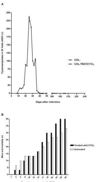
Parasitaemia - In the acute phase of infection with the Colombian strain, parasitaemia is indicative of the Biodeme Type III (T. cruzi I) infection pattern. A peak in parasitaemia at the 29th day post-infection was ob-served, followed by a progressive decrease until the chronic phase (60 days), when parasitaemia became negative (Fig. 1A).
Cumulative mortality - The cumulative mortality from the acute phase until 180 days post-infection was 40%. The cumulative mortality of these groups from the first day until the 30th day of treatment was also evalu-ated, which totalled 33% of the untreated controls and 46.6% in CYCL-treated group (Fig. 1B).
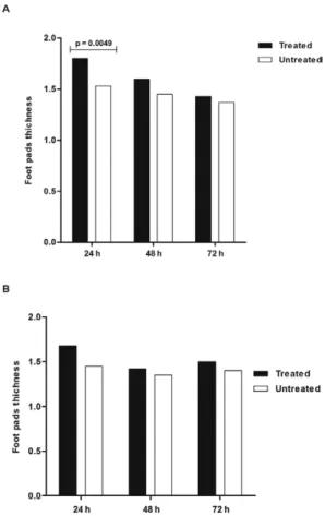
Skin test (DTH) - The evaluation of the results of the skin test was performed at two time points after the end of the treatment: the first evaluation occurred at 24 h and the second at 15 days after the end of treatment. In both cases, measurement of footpad thickness was per-formed at 24 h, 48 h and 72 h after antigen injection (Fig. 2A, B). The footpads injected with PBS did not pres-ent alterations when compared with normal controls. In
contrast, the footpads injected with antigen displayed increased thicknesses in both the untreated and CYCL-treated mice. The maximum reaction was observed 24 h after the antigen injection, decreased progressively and disappeared by 72 h. Footpad measurement revealed a predominance of the revealed greater thickness in the group treated with CYCL at the three time points. Sta-tistical analysis using the Mann-Whitney U test showed significant differences at 24 h after antigen injection in both the group injected 24 h after treatment with CYCL (p = 0.0049) (Fig. 2A) and the group injected 15 days after CYCL treatment (p = 0.0117) (Fig. 2B).
Histopathological evaluation of the skin test - First group (infected untreated mice): 24 h after antigen injec-tion, a diffuse mononuclear infiltration involving small vessels was present in the dermis (Fig. 3A, B); second group (infected mice treated with CYCL): necrotic areas were present in the perimuscular subdermic conjunc-tive tissue, along with polymorphonuclear neutrophils, diffuse mononuclear infiltration and arteritis (Fig. 3C). In the dermis, the presence of diffuse mononuclear cell
infiltration with accumulation around small vessels and cutaneous annexes was observed (Fig. 3D-F); third group (uninfected control mice): 24 h after antigen in-jection in the footpad, mild perivascular and subdermal mononuclear cell infiltration was observed. This infil-tration involved the muscles, with an absence of inflam-matory infiltrations in the dermis.
Histopathological evaluation of the myocardium and skeletal muscles - First group (infected untreated mice) myocardium: moderate, diffuse and focal mononuclear infiltrates in the atria, sub-epicardial and perivascular spaces was observed, along with the presence of mild, interstitial fibrosis (Fig. 4A). Skeletal muscles: inflam-matory lesions varied from mild to moderate and were limited to focal perivascular mononuclear cell infiltra-tion with involvement of the small arteries (Fig. 4B); sec-ond group (infected mice treated with low-dose CYCL) myocardium: the presence of moderate necrotic-inflam-matory lesions with focal and diffuse mononuclear infil-trations was present in most of the cases (10/16). Intense lesions in the myocardium were present in three/16 cases, with large areas of necrosis of cardiac myocells, diffuse
Fig. 3: histopathology of the delayed type hypersensitivity (DTH) skin tests. Untreated mouse chronically infected with Colombian strain of
Trypanosoma cruzi. A, B: sections of the skin with mild interstitial diffuse and perivascular mononuclear infiltrations; C: mouse treated with cyclophosphamide. Necrosis of the intradermal connective tissue and intense perivascular infiltration with mononuclear cells. Sections of the DTH skin test in untreated mouse; D-F: diffuse mononuclear infiltration in the dermis and involvement of the small vessels and cutaneous annexes. H&E 400X.
mononuclear infiltration and matritial fibrillar deposits (Fig. 4C, D). The atrial wall was predominantly involved, showing myocellular destruction with mononuclear cell infiltration and arteriolar involvement (Fig. 4E, F).
Skeletal muscle - Lesions were mild in most of cases, represented by focal perivascular mononuclear infil-trates and arteritis. Diffuse interstitial infiltration was present in some cases.
Morphometric evaluation of inflammatory cells - The numbers of mononuclear inflammatory cells in the heart and skeletal muscle sections were significantly higher in the CYCL-treated group as compared with those in the untreated group, as assessed by H&E staining (Fig. 5). The statistical analysis indicated that the differences in mononuclear cell numbers were significant in both the heart (p = 0.0191) and the skeletal muscle (p = 0.0149).
Immunohistochemical labelling of TCD4 and TCD8 cells in the myocardium - TCD4 and TCD8 cells were observed in both the interstitium and the perivascular areas (Fig. 6A, B).
Morphometric evaluation of the numbers of TCD4 and TCD8 cells - Assessment of the CD4/CD8 relationship in both untreated and treated mice revealed a predominance of CD8 cells; this difference was not significant for the untreated mice (p = 0.0809), but was significant for those treated with CYCL (p = 0.0495) (Fig. 7A, B).
Fig. 4: histopathology of the myocardium in mice chronically infect-ed with the Colombian strain of Trypanosoma cruzi. A, B: untreated controls.Focal and diffuse mononuclear inflammatory infiltration and moderate collagen deposits in the interstitium; C-F: treated with cyclophosphamide;C: perivascular area of cardiac cells disintegra-tion. Focal and interstitial mononuclear cells infiltration and fibrosis; D: myocardium with extensive areas of myocells destruction, diffuse mononuclear infiltration and fibrillar matritial deposits; E: focal ne-crosis of cardiac myocells with macrophages and lymphocytes infil-tration; F: epicardiac and myocardiac mononuclear cells infiltration with cardiac myocells dissociation. H&E 400X.
Fig. 5: morphometric evaluation of the number of inflammatory cells in the myocardium and skeletal muscles sections, stained with H&E. The media of the number of mononuclear inflammatory cells was significantly higher in the group treated with cyclophosphamide, as compared with the media in the untreated group. The statistical analy-sis have shown in the heart a significance of p = 0.0191 and in the skeletal muscle of p = 0.0149.
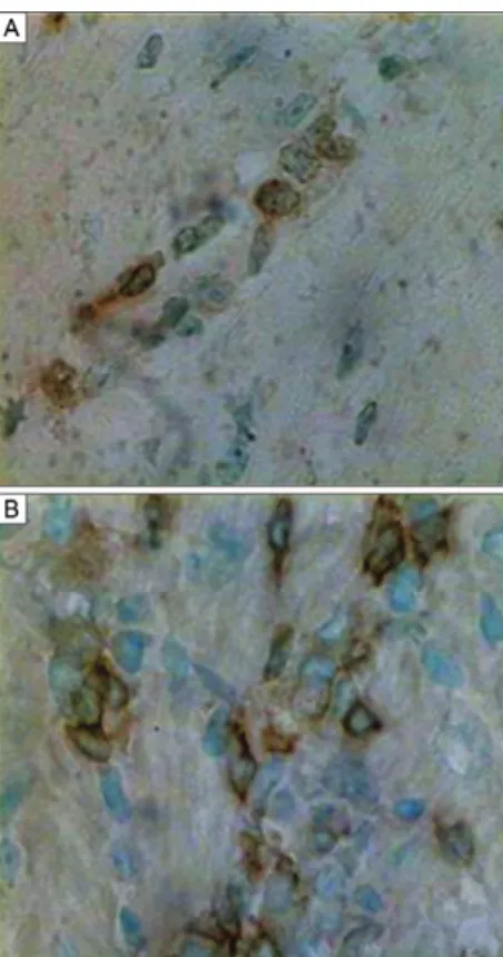
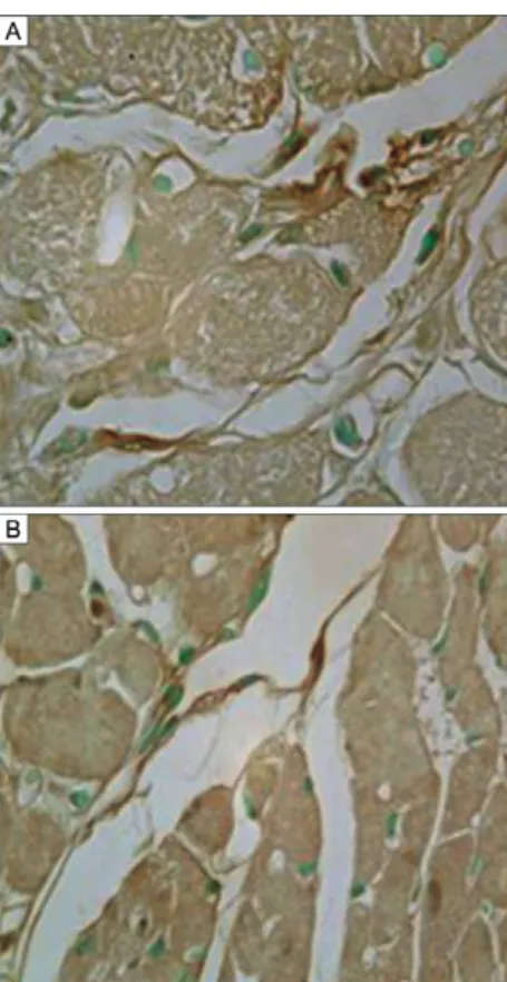
Immunolabelling of heart IDCs - Immunolabelling was performed using the monoclonal antibodies anti-CD11b and anti-CD11c, as shown inFig. 8A, B. The IDCs (arrows) appeared elongated and isolated, with compact cytoplasm, a central round pale nucleus and two or three fine cytoplasmic processes. For identification of T. cruzi
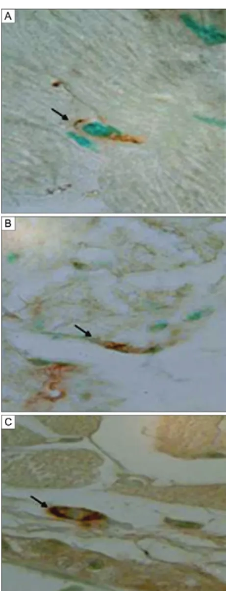
antigens in IDCs, immunolabelling was performed us-ing an anti-T. cruzi antibody in the heart tissue of chroni-cally infected mice not treated with CYCL (Fig. 9A, B) and in the heart tissue of mice treated with low-dose CYCL (Fig. 10C, D). Parasite antigens were visible as dense granular deposits in the IDC membranes.
Morphometric evaluation of IDCs - As seen in Fig. 10, a significant increase (p < 0.05) in the IDC numbers in the myocardia of CYCL-treated mice were significant-ly increased compared to the numbers in untreated mice.
DISCUSSION
In this study, low-dose treatment with CYCL de-termined the development of a DTH response in mice chronically infected with T. cruzi. The DTH response was characterised by the presence of a positive skin test following intradermal injection of T. cruzi antigens, which showed significant enlargement of the injected footpad and focal, perivascular, intradermic tory lesions. An exacerbation of the chronic inflamma-tory response in the heart, with predominance of TCD8 cells, was observed concomitantly with significantly
Fig. 8: histochemistry. Sections of the heart of mouse chronically in-fected with Trypanosoma cruzi treated with cyclophosphamide in low doses. Immunolabelling of the interstitial dendritic cells. A: with anti-CD11c monoclonal antibody; B, C: sections of the heart of untreated mice immunolabelled with anti-T. cruzi antibody. H&E: 1,000X. Fig. 7: morphometric evaluation of the number of TCD4 and TCD8
increased IDC numbers. Dendritic cells (DCs) are a major accessory cell for the activation of both T-cell subpopulations. Antigen-specific CD8+CD4- cells can
be induced to proliferate and become killer cells (Inaba et al. 1987). In this study, we demonstrated IDC stimu-lation in the myocardium by quantitative evaluation and observed a significant increase in the number of these cells expressing T. cruzi antigens in the hearts of treated mice. Previous studies based on a canine model of experimental Chagas disease (Andrade et al. 2000) have shown the evolution of the indeterminate form of the disease to chronic diffuse myocarditis after the use of low-dose CYCL, which was characterised by intense inflammatory infiltration and focal damage of myocardiocytes. The presence of a significant num-ber of T. cruzi antigen-expressing IDCs upon CYCL treatment confirms the importance of these cells as APCs. With this function, these cells can migrate to the T zone of the spleen and can stimulate the CD4/CD8 response in the heart.
In low doses, CYCL functions as an immunostimu-latory drug, as has been shown in studies in the field of cancer immunotherapy (Sistigu et al. 2011). CYCL markedly influences IDC homeostasis and promotes
interferon (IFN)γ secretion, contributing to the induc -tion of anti-tumour cytotoxic T-lymphocytes and the
proliferation of CD4 T-cells. This eventually affects the T-reg/T effector ratio in favour of tumour regression.
The immune response to infection with T. cruzi is dependent on several factors and is initiated by the DCs (Van Overtvelt et al. 2002, Chaussabel et al. 2003). T. cruzi downregulates lipopolysaccharide-induced major histocompatibility (MHC) class I presentation in human DCs and impairs antigenic presentation to specific TCD8 lymphocytes (Van Overtvelt et al. 2002, Chaussabel et al. 2003, Soto et al. 2003). Splenic white pulp was se-verely depleted of both CD4 and CD8 T-cells at the peak of T. cruzi infection. CYCL acts directly on IDCs to ini-tiate maturation, to process the antigens and to present to the CD4+ and CD8+ lymphocytes in the spleen. In vivo
infection with T. cruzi modulates APC functionality in a strain-dependent manner (Steinman 1991); highly viru-lent strains downregulate MHC II expression on splenic DCs and inhibit its induction on peritoneal macrophages and splenic B cells (Soto et al. 2003).
During the indeterminate phase of Chagas disease, development of DTH is inhibited. Several factors are involved in this process, including parasite direct ac-tion and the inhibiac-tion of heart IDCs. DCs mediate
T-helper 1 development and IFNγ production (Heufler et
al. 1996). These cells reside in the tissues in immature forms and respond to various chemo-attractants from sites of inflammation. The cells can be activated by microorganisms and inflammatory stimuli, after which they complete their maturation process and become po-tent stimulators of T-cells. Specific cytokines upregulate both the presentation and sensitisation functions of ac-cessory cells, which require the action of interleukin-1, a DC activator (Heufler et al. 1996). The impairment of the DTH determined by suppressor cells is responsible for the persistence of the infection until the chronic in-determinate form.
Fig. 9A, B: histochemistry. Sections of the heart of mouse chronically infected with T. cruzi, treated with cyclophosphamide in low dose. immunolabelling of interstitial dendritic cells with anti-Trypanosoma cruzi antibody. H&E: 1,000X.
Fig. 10: morphometric evaluation of interstitial dendritic cells (IDCs) in mice infected with the Colombian strain of Trypanosoma cruzi
A murine model has recently been used (Portella & Andrade 2009) to investigate the participation of heart IDCs carrying T. cruzi antigens in the maintenance of residual inflammatory infiltrates in chronically infected mice treated with benznidazole. The elimination of par-asites upon benznidazole treatment led to a regression of inflammatory lesions and a decrease in IDCs. Residual inflammatory infiltrates persisted in treated mice and were correlated with the presence of IDCs carrying par-asite antigens (Portella & Andrade 2009). This observa-tion raised the possibility that regression of the cellular immune response after specific chemotherapy with ben-znidazole, followed by parasitological cure, may depend on the clearance of the processed antigens expressed on the membranes of IDCs.
REFERENCES
Andrade SG, Magalhães JB 1997. Biodemes and Zymodemes of Try-panosoma cruzi strains: correlations with clinical data and ex-perimental pathology. Rev Soc Bras Med Trop30: 27-35.
Andrade SG, Pimentel AR, Souza MM, Andrade ZA 2000. Interstitial dendritic cells of the heart harbor Trypanosoma cruzi antigens in experimentally infected dogs: importance for the pathogenesis of chagasic myocarditis. Am J Trop Med Hyg63: 64-70.
Andrade ZA 1991. Pathogenesis of Chagas disease. Res Immunol142: 126-129.
Andrade ZA 1999. Immunopathology of Chagas disease. Mem Inst OswaldoCruz94 (Suppl. I): 71-80.
Andrade ZA, Andrade SG, Sadigursky M 1987. Enhancement of chronic Trypanosoma cruzi myocarditis in dogs treated with low doses of cyclophosphamide. Am J Pathol 127: 467-473.
Andrade ZA, Andrade SG, Sadigursky M, Wenthold Jr, Hilbert SL, Ferrans VJ 1997. The indeterminate phase of Chagas disease: ultrastructural characterization of cardiac changes in the canine model. Am J Trop Med Hyg 57: 328-336.
Anonymous 1999. Recommendations from a Satellite Meeting. Mem Inst Oswaldo Cruz94 (Suppl. I): 429-432.
Askenase PW, Hayden BJ, Gerson RK 1975. Augmentation of delayed type hypersensitivity by doses of cyclophosphamide which does not affect antibody response. J Exp Med141: 698-702.
Chaussabel D, Pajak B, Vercruysse V, Bisseyé C, Garzé V, Habib M, Goldman M, Moser M, Vray B 2003. Alteration of migra-tion and naturamigra-tion of dendritic cells and T-cell deplemigra-tion in the course of experimental Trypanosoma cruzi infection. Lab Invest 83: 1373-1382.
dos Reis GA 1997. Cell-mediated immunity in experimental Trypano-somacruzi infection. Parasitol Today13: 335-342.
Heufler C, Koch F, Stanzl U, Topar G, Wysocka M, Trinchieri G, Enk A, Steinman RM, Romani N, Schuler G 1996. Interleukin-12 is produced by dendritic cells and mediates T helper I development as well as interferon-gamma production by T helper I cells. Eur J Immunol26: 659-668.
Inaba K, Young JW, Steiman RM 1987. Direct activation of CD8+
cyto-toxic T lymphocytes by dendritic cells. J Exp Med166: 182-194.
Lutsiak CME, Semnani RT, Pascalis R, Kashmiri SVS, Schlom J, Sabzevari H 2005. Inhibition of CD4+25 T regulatory cell
func-tion implicated in enhanced immune response by low-dose cyclo-phosphamide. Blood105: 2862-2868.
Murata M, Suzuki T, Midorikawa K, Oikawa S, Kawanishi S 2004. Oxidative DNA damage induced by a hydroperoxide derivative of cyclophosphamide. Free Radic Biol Med 37: 793-802.
Portella RS, Andrade SG 2009. Trypanosoma cruzi: parasite anti-gens sequestered in heart interstitial dendritic cells are related to persisting myocarditis in benznidazole-treated mice. Mem Inst Oswaldo Cruz104: 1023-1030.
Schwartz A, Askenase PW, Gershon RK 1978. Regulation of delayed type hypersensitivity reactions by cyclophosphamide-sensitive T cells. J Immunol121: 1573-1577.
Shevach EM, McHugh RS, Piccirillo CA, Thornton AM 2001. Con-trol of T-cell activation by CD4+CD25+ suppessor T cells.
Immu-nol Rev182: 58-67.
Sistigu A, Viaud S, Chaput N, Bracci L, Proietti E, Zitvogel L 2011. Immunomodulatory effects of cyclophosphamide and implemen-tations for vaccine design. Semin Immunopathol33: 369-383.
Soto CDA, Mirkin GA, Solana ME, Cappa SMG 2003. Trypanosoma cruzi infection modulates in vivo expression of major histocom-patibility complex class II molecules on antigen-presenting cells and T-cell stimulatory activity of dendritic cells in a strain-de-pendent manner. Infect Immun71: 1194-1199.
Steinman RM 1991. The dendritic cell system and its role immunoge-nicity. Annu Rev Immunol9: 271-296.
Tarleton RL 1995. The role of T cells in Trypanosoma cruzi infec-tions. Parasitol Today 11: 7-9.
Tarleton RL 2001. Parasite persistence in the aetiology of Chagas dis-ease. Int J Parasitol31: 550-554.
Tarleton RL, Grusby MJ, Postan M, Glimcher LH 1996. Trypanoso-ma cruzi infection in MHC-deficient mice: further evidence for the role of both class I and class II-restricted T cells in immune resistance and disease. Int Immunol 8: 13-22.
Turk JL, Parker D, Poulter LW 1972. Functional aspects of the selec-tive depletion of lymphoid tissue by cyclophosphamide. Immu-nology23: 493-501.
Van Overtvelt L, Andrieu M, Verhasselt V, Connan JC, Choppin J, Vercruysse V, Goldman M, Hosmalin A, Vray B 2002. Trypano-soma cruzi down-regulates lipopolysaccharide-induced MHC class I on human dendritic cells and impairs antigen presentation to specific CD8+ T lymphocytes. Int Immunol 14: 1136-1144.
Zingales B, Andrade SG, Briones MRS, Campbell DA, Chiari E, Fer-nandes O, Guhl F, Lages-Silva E, Macedo AM, Machado CR, Miles MA, Romanha AJ, Sturm NR, Tibayrenc M, Schijman AG 2009. A new consensus for Trypanosoma cruzi intraspecific no-menclature: second revision meeting recommends TcI to TcVI.