Ananthi Radhakrishnan1, Ananthasubramanian M 2*
1
Department of Biotechnology, PSG College of Technology, Coimbatore, India; 2Corresponding Author: Ananthasubramanian.M, Associate Professor, PSG College of Technology, Coimbatore, India.
Submitted: December 04, 2010; Returned to authors for corrections: May 12, 2011; Approved: August 30, 2011.
ABSTRACT
Pseudomonas fluorescens phages from sewage were tested against P. fluorescens isolates of soil and
sewage. The phages were characterized as to host range, morphology, structural proteins and genome
fingerprint. Of the seven phages isolated, one was found to be abundant in sewage (5.9×10⁷ pfu/mL), having broad host range, and distinct protein and DNA profile when compared to the other six phages. DNA
restriction and protein profiles of the phages and their morphology indicate the diversity in the sewage
environment. None of the isolates from the rhizosphere regions of various cultivated soils were susceptible
to phages isolated from sewage.
Key words: Pseudomonas fluorescens, phages, RFLP, AFM, Protein analysis.
INTRODUCTION
Pseudomonas fluorescens is a diverse community of
bacteria, found in wide range of habitats including soil, water
and in industrial environments (34). Bacteriophages, the most
abundant life forms on earth play major roles in bacterial
ecology, adaptation to novel environments, and in bacterial
evolution and pathogenesis (23, 10). They have shown good
sensitivity and specificity in detecting bacterial pathogens (15).
In soil environment P. fluorescens is known to improve plant
growth and health. (8,17,24). P. fluorescens phages are thought
to be ecologically important in controlling bacterial number
and activity, affecting composition and diversity of beneficial
bacterial populations thereby affecting agricultural
productivity. Earlier studies have reported isolation of
fluorescent Pseudomonas phages from different environments
(1,5,9). According to the first ever global survey of wastewater
irrigation untreated sewage is used to irrigate 10 percent of the
world’s crops. Sewage should be treated to remove pathogens
to make it safe for irrigation (33). This study reports the
diversity of P. fluorescens phages in sewage with reference to
genome size, host range, structural proteins and morphology
and their lytic activity on soil bacteria.
MATERIALS AND METHODS
Sample Collection
Sewage sample was obtained from the inlet of the sewage
treatment plant located in Sulur, Coimbatore, India. The
samples were immediately transported to the laboratory in
collection bottles within 2 hrs and kept at 4°C prior to
processing. The samples were clarified by filtration through
cheese cloth and used for further analysis. Soil samples were
collected at the depth of 2m from the cultivated lands nearby
the sewage treatment plant and stored at 4°C.
Isolation of bacteria from soil and sewage
One mL of sewage sample was added to 9 mL sterile water
which makes 10-1 dilution and subsequently diluted up to 10-6. Serial dilutions of soil samples were also prepared by adding
one gram of soil into 9 mL of sterile water (10-1 dilution) and dilutions were made up to 10-6. The diluted samples were plated on King’s B agar medium and incubated at 30°C for 48
hrs. Distinct colonies showing fluorescence under UV light at
360 nm were picked and streaked on KB agar medium to check
the purity.
P. fluorescens isolated from sewage and from each of the
twelve soil samples were subjected to biochemical tests as
reported earlier Goszozynska et al (12) and Pickett et al (28).
P. fluorescens MTCC 103 and P.aeruginosa MTCC 2474 were
used as a referral strains. The bacterial cultures were grown at
30°C and 42°C with King’s B agar medium.
Bacteriophage isolation
P. fluorescens phages isolated from sewage with sewage
bacterial isolate as host was enriched by the modified method
of Smith and Huggins (35). Briefly, 50 mL of LB broth and 50
mL of tryptone broth were added to 150 ml of the sewage
water and incubated for 1 hr at 30°C. To this 5 ml of 24 hr P.
fluorescens broth culture was added and grown for 16 hours at
30°C. After centrifugation the supernatant was filtered through
0.22 µm membrane filter and stored at 4°C. 100µl of diluted
lysate mixed with 100µl of overnight bacterial culture were
mixed with 0.7% tryptone soft agar and overlaid on hard agar
and plaque forming units (PFU) were observed after incubated
overnight at 30°C. Isolated plaques were pooled and
concentrated using PEG (8000) precipitation method (30).
Susceptibility Analysis
Phage host range was examined using the spot test method
(3). Soft agar (3-5 mL) with 100 µl of an overnight bacterial
culture and equal volume of CaCl2 (300 mM) were gently vortexed and spread on the surface of hard agar. Single drops
of each phage lysate were spotted on the inoculated hard agar
plates, and the plates were incubated overnight at 30°C.
Phage DNA isolation
Phage DNA was extracted as reported earlier (16). Briefly,
0.5% of SDS and proteinase K (0.05mg/mL) were added to
0.5ml of phage lysate and heated for 15-30 minutes in a 65°C
water bath. A 1:1 phenol:chloroform extraction was performed
4 times followed by incubation with 0.3M Sodium acetate and
3 volumes of 100% cold ethanol at -20°C for 30 minutes. Then
70% ethanol wash was performed 3 times and then the DNA
was air dried and resuspended in TE buffer.
Restriction analysis of phage DNA
The Phage DNA isolates were digested with restriction
enzymes using a standard restriction digestion assay (32) as per
the manufacturer’s instructions (Fermentas). Of the five
enzymes tested (Eco RI, Hind III, Nhe I, Kpn I and Alu I)
two were selected (Eco RI, Hind III) based on the observation
of clear multiple bands on agarose gels. The restriction digests
were electrophoresed on 0.8% agarose gels in 1 X TAE buffer
stained with ethidium bromide and visualized by UV
photography. The phage genome sizes were determined by
using semi-log plotting method where, a logarithmic scale is
used to graph the results in semi-log paper. (11). Restriction
digested samples run on the agarose gel along with the 1 kb
ladder marker was used for semi-log plotting. The sum of the
size of the bands present in each sample is the approximate size
of the bacteriophage genome.
SDS-PAGE analysis of phage proteins
dodecyl sulfate-polyacrylamide gel electrophoresis (SDS
PAGE) as described by Laemmli (20). Samples were heated at
95°C for 15 min and subjected to electrophoresis with a 10%
polyacrylamide gel at 150 V for 2 hrs. Proteins were visualized
on gels stained with silver nitrate (7).
AFM analysis
The sample was prepared as described by Archer et al.,
(4). The virus particles were deposited for adsorption on
preliminary cleaned, glass cover slips. After adsorption of the
phage particles onto the surface, the substrates were allowed to
dry at room temperature and then carefully rinsed with Milli-Q
water. Imaging was performed using NTMDT-Atomic Force
Microscopy.
RESULTS
A total of 15 fluorescent bacteria were isolated from both
soil and sewage samples. Each soil sample was enriched with
soil isolates as their isolation host. Plaques were observed only
with phage lysates obtained from sewage. In total, 7 lytic
phages were isolated from sewage enrichment process. They
were selected based on their size and morphology of the
plaques. The size and plaques produced by the isolated phages
in all three months are listed in Table 1. The bacteriophage
load on both inlet and outlet of sewage treatment plant was also
assessed. Ninety percent of bacteriophage reduction was found
in outlet sample (data not shown).
The susceptibility of the bacterial isolates to infection by 7
phages was examined. All the 7 phages isolated, individually
lysed the sewage bacterial isolates Pfsw-13 and Pfsw-15 by the
spot test method. Phage #5 showed lytic activity against several
fluorescent isolates and also against P. aeruginosa MTCC
2474 standard strain. The susceptibility of the bacterial isolates
to these phages is shown in Table 2.
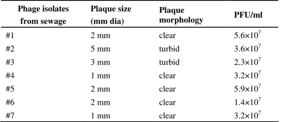
Table 1. Morphology and size of the plaques
Phage isolates from sewage
Plaque size (mm dia)
Plaque
morphology PFU/ml
#1 2 mm clear 5.6×10⁷
#2 5 mm turbid 3.6×10⁷
#3 3 mm turbid 2.3×10⁷
#4 1 mm clear 3.2×10⁷
#5 2 mm clear 5.9×10⁷
#6 2 mm clear 1.4×10⁷
#7 1 mm clear 3.2×10⁷
Table 2. Bacterial susceptibility to Pseudomonas phages
PHAGE ISOLATES
HOST SOURCE
#1 #2 #3 #4 #5 #6 #7
Pfsw-13 Sewage treatment plant S S S S S S S
Pfsw-14 Sewage treatment plant R R R R R R R
Pfsw-15 Sewage treatment plant S S S S S S S
P.fluorescens Reference strain S R S S S R S
P.aeruginosa Reference strain R S R R S S R
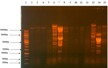
Genome fingerprinting analysis
The DNA isolated from all the phages were double
stranded and were sensitive to EcoRI and Hind III. Figure 1
shows the restriction pattern of the phage DNA. The estimated
genome sizes ranged from 26 to 82 Kb. The genome size of
each phage isolates was found to be similar when tested by the
two enzymes used. The genome fingerprint of the phage DNA
shows the diversity among the phage isolates.
Analysis of phage proteins
Phage lysates were examined using SDS-PAGE, stained
with silver nitrate. Multiple bands were present in each of the
lysates. At least 5 bands can be clearly distinguished in the gel
ranging from approximately 97 kDa to 14 kDa (Figure 2).
Phage #5 isolate has a distinct band at a size of approximately
35 kDa with high concentration.
Figure 1. Restriction profile of the isolated
bacteriophage DNA
Lane 1- 1Kb ladder
Lane 2- 8: EcoRI digestion of the phage DNA
Lane 9-15: HindIII digestion of phage DNA
samples
(Phage #1 to #7 respectively)
Figure 2. Protein profile of the bacteriophages on SDS
PAGE, stained with silver nitrate
Lane 1: Standard protein marker
Lane 2-8: Whole cell protein of the bacteriophage
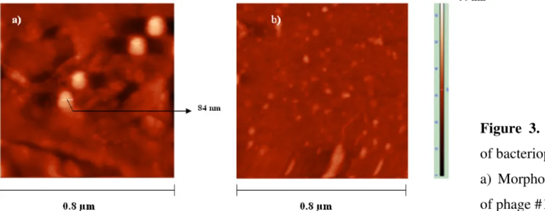
AFM analysis
Fig 3 shows atomic force microscopy topological images
of bacteriophage particles on aminosilanized glass. Phage size
measurements were performed directly on micrographs at 1µ m
magnification. According to the head diameter, viral particles
were classified into three size classes (>= 85 nm, >40nm and <
15nm). Bacteriophages could not be clearly differentiated
according to their fine structures.
DISCUSSION
Seven phages were isolated from sewage enrichment
process, whereas isolation of phages from soil samples was
found to be unsuccessful. The results are in concordance with
an earlier study wherein the phages of Pseudomonas were
successfully isolated only from water samples, while in soil
and feces no Pseudomonas specific phages were detected
(22).Earlier studies attribute similar results to relatively low
viable counts of phage capable of infecting specific bacteria
(6), relatively low rates of phage diffusion within soil,
particularly under drier conditions (18) and relatively high rates
of free-phage inactivation within soil (27). There have been a
number of reports of phage presence, in various environments
(14,2). However, it is uncertain how those phages from sewage
have impact on bacterial populations in soil environment. The
susceptibility analysis of soil bacterial isolates in this study
shows the soil bacterial population was resistant to these phage
isolates. It is interesting to note that soil bacterial isolates
obtained within the vicinity of sewage treatment plant showed
resistance to the phages isolated from sewage. Each of the 7
phages was able to individually lyse the isolation host. None of
the P. fluorescens bacteria isolated from the cultivated soils
were susceptible to seven phages. In one study, phages of
human enteric bacteria were also reported to be associated with
plants displaying broad host range (29). Similarly, phage
infecting phytopathogenic Pseudomonas have been isolated
from sewage (21, 36) while Agrobacterium-infecting phage
have been isolated from feces (25, 31). But from our study, it
was found that the bacteriophage having broad host range
activity isolated from sewage environment showed no lytic
activity among cultivated soil isolates. However, further
studies need to be carried out to understand the impact on
natural bacterial population of cultivated soils. The phage #5 in
our study was able to completely lyse the host bacterium P.
fluorescens and P. aeruginosa. This indicated the broad range
of host specificity of the bacteriophage. Broad host range
phages play a key role in phage ecology and gene transfer in
nature (14). The restriction pattern of the phage DNA shows
the genomic diversity among the isolates. All the phages Figure 3. Atomic force microscopy images
of bacteriophages
a) Morphology of phage #5, b) morphology
isolated were double stranded and sensitive to EcoRI and
HindIII (Figure 1). The restriction analysis indicated that the
phages were genetically distinct. The phage #6 has a genome
size of about 37 kb which is very close to the P. aeruginosa
phage MP22 (12) and also phage #2 has approximately 58 kb
genome size, similar to D3 phage which is a P. aeruginosa
phage (19). The host range of these two phages also suggests
that they are lytic against P. aeruginosa (Table 1). The phage
#5 has a genome size of about 80 kb, which showed broad
range host specificity and did not have match to any sequences
of the phage genome databases. A major protein with the same
molecular weight (above 97 kDa) was present in all the seven
phages analyzed, which is consistent with the results obtained
by Alonso et al., (2). It suggested that the molecular weights of
the major proteins do not vary among phage lysates specific to
the same bacterial strains. The morphological feature of the
phage #5 was also different from other six phage isolates. The
average size of the capsid for bacteriophage #5 was
approximately 84nm. This phage has morphological
characteristics somewhat similar to T4 bacteriophage (4).
However, the fine structure like tail length was not observed.
The ecological importance of this phage must be further
studied. Though only representative part of the Pseudomonas
phages was investigated, a great variability has been observed.
REFERENCES
1. Ackermann, H.W. (2001). Frequency of morphological phage descriptions in the year 2000. Arch. Virol., 146; 843-857.
2. Alonso, M.D.; Rodriguez J.; Borrego J.J. (2002). Characterization of marine bacteriophages isolated from the Alboran Sea; Western Mediterranean. J. Plankton Res., 24; 1079-1087.
3. Armon, R.; Kott, Y. (1993). A simple, rapid and sensitive presence/ absence detection test for bacteriophage in drinking water. J. Appl. Bact., 74; 490-496.
4. Archer, J.M.; Jinny, L. Liu. (2009). Bacteriophage T4 Nanoparticles as Materials in Sensor Applications: Variables That Influence Their Organization and Assembly on Surfaces’ Sensors. 9(8); 6298-6311. 5. Ashelford, K.E.; Day, M.J.; Bailey, M.J.; Lilley, A.K.; Fry, J.C. (1999).
In situ population dynamics of bacterial viruses in a terrestrial environment. Appl. Environ. Microbiol., 65; 169-174.
6. Ashelford, K. E.; Day, M. J.; Fry, J. C. (2003). Elevated abundance of bacteriophage infecting bacteria in soil. Appl. Environ. Microbiol., 69; 285-289.
7. Bassom B.J.; Caetano-Annolles G.; Gresshoff P.M. (1991). Fast and sensitive silver staining of DNA in polyacrylamide gels, Analytical Biochemistry, 196; 80-83.
8. Brazil G.M.; Kenefick L.; Callanan M.; Haro A.; de Lorenzo V.; Dowling D.N.; O'Gara F. (1995). Construction of a rhizosphere Pseudomonad with potential to degrade polychlorinated biphenyls and detection of bph gene expression in the rhizosphere, Appl. Environ. Microbiol., 61;1946-1952.
9. Campbell, J.I.A.; Albrechtsen, M.; Sorensen, J. (1995). Large Pseudomonas phages isolated from barley rhizosphere. FEMS (Fed. Eur. Microbiol. Soc.)Microbiol. Ecol., 18; 63-74.
10. Dobrindt, U.; Reidl J. (2000). Pathogenicity islands and phage conversion: evolutionary aspects of bacterial pathogenesis. Int J Med Microbiol., 6; 519-27.
11. Dooley, M. (2007). Restriction Endonuclease Digestion of a Plasmid, Association for Biology Laboratory Education Proceedings, 29; 389-392 12. Goszczynska, T.; Serfontein, J.J.; Serfontein, S. (2000). Introduction to Practical Phytobacteriology. Bacterial Disease Unit. ARC-Plant Protection Research Institute Pretoria. South Africa.
13. Heo,Y.J.; Chung, I.Y.; Choi, K.B.; Lau,G.W.; Cho, Y.H. (2007). Genome sequence comparison and superinfection between two related Pseudomonas aeruginosa phages, D3112 and MP22. Microbiology., 153l; 2885-2895.
14. Jensen, C.E.; Schrader S.H.; Rieland B.; Thompson T.L.; Lee K.W.; Nickerson K.W.; Kokjohn T.A. (1998). Prevalence of Broad Host Range lytic Bacteriophages of Sphaerotilus natans, Escherichia coli and Pseudomonas aeruginosa. Appl.Environ. Microbiol., 64(2); 333-340. 15. Joas, L.; Da Silva.; Rosario D.C. Hirata.; Mario H. Hirata. (2009).
Bacteriophage: laboratorial diagnosis and phage therapy Braz. J. Microbiol., 40 (3).
16. Jonathan, M.B. (2003). Phage Isolation and Investigation Dartmouth Undergraduate. J. of Science., 3(1).
17. Karlson, U.; Dowling, D.; O’Gara, F.; Rivilla, R.; Bittens, M.; Francesconi, S.; Pritchard, H.;Pedersen, H.C. (1998). Development of self-contained plant/GMM systems for soilbioremediation, In de Vries G.E. (ed.), Past, present and future risk assessment when using GMOs, Overschild, The Netherlands, 23.
18. Knezevic, P.; Kostanjsek, R.; Obreht, D.; Petrovic, O. (2009). Isolation of Pseudomonas aeruginosa specific phages with broad activity spectra. Curr Microbiol., 59; 173-80.
19. Kwan, T.; Liu, J.; DuBow, M.; Gros, P.; Pelletier, J. (2006). Comparative Genomic Analysis of 18 Pseudomonas aeruginosa Bacteriophages. Journal of Bacteriology. 188(3); 1184-1187.
assembly of the head of bacteriophage T4. Nature, 227; 680-685. 21. Lee, I. F.; Boezi, J. A. (1966). Characterisation of bacteriophage gh- 1
for Pseudomonas putida. J.Bacteriol., 92; 1821-1827.
22. Lu, Z.; Breidt, F.; Plengvidhya, V.; Fleming, H.P. (2003). Bacteriophage ecology in commercial sauerkraut fermentations. Appl. Environ. Microbiol., 3192-3202.
23. Miao, E.A.; Miller, S.I. (1999). Bacteriophages in the evolution of pathogen-host interactions. Proc. Natl. Acad. Sci. USA, 96; 9452-9454. 24. Nourozian J.; Etebarian H.R.; Khodakaramian G. (2006). Biological
control of Fusarium graminearum on wheat by antagonistic bacteria. Journal of Science and Technology, 28; 29-38.
25. Okabe, N.; Goto, M. (1963). Bacteriophages of plant pathogens. Ann. Rev. Phytopathol., 1; 397-418.
26. O’Sullivan, D.J.; O’Gara, F. (1992). Traits of fluorescent Pseudomonas spp. involved in suppression of plant root pathogens. Microbiol. Mol. Biol. Rev.,56 (4); 662-676.
27. Pantastica, C.M.; Duncan, K. E.; Istock, C. A.; Bell, J. A. (1992). Population dynamics of bacteriophage and Bacillus subtilis in soil. Ecology. 73; 1888-1902.
28. Pickett, M.J.; Goodneer, J.R.; Harvey, S.M. (1991). Test for detecting degradation of gelatin: Comparison of five methods. J. Clinical Microbiol., 29; 2322-2325.
29. Pirhonen, M.; Palva, E. T. (1988). Occurence of bacteriophage T4 receptor in Erwinia carotovora. Mol. Gen. Genet., 214; 170-172. 30. Prescott, L.M.; Harley, J.P. (1993). Laboratory Exercises in
Microbiology. 2nd edition. Chapters 58 and 59.
31. Roslycky, E. B. (1962). Phages for Agrobacterium radiobacter with reference to host range. Can. J. Microbiol., 8; 71-78.
32. Sambrook, J.; Maniatis, T.; Fritsch E.F. (1989). Molecular Cloning: A Laboratory Manual, 2nd edition, Cold Spring Harbor Laboratory Press, Cold Spring Harbor, NY.
33. Scott, C.A.; Faruqui, N.I.; Raschid-Sally, L. (2004) Wastewater use in irrigated agriculture: confronting the livelihood and environmental realities. Wallingford: Cabi Publishing, 1–10.
34. Sillankorva, S.; Neubauer P.; Azeredo J. (2008). Isolation and characterization of a T7-like lytic phage for Pseudomonas fluorescens, BMC Biotechnology, 8(80).
35. Smith, H.W.; Huggins, M.B. (1982). Successful treatment of experimental Escherichia coli infections in mice using phage: its general superiority over antibiotics, Journal of General Microbiology, 128; 307-318.
36. Thomas, M.D.; Leary, J.V. (1983). Bacteriophages from sewage specific for fluorescent phytopathogenic pseudomonads. Phytopathology, 73; 403-406.