Use of newly isolated phages for control of
Pseudomonas aeruginosa
PAO1
and ATCC 10145 biofilms
Diana Pires
a, Sanna Sillankorva
a, Alberta Faustino
b, Joana Azeredo
a,*
aIBBeInstitute of Biotechnology and Bioengineering, Center of Biological Engineering, University of Minho, Campus de Gualtar 4710-057, Braga, Portugal bSa˜o Marcos Hospital, P.O. box 2242, 4701-965 Braga, Portugal
Received 25 June 2010; accepted 9 May 2011 Available online 5 July 2011
Abstract
Pseudomonas aeruginosa is a relevant opportunistic pathogen involved in nosocomial infections that frequently shows low antibiotic susceptibility. One of its virulence factors is associated with the ability to adhere to surfaces and form virulent biofilms. This work describes the isolation and characterization of lytic phages capable of infecting antibiotic-resistantP. aeruginosa strains. In addition, characterization of
P. aeruginosabiofilms and the potential of newly isolated phages for planktonic and biofilm control was accessed. According to the results, the isolated phages showed different spectra of activity and efficiency of lysis. Four broad lytic phages were selected for infection of planktonic cells; however, despite their broad range of activity, two of the selected phages failed to efficiently control planktonic cultures. Therefore, only two phages (phiIBB-PAA2 and phiIBB-PAP21), highly capable of causing strong biomass reduction of planktonic cells, were tested against 24 h biofilms using a m.o.i. of 1. Both phages reduced approximately 1e2 log the biofilm population after 2 h of infection and reduction was further enhanced after 6 h of biofilm infection. However, biofilm cells ofP. aeruginosaPAO1 acquired resistance to phiIBB-PAP21; consequently, an increase in the number of cells after 24 h of treatment was observed. Conversely, phage phiIB-PAA2 forP. aeruginosaATCC10145 continued to destroy biofilm cells, even after 24 h of infection. In these biofilms, phages caused a 3 log reduction in the number of viable counts of biofilm cells.
Ó2011 Institut Pasteur. Published by Elsevier Masson SAS. All rights reserved.
Keywords: Pseudomonas aeruginosa; Bacteriophages; Biofilms; Control
1. Introduction
Pseudomonas aeruginosais an ubiquitous organism which has emerged as a major threat in the hospital environment. This bacterium is the most frequently isolated Gram-negative organism in bloodstream and wound infections, pneumonia and intra-abdominal and urogenital sepsis, and is a serious problem, infecting immunocompromised patients with cystic fibrosis (CF), severe burns, cancer, AIDS, etc. (Driscoll et al., 2007; Page and Heim, 2009). One of the most worrying characteristics of this bacterium is its low antibiotic
susceptibility, which can be attributed to a concerted action of multidrug efflux pumps with chromosomally-encoded antibi-otic resistance genes and the low permeability of the bacterial cellular envelopes (Lambert, 2002).
Overuse of antibiotics has also significantly increased the emergence of antimicrobial multiresistant bacteria; conse-quently, treatment of most chronic P. aeruginosa infections with antibiotics is notoriously difficult (Cunha, 2002; Lambert, 2002). Additionally, P. aeruginosa has an innate ability to adhere to surfaces and form virulent biofilms particularly difficult to eradicate (Drenkard, 2003; Mah et al., 2003; Stewart and Costerton, 2001). Biofilm formation is an important bacterial survival strategy and, in humans, biofilms are responsible for numerous pathologies usually associated with use of medical devices (Azeredo and Sutherland, 2008; Donlan, 2002; O’Toole et al., 2000). Thus, new alternative * Corresponding author.
E-mail addresses:[email protected](D. Pires),s.sillankorva@ deb.uminho.pt (S. Sillankorva), [email protected] (A. Faustino), [email protected](J. Azeredo).
Research in Microbiology 162 (2011) 798e806
www.elsevier.com/locate/resmic
strategies to antibiotherapy are highly in demand by the worldwide medical and scientific community. Bacteriophages (phages) are the natural enemies of bacteria and might represent one attractive solution to this problem (Clark and March, 2006; Joerger, 2003; Sulakvelidze, 2005).
Phage therapy is based on the use of lytic phages to combat bacterial infections, including multidrug-resistant bacteria, and has many advantages comparated to antibiotics: they are very specific and efficient for their target bacteria, which reduces destruction of the host’s natural flora; they are not pathogenic for man; and they persist only as long as the tar-geted bacteria are present (Azeredo and Sutherland, 2008; Clark and March, 2006; Matsuzaki et al., 2005; Skurnik and Strauch, 2006).
Here we describe the isolation and characterization of novel phages for P. aeruginosa and their application to planktonic cultures and biofilms. The main goal is to determine phage potential to control these two types of cells.
2. Materials and methods
2.1. Bacterial strains and culture conditions
Thirty-five strains ofP. aeruginosawere used in this work: 4 strains for isolation of phageseATCC 10145, CECT 111, PAO1 and a clinical isolate (CLIN) (isolated from an endo-scope, Rouen, France)eand 31 strains for evaluating the lytic spectra of the isolated phages. These 31 strains were clinical isolates provided by the Hospital de Braga (Braga, Portugal). Bacteria were grown at 37
C in tryptic soy broth (TSB, Merck) or in tryptic soy agar (TSA, Merck). Biofilm assays were performed using yeast peptone dextrose (YPD - 10 g l 1 yeast extract, 20 g l 1peptone, 20 g l 1dextrose).
2.2. Isolation of phages
Bacteriophages were isolated from 2 hospital effluents provided by the Hospital de Sa˜o Joa˜o (Porto, Portugal). These effluents were enriched with 4 bacterial hosts (ATCC 10145, CECT 111, PAO1 and a clinical isolate) in 2TSB medium. This solution was incubated (37
C, 120 rpm) for 48 h and then centrifuged to collect supernatant for spot tests indicative of the existence of phage(s). Each inhibition halo was further purified with toothpicks and paper to isolate all different phages. All morphologically different phage plaques were purified until de Petri plates presented a single plaque morphology. Five similar plaques of each isolated phage were measured and characteristics are presented inTable 1.
2.3. Production of phages
The production of phages in Petri plates consists of immersing a paper strip in a solution containing phage and passing it on a top-agar layer containing the proper bacterial strain. The plates were incubated for 16e18 h at 37C; then,
3 ml of SM buffer (5.8 g l 1 NaCl, 2 g l 1 MgSO4.7H2O, 50 ml l 11M-tris-HCl pH 7.5) were added. The plates were
placed under agitation (120 rpm) at 4C for 16e18 h;
after-wards, liquid and top-agar were collected, centrifuged and the supernatant was filtrated (0.2mm).
2.4. Selection of phages
The selection of the 4 best phages (one for each host) was done according to results of lytic spectra. To evaluate lytic spectra of all isolated phages, they were tested against 35 strains ofP. aeruginosa. One drop of each serial diluted phage suspension, with titers of about 104 to 107 PFU ml 1, was placed on the different bacterial lawns and incubated overnight at 37
C. The following day, the susceptibility of each host to the different phages was evaluated.
2.5. Infection of planktonic cells
Infection of planktonic cells was done at two different stages of bacterial growth e exponential and stationary pha-ses. To induce planktonic cell infections at the stationary stage, the 4 hosts were inoculated overnight in TSB medium at 37
C (120 rpm). The resulting cell suspensions were diluted with TSB medium to obtain an optical density (OD600nm) of approximately 0.75, which corresponds to approximately 1.32109, 1.11109, 1.28109and 8.71108CFU ml 1 of P. aeruginosaATCC 10145, CECT 111, CLIN and PAO1, respectively. To carry out planktonic cell infection at expo-nential stage, an overnight pre-inoculum was used to inoculate 100 ml of fresh TSB medium, which were allowed to grow to an optical density (OD600nm) of 0.5, which corresponds to approximately 8.63 108, 7.42 108, 8.67 108 and 6.21108CFU ml 1ofP. aeruginosa ATCC 10145, CECT 111, CLIN and PAO1, respectively. The suspensions were centrifuged and resuspended in fresh TSB media in obtain an
OD600nm of 0.75 which allows better comparison of the two
different assays performed. In all experiments, 125 ml of Table 1
Characterization of the differentP. aeruginosaphage plaques.
Host Phage (phiIBB-.) Diameter (mm)
a Turbidity Halo
ATCC 10145 PAA2 1 T e
CECT 111 PAC222 2 T e
PAC2211 1.5 T e
PAC2212 1 C þ
PAC111 2.5 C þ
PAC23 1.5 T e
PAC24 4 C e
PAO 1 PAP2222 2 C þ(large)
PAP21 1 T e
PAP2221 2.5 C þ(large)
PAP221 1.5 T e
PAP121 2 C e
CLIN PACL212 1 C þ
PACL211 3 C þ
PACL22 3 C þ
PACL11 1 T e
PACL12 2 T e
phages were added to 125ml of suspensions of the respective hosts in order to obtain a multiplicity of infection (m.o.i.) of 1. The control experiments were performed with SM buffer instead of phages. The infection of planktonic cell assays were performed in 96-well microtiter plates. The plates were placed into an orbital incubator (120 rpm) at 37 C and the optical
density (OD600nm) was regularly read. Three independent experiments were performed in duplicate.
2.6. Biofilm formation
Based on phage infection experiments on planktonic cells, onlyP. aeruginosaATCC 10145 and PAO1 strains were used in biofilm assays. Biofilm formation was carried out in 24-well microplates containing 1 ml of YPD medium and 10 ml of cellular suspension with an OD600nmof 1.0 which corresponds to approximately 1.9 109 and 1.1 109 CFU ml 1, respectively. Biofilms were formed for 24 and 48 h with medium renewal every 12 h. The plates were incubated at 37 C in an orbital shaker (120 rpm). Three independent
experiments were performed in duplicate.
2.7. Crystal violet assay
Total biomass attached to each well was measured by crystal violet assay. First, the wells were washed twice with a saline solution (0.9% NaCl (Merck) in distilled water)) and then biofilms were fixed with 1 ml of methanol (Merck) for 15 min. After this time, methanol was removed and to each well was added 1 ml of crystal violet (1% v/v, Merck) for 5 min. The wells were then washed with water, and 1 ml of acetic acid (33% v/v, Merck) was added to dissolve the stain. The eluted stain was placed in a 96-well microtiter plate and its absorbance was read by an ELISA reader at 570 nm. Two independent experiments were performed in duplicate.
2.8. XTT reduction assay
The determination of biofilm cellular activity was measured by the XTT reduction assay. Biofilms were washed twice with a saline solution (0.9% NaCl (Merck) in distilled water)) and then 1 ml of XTT (200 mg l 1, Sigma) solution plus PMS (20 mg l 1, Sigma) was added to each well. The plates were incubated in the dark at 37
C for 3 h. After this time, the solution was removed from each well and placed in a 96-well microtiter plate to determine its absorbance at 490 nm. Two independent experiments were performed in duplicate.
2.9. Biofilm infection
Biofilm infection was done with two different phages e phiIBB-PAA2 and phiIBB-PAP21. After 24 h of biofilm formation, all medium and planktonic bacteria were removed from each well and washed with fresh YPD medium. Following that, 500 ml of fresh YPD medium and 500 ml of phage solution or 500ml of SM buffer in the case of control experiments were added. The m.o.i. used for both phages
was 1. The plates were incubated in an orbital shaker (120 rpm) at 37
C and samples were taken after 2, 6 and 24 h of infection for CFU and PFU counts. Three independent experiments were performed in duplicate.
2.10. CFU and PFU counts
To determine the amounts of bacteria present in biofilms, CFU counts was performed using the microdrop technique. Briefly, wells of the microplates were washed once with saline solution (0.9% NaCl) to remove unattached bacteria and then 1 ml of fresh saline solution was added to each well and the biofilm scraped with a cell scraper prior to sonication (5 min). After this, the cellular suspension of each well was removed, centrifuged (5 min, 9000g, 4
C) and the pellet resuspended in 1 ml of saline solution (0.9%). The samples were diluted in saline solution (0.9%) and one drop (10 ml) was placed in a Petri plate containing YPD solid medium and allowed to run down the plate. Plates were incubated at 37
C for 16e18 h and after CFUs were counted. For PFU counting, the small drop method described by Mazzoco et al. with some modifi-cations was used (Mazzocco et al., 2008). Briefly, 20 ml of diluted phage solution were added to 20ml of the overnight-grown host and incubated for 15e20 min to allow phage binding to the host. After this, 20ml of sample was placed in an agar plate and allowed to dry. Plates were incubated overnight at 37
C. Three independent experiments were performed in duplicate.
2.11. Statistical analysis
To compare the amounts of viable cells present in biofilms, analysis of variance (ANOVA single factor from MS Office) was used. In all analyses performed, the confidence interval used was 95%.
3. Results
3.1. Phage characterization
Initially, 17 phages were isolated from hospital effluents by enrichment with 4 strains ofP. aeruginosa. Characteristics of these phages were determined by the diameter and turbidity of phage plaques and the presence of a halo (Table 1). From the group of isolated phages, there were approximately the same number of phages responsible for clear (9 phages) and turbid (8 phages) plaques. Furthermore, some phages formed large plaques (ex: phiIBB-PAC24) while others had more pinhole characteristic (ex. phiIBB-PAP21) plaque morphology. Furthermore, most isolated phages were non halo-generating ones.
After characterizing the morphology of phage plaques, newly isolated phages were also tested against altogether 35 antibiotic-resistant clinical and reference strains ofP. aerugi-nosain order to study their lytic spectra (Table 2). Most phages showed lytic activity against several isolates, even against strains resistant to most of the 7 antibiotics tested, such as
Lytic spectra of newly isolated phages against reference and clinical strains ofP. aeruginosa.
Phage (phiIBB-.)
Host Antibioga PAP21 PAP121 PAP221 PAP2221 PAP2222 PAA2 PAC24 PAC23 PAC111 PAC222 PAC2211 PAC2212 PACL11 PACL12 PACL22 PACL211 PACL212
PAO 1 nd C T T T T e T C e C e T e T e e e
ATCC 10145 nd C C T C C T C C C T T C e e e e e
CECT 111 nd T e e e e T C C C T T C e e e e e
CLIN nd T T e T e T T T T T e e T T T T T
1 54 T C T C T C C C C C C C e C e e e
2 0 e T e e e C (RB) T T T T T T e e e e e
3 14 C e e e e C C C C C C C e e e e
4 0 C e C C T T T T T T e e e e e e e
5 14 C T C T T T C (RB) C (RB) C (RB) T T C (RB) T T T T T
6 0 C C C T T C (RB) C (RB) C (RB) C (RB) T T T C C e C C
7 29 C (RB) e e e e C (RB) C C C C T C (RB) e C e e e
8 0 C C C T e C C C (RB) C (RB) C (RB) e C e C e e e
9 0 e e e e e T T T T T T T e e e e e
10 43 e T T T e T T T e e T e T T T T e
11 57 C C C C C T T T T C C C e C e e e
12 86 C T T T T T T T T e e T C e C (RB) T T
13 14 e e e e T T C C C C C C e e e e e
14 14 e e e e e e T T e e e e e e e e e
15 0 e e e e e T T T e T T T e e e e e
16 0 C C C C C C (RB) C C T C T e e C e e T
17 86 C T e T C C (RB) C C C (RB) C e e e e e e e
18 29 C C C C (RB) C T e e T e e e e C (RB) e e e
19 71 e e e e e T T T e T e e e e e e e
20 0 T T T T T e e T e T T e T T T T T
21 0 T T T T C T T e T e T T C C e e T
22 0 T C C C T e e e e e T e e C e e e
23 14 T e T e T T T T T T e e e e e e e
24 14 T e e e e T T T e T e e e e e e e
25 86 C C C C C C C C T C T T e C e e e
26 0 C C C C C T C C (RB) T T T T e C e e e
27 0 T C T C C C C C T C T T e C e e e
28 14 T e e e e T T T T T T T e e e e e
29 0 T C T C C C (RB) T C (RB) T C (RB) e e e T e e e
30 nd T C C C C e e e e e e e e C e e e
31 nd T T T e T T T T T e T T T e T e T
% Infection 80.00 65.71 62.86 62.86 62.86 85.71 88.57 88.57 74.29 77.14 62.86 60.00 22.86 54.29 17.14 17.14 22.86
T: Turbid; C: Clear; C(RB): Clear with resistant bacteria; nd: not determined.
a Percentage of resistance of the bacteria to 7 antibiotics used in the antibiogram - imipenem, ciprofloxacin, ceftazidime, gentamicin, amikacin, tazobactam, tobramycin.
801
D.
Pir
es
et
al.
/
Resear
ch
in
Micr
obiology
162
(2011)
798
e
isolate numbers 12, 17, 19 and 25, respectively. Phages isolated for strain CECT 111 were shown to infect the highest number of strains used in this study. Nevertheless, some phages, and especially those isolated for the clinical strain ofP. aeruginosa, had a narrower spectrum of activity (ex. phiIBB-PACL22 and phiIBB-PACL211) towards the tested isolates and therefore were considered less efficient. Although only one phage was isolated for the ATCC 10145 strain, phiIBB-PAA2 respectively, this phage proved to have a broad spectrum of activity, infecting 85.71% of the tested strains (seeTable 2).
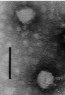
After lytic spectrum assays, one phage for each host was selected for use in planktonic and biofilm infection experi-ments. The criterion used for this selection was the number of strains that each phage was able to infect. Accordingly, the selected phages were: PAA2, PAC23, phiIBB-PACL12 and phiIBB-PAP21, respectively. These phages were analysed by transmission electron microscopy (TEM) and presented a head size of 58 nm and a tail of 12 nm 8 nm characteristic of thePodoviridaefamily of phages (Fig. 1).
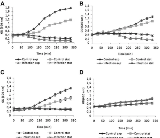
3.2. Infection of planktonic cells
After phage selection and characterization, phage infection experiments were performed in exponentially growing and stationary cells (Fig. 2). The growth of the 4 different Pseu-domonas strains varied considerably. Since this experiment was performed using fresh media, an increase in optical density of stationary phase cells was also observed. The 4 phage-host systems showed a clear difference in the infection patterns of stationary and exponentially growing cells. Both phages phiIBB-PAA2 and phiIBB-PAP21 infected exponen-tially growing cells. Unlike PAP21, phage phiIBB-PAA2, besides infecting exponential phase cells, was also
capable of lysing stationary phase cells. The infection of both stationary and exponential growth phase cells with phage phiIBB-PAC23 caused only a minor reduction in OD. Furthermore, strain CLIN continued to grow slowly even in the presence of phage phiBB-PACL12. Despite the last two mentioned phages, which were less efficient than phages phiIBB-PAA2 and phiIBB-PAP21, there was a significant difference in the infected cultures compared to the exponential and stationary controls, performed with SM buffer instead of the different phages.
3.3.P. aeruginosabiofilm characteristics
24- and 48-h-oldP. aeruginosabiofilms were characterized in terms of biomass quantity by CV staining, CFU counts, and also through evaluation of metabolic activity by XTT reduction. The medium in both biofilms was renewed every 12 h. Since phages phiIBB-PACL12 and phiIBB-PAC23 were less effective in lysing cells, only biofilms ofP. aeruginosaATCC 10145 and PAO 1 were characterized for further infection experiments with phages phiIBB-PAA2 and phiIBB-PAP21, respectively.
Biofilms ofP. aeruginosaATCC 10145 were characterized by having a higher amount of biomass attached to the surface of the 24 well-plate (Fig. 3A) than biofilms ofP. aeruginosa PAO 1. There were no significant differences in the amount of total biomass existing in 24- and 48-h-old biofilms. In terms of metabolic activity, XTT results showed that 24-h-old biofilms ofP. aeruginosaPAO 1 were more active than 48-h-old bio-films of this strain; however, 24 and 48 h PaeruginosaATCC 10145 biofilms resulted in similar values (Fig. 3B). The viable counts of both strains in the studied biofilms were statistically similar (p ¼0.15). Furthermore, 24 h biofilms of P. aerugi-nosa ATCC 10145 had a significantly higher (p ¼ 0.025) number of viable cells than biofilms formed for 48 h (Fig. 3C). Although, 24 h biofilms of PAO1 presented more viable cells, values compared to 48 h biofilms were not statistically different (p ¼0.29).
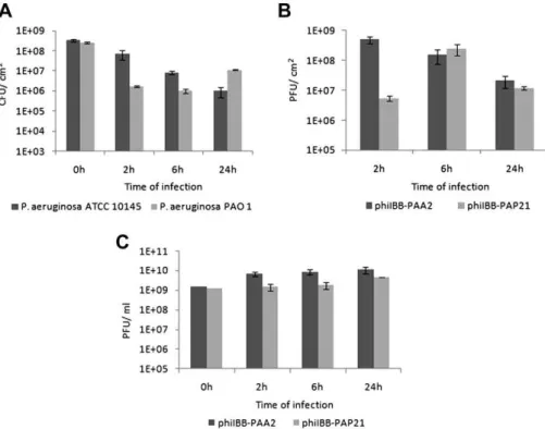
3.4. Biofilm infection
Phage infection of P. aeruginosa ATCC 10145 and PAO1 was performed in 24 h biofilms (Fig. 4) due to the fairly similar amounts of viable cells present in 24 and 48 h PAO1 and ATCC 10145 biofilms (as described above). According to infection results, phage phiIBB-PAA2 gradually decreased the numbers of viable cells present in the biofilms; already after 2 h, there was a statistically significant (p¼0.025) reduction in viable cells, which was further enhanced after 6 and 24 h of biofilm infection, achieving an almost 3-log reduction. Phage phiIBB-PAP21 caused a considerably greater reduction (p¼0.0008) inP. aeruginosaPAO1 biofilm cells after 2 h of infection compared to reduction ofP. aeruginosaATCC 10145 infected with phiIBB-PAA2. The reduction of P. aeruginosa PAO1 biofilms due to phiIBB-PAP21 continued until 6 h of biofilm treatment; however, biofilm cells acquired resistance to the phage and consequently, the numbers of cells after 24 h of infection increased.
Fig. 1. Phage phiIBB-PAA2 observed by TEM. (Bar corresponds to 100 nm).
One unexpected result occurred in the number of phages attached to the biofilms (and the surface of the well) as measured by plaque-forming unit (PFU) counts. After 2 h of infection, higher amounts of phage phiIBB-PAA2 than phage phiIBB-PaP21 were observed entrapped in the biofilms. Experiments performed in 24-well microtiter plates in the absence of P. aeruginosa cells, showed that phiIBB-PAA2 attached more rapidly to the well substrata (data not shown). This can explain the weaker reduction in the amount of viable cells ofP. aeruginosa ATCC 10145 by phage phiIBB-PAA2, since phages attached to the substrata far from biofilm cells will not cause their lysis. The number of phage phiIBB-PAA2 entrapped in the biofilms (and substrata) decreased throughout the 24 h experiment by approximately 1.7 logs. Unlike phage
phiIBB-PAA2, the amount of phiIBB-PAP21 phages adsorbed increased until 6 h of infection. Nevertheless, as in phiIBB-PAA2 experiments, also after 24 h of biofilm cell infection, the number of phages attached was significantly lower.
The time for biofilm cell resistance toward phages was compared with the resistance acquired by planktonic cells (Fig. 5). After approximately 9e10 h of infection, planktonic P. aeruginosa PAO1 cell cultures began to grow again. Both colonies of planktonic and biofilm experiments were tested with phage stock solution, and all showed resistance to phiIBB-PAP21. Despite the lack of resistance ofP. aeruginosa ATCC 10145 biofilms after 24 h of exposure to phage phiIBB-PAA2, acquisition of resistance by planktonic cultures to the phage was observed.
Fig. 2. Infection of planktonicP. aeruginosacultures with phages: A) phiIBB-PAA2 (ATCC 10145), B) phiIBB-PAP21 (PAO1), C) phiIBB-PAC23 (CECT 111), and D) phiIBB-PACL12 (CLIN) in exponential and stationary growth using a MOI 1. Error bars represent standard deviations from 3 independent experiments performed in duplicate.
4. Discussion
The development of new alternatives to antibiotherapy for eradication and control of virulent biofilms from surfaces, mainly of medical devices, has become a great challenge in the scientific community. Several studies have shown the potential of use of phages to treat infectious diseases in animals (Loc Carrillo et al., 2005; McVay et al., 2007; Nakai and Park, 2002; Wagenaar et al., 2005) and humans ( Weber-Dabrowska et al., 1987, 2001), even those caused by antibiotic-resistant bacteria (Weber-Dabrowska et al., 2003).
In the present study, 17 new phages forP. aeruginosawere isolated from hospital effluents using three collection strains
and a clinical strain and they were tested for efficiency against 35 clinical strains. The least efficient isolated phages were those with the clinicalP. aeruginosaisolate as host, where the percentages of strains infected varied between 17 and 54 percent, while the most efficient phages, those with CECT 111 as propagating strain, were capable of lysing approximately 60e89 percent of the clinical strains tested. These percentages are significantly above those observed by Knezevic et al. for 19 newly isolatedP. aeruginosaphages which were found to be capable of killing up to 75.8 percent of 33 strains isolated from humans (21), animals (1), the environment (10) and 1 culture strain (ATCC 9027) (Knezevic et al., 2009).
Based on the efficiency of killing clinical strains, four lytic phages were selected for further characterization and used in infection assays against planktonic and biofilm cells. Despite the broad spectra of activity of the selected phages against clinical P. aeruginosa isolates, infection experiments on planktonic cultures showed, surprisingly, that two of them failed to infect their hosts even at the ideal bacterial growth phasedthe exponential phase. This suggests that, although phages can be selected based on their broad activity, this does not necessarily guarantee their effectiveness in controlling their respective host population. Nevertheless, two of the used phages, phages phiIBB-PAA2 and phiIBB-PAP21, were both efficient towards exponential as well as stationary phase cells and caused over a 50% reduction in initial OD. Yet phage phiIBB-PAP21 revealed a slightly weaker ability to infectP. aeruginosa PAO1 planktonic cells than phage phiIBB-PAA2 for strain ATCC 10145. It has been frequently reported, with different phage-host systems, that there is reduced lysis when Fig. 5. Infection ofP. aeruginosaATCC 10145 and PAO1 planktonic cultures
with phages phiIBB-PAA2 and phiIBB-PAP21, respectively, using an m.o.i. of 1 for 25 h.
Fig. 4. Infection ofP. aeruginosaATCC 10145 and PAO1 biofilms with phages phiIBB-PAA2 and phiIBB-PAP21, respectively. A) Number of viable cells before and after infection; B) number of phages (PFU) present in the biofilms; C) number of phages (PFU) released from infected biofilms. The error bars represent standard deviations from 3 independent experiments performed in duplicate.
cells are near the stationary phase (Abedon and Yin, 2009; Burch and Chao, 2004; Middelboe, 2000; Ricciuti, 1972; Sillankorva et al., 2004). For instance, the T4 phage cannot even produce a burst if Escherichia coli cells are in the stationary phase (Delbru¨ck, 1940). The main factors accounting for reduction of lysis in stationary cells are: fewer phage-adsorption sites, lower phage progeny per infection and reduced cell lysis due to cell wall thickness or increased non-viable infections (Weitz and Dushoff, 2008). Thus, our results are extremely interesting, as they demonstrate that some, but not all “T7-like” phages can be effective against stationary phase cells.
It is not surprising that phage infection in planktonic cells is more efficient than in biofilms due to biofilm architecture, which prevents easy access by phages to the bacteria (Hanlon et al., 2001). Due to the inefficient activity of phages phiIBB-PACL12 and phiIBB-PAC23 against planktonic cultures, only two phages, phiIBB-PAA2 and phiIBB-PAP21, were chosen for P. aeruginosa biofilm control experiments. Both phages, tested against 24 h biofilms, caused significant reduction on biofilm cells already after 2 h of infection and the reduction was further enhanced after 6 h of biofilm treatment, reaching a reduction of almost 3 log by phage phiIBB-PAP21 and a 2 log reduction by phage phiIBB-PAA2. Although our approach was different, viable cell reductions achieved were similar to those reported by Fu et al., whereP. aeruginosa phages were used to prevent biofilm formation on hydrogel-coated catheter surfaces. Accordingly, Fu et al., after 24 h, achieved a 2.84-log lower number of viable cells on phage pre-treated catheters than on untreated catheters (Fu et al., 2010).
Despite lower activity against planktonic cells, phage phiIBB-PAP21 caused a stronger reduction in viable cells from P. aeruginosaPAO1 biofilms after 2 and 6 h of treatment than phage phiIBB-PAA2 inP. aeruginosaATCC 10145 biofilms. Similar reductions in viable cells have been observed for Pseudomonas fluorescensphages (Sillankorva et al., 2004) and there is only one work in the literature where complete erad-ication of 24 hEnterobacter cloacebiofilms by a combination of 3 different phages was achieved (Tait et al., 2002). The main difference between the two phages used in our work for P. aeruginosabiofilm control experiments, concerns resistance to phages. The interaction of phages and bacteria during long periods of time can result in the emergence of phage-resistant bacteria (Bohannan and Lenski, 2000; Buckling and Rainey, 2002). Biofilm cells of P. aeruginosa PAO1, the host of phage phiIBB-PAP21, acquired resistance to the phage, resulting in an increase in the number of cells after 24 h of biofilm treatment. To evaluate the period when resistance starts appearing in planktonic cultures, P. aeruginosa PAO1 planktonic cells infected with phage phiIBB-PAP21 showed that resistance appeared after approximately 9e10 h of infection, which is in accordance with biofilm results showing the emergence of resistance between 6 and 24 h. On the other hand, phage phiIBB-PAA2 continued to destroy biofilm cells of P. aeruginosaATCC 10145 and there was no evidence of biofilm cells becoming resistant even after 24 h of phage infection. Unlike experiments with P. aeruginosa ATCC
10145 biofilms, assays performed with planktonic cells and phage phiIBB-PAA2 resulted in increased resistance to this phage. In 24 h P. aeruginosa ATCC 10145 biofilms, most likely there are already some resistant phenotypes; however, their growth is possibly slower and thus, non-randomly selected and tested colonies showed resistance to phage phiIBB-PAA2. Further studies will be performed to under-stand and confirm the phenomenon of increased resistance of planktonic cells, compared to biofilm cells.
Thus, this study reveals that both novel isolated phages were capable of controlling P. aeruginosabiofilms; however, short periods of treatment seem to be a better solution for avoiding the emergence of phage-resistant hosts. In addition, use of phage cocktails presented advantages over use of single cells, since phages can be selected in such a way as to over-whelm host resistance mutations.
Acknowledgements
The authors gratefully acknowledge the Hospital de Braga for providing P. aeruginosa clinical strains. The authors are also grateful to Dr. Hans Ackermann for morphological characterization of the phages.
References
Abedon, S.T., Yin, J., 2009. M.R. Clokie and A.M. Kropinski. Bacteriophage Plaques: Theory and Analysis. Bacteriophages Methods and Protocols, 1. Springer-Verlag, New York. 161e174.
Azeredo, J., Sutherland, I.W., 2008. The use of phages for the Removal of infectious biofilms. Curr. Pharm. Biotechnol. 9, 261e266.
Bohannan, B.J.M., Lenski, R.E., 2000. Linking genetic change to community evolution: insights from studies of bacteria and bacteriophage. Ecol. Lett. 3, 362e377.
Buckling, A., Rainey, P.B., 2002. Antagonistic coevolution between a bacte-rium and a bacteriophage. Proc. Biol. Sci. 269, 931e936.
Burch, C.L., Chao, L., 2004. Epistasis and its Relationship to Canalization in the RNA virus f6. Genetics 167, 559e567.
Clark, J.R., March, J.B., 2006. Bacteriophages and biotechnology: vaccines, gene therapy and antibacterials. Trends Biotechnol. 24, 212e218. Cunha, B.A., 2002.Pseudomonas aeruginosa: resistance and therapy. Semin.
Respir. Infect. 17, 231e239.
Delbru¨ck, M., 1940. Adsorption of bacteriophage under various phisiological conditions of the host. J. Gen. Physiol. 23, 631e642.
Donlan, R.M., 2002. Biofilms: microbial life on surfaces. Emerg. Infect. Dis. 8, 881e890.
Drenkard, E., 2003. Antimicrobial resistance of Pseudomonas aeruginosa biofilms. Microbes Infect. 5, 1213e1219.
Driscoll, J.A., Brody, S.L., Kollef, M.H., 2007. The epidemiology, patho-genesis and treatment ofPseudomonas aeruginosainfections. Drugs 67, 351e368.
Fu, W., Forster, T., Mayer, O., Curtin, J.J., Lehman, S.M., Donlan, R.M., 2010. Bacteriophage cocktail for the Prevention of biofilm formation by Pseu-domonas aeruginosaon catheters in an in vitro model System. Antimicrob. Agents Chemother. 54, 397e404.
Hanlon, G.W., Denyer, S.P., Olliff, C.J., Ibrahim, L.J., 2001. Reduction in Exopolysaccharide Viscosity as an Aid to bacteriophage penetration throughPseudomonas aeruginosabiofilms. Appl. Environ. Microbiol. 67, 2746e2753.
Knezevic, P., Kostanjsek, R., Obreht, D., Petrovic, O., 2009. Isolation of Pseudomonas aeruginosaspecific phages with broad activity spectra. Curr. Microbiol. 59, 173e180.
Lambert, P.A., 2002. Mechanisms of antibiotic resistance inPseudomonas aeruginosa. J. R. Soc. Med. 95, 22e26.
Loc Carrillo, C., Atterbury, R.J., El-Shibiny, A., Connerton, P.L., Dillon, E., Scott, A., Connerton, I.F., 2005. Bacteriophage therapy to reduce Campylobacter jejunicolonization of Broiler Chickens. Appl. Environ. Microbiol. 71, 6554e6563.
Mah, T.F., Pitts, B., Pellock, B., Walker, G.C., Stewart, P.S., O’Toole, G.A., 2003. A genetic basis for Pseudomonas aeruginosa biofilm antibiotic resistance. Nature 426, 306e310.
Matsuzaki, S., Rashel, M., Sakurai, S., Ujihara, T., Kuroda, M., Ikeuchi, M., Tani, T., Fujieda, M., Wakiguchi, H., Imai, S., 2005. Bacteriophage therapy: a revitalized therapy against bacterial infectious diseases. J. Infect. Chemother. 11, 211e219.
Mazzocco, A., Waddell, T.E., Lingohr, E., Johnson, R.P., 2008. Enumeration of Bacteriophages Using the Small Drop Plaque Assay System. Bacte-riophages: Methods and Protocols. In: Isolation, Characterization and Interactions, vol. 1. Humana Press. 81e85.
McVay, C.S., Velasquez, M., Fralick, J.A., 2007. Phage therapy of Pseudo-monas aeruginosainfection in a mouse burn wound model. Antimicrob. Agents Chemother. 51, 1934e1938.
Middelboe, M., 2000. Bacterial growth Rate and Marine ViruseHost dynamics. Microb. Ecol. 40, 114e124.
Nakai, T., Park, S.C., 2002. Bacteriophage therapy of infectious diseases in aquaculture. Res. Microbiol. 153, 13e18.
O’Toole, G., Kaplan, H., Kolter, R., 2000. Biofilm formation as microbial development. Annu. Rev. Microbiol. 54, 49e79.
Page, M.G.P., Heim, J., 2009. Prospects for the next anti-Pseudomonasdrug. Curr. Opin. Pharmacol. 9, 558e565.
Ricciuti, C.P., 1972. Host-virus interactions inEscherichia coli: effect of stationary phase on viral release from MS2-infected bacteria. J. Virol. 10, 162e165.
Sillankorva, S., Oliveira, R., Vieira, M.J., Sutherland, I., Azeredo, J., 2004. BacteriophageFS1 infection ofPseudomonas fluorescensplanktonic cells versusbiofilms. Biofouling 20, 133e138.
Skurnik, M., Strauch, E., 2006. Phage therapy: facts and fiction. Int. J. Med. Microbiol. 296, 5e14.
Stewart, P.S., William Costerton, J., 2001. Antibiotic resistance of bacteria in biofilms. Lancet 358, 135e138.
Sulakvelidze, A., 2005. Phage therapy: an attractive option for dealing with antibiotic-resistant bacterial infections. Drug Discov. Today 10, 807e809. Tait, K., Skillman, L.C., Sutherland, I.W., 2002. The efficacy of bacteriophage
as a method of biofilm eradication. Biofouling 18, 305e311.
Wagenaar, J.A., Bergen, M.A.P.V., Mueller, M.A., Wassenaar, T.M., Carlton, R.M., 2005. Phage therapy reducesCampylobacter jejuni colo-nization in broilers. Vet. Microbiol. 109, 275e283.
Weber-Dabrowska, B., Dabrowski, M., Slopek, S., 1987. Studies on bacte-riophage penetration in patients subjected to phage therapy. Arch. Immunol. Ther. Exp. 35, 563e568.
Weber-Dabrowska, B., Mulczyk, M., Go´rski, A., 2001. Bacteriophage therapy for infections in cancer patients. Clin. Appl. Immunol. Rev. 1, 131e134. Weber-Dabrowska, B., Mulczyk, M., Go´rski, A., 2003. Bacteriophages as an efficient therapy for antibiotic-resistant septicemia in man. Transplant Proceed 35, 1385e1386.
Weitz, J., Dushoff, J., 2008. Alternative stable states in hostephage dynamics. Theor. Ecol. 1, 13e19.