VIRULENCE AND CYTOTOXICITY OF SEAFOODBORNE Aeromonas hydrophila
Seethalakshmi Illanchezian, SathishKumar Jayaraman*, Muthu Saravanan Manoharan, Saritha Valsalam
Life Teck Research Centre, Vadapalani, Chennai – 600026, India.
Submitted: April 17, 2009; Returned to authors for corrections: April 23, 2010; Approved: May 13, 2010.
ABSTRACT
The present study was conducted to determine the virulence and cytotoxicity of Aeromonas hydrophila
strains isolated from seafood samples collected from 5 major fish markets in Chennai, Tamil Nadu, India.
Among 73 A. hydrophila strains isolated from fish and shrimp samples, 86.3% exhibited haemolysis,
78.1% produced slime, 98.63% produced protease and also demonstrated cytotoxicity on Vero cells. Cell
shrinkage, detachment and rounding of Vero cells were recorded as cytotoxic changes. Only one strain did
not show haemolysis, slime production, proteolytic activity and cytotoxicity on treatment with Vero cells.
Positive correlation was observed between proteolytic activity and cytotoxicity irrespective of haemolytic
activity of the strains. These results demonstrated the presence of wide spread, pathogenically
characterized, cytotoxic seafood borne A. hydrophila in Chennai.
Key words: Aeromonas hydrophila; seafood; virulence; cytotoxicity; Vero cells
INTRODUCTION
Fish and fishery products are of great importance
worldwide due to their nutritional value, clear health benefits
and wholesome properties (5). Though seafood are nutritive,
they are highly prone to contamination. They act as a vehicle
for pathogenic bacteria naturally occurring in the aquatic
environment referred as indigenous or derived from post
harvest contamination (7) may lead to cause human morbidities
and mortalities worldwide.
Aeromonads are more commonly isolated from wide
variety of sources including seafood (21) and associated with
economic loss in fish culture world wide. These bacteria are
opportunistic pathogens though they are part of the normal
intestinal micro flora of the healthy fish (25).
Aeromonas hydrophila inhabit a wide variety of sources
and has been implicated in a variety of infections in humans
such as gastroenteritis, wound infections, septicemia and
occasionally others including urinary tract infection,
meningitis, and peritonitis. A. hydrophila is capable of
expressing a number of virulence factors such as haemolysin,
aerolysin, cytotoxin, enterotoxin, cytotonic enterotoxin,
endotoxin lipopolysaccharide, outer membrane proteins and
enzymes such as proteases, lipases, DNases, elastase and
gelatinase (3, 12, 14). The increasing antibiotic resistance
among them also causes health problems in human beings.
These characteristics make it to be an emerging pathogen
posing several threats to humans.
The prevalence and multiple antibiotic resistance of A.
hydrophila in seafood samples of Chennai, Tamil Nadu, India
have been reported (20). The present study was performed to
determine the virulence and cytotoxicity of the Aeromonas
hydrophila isolated from seafood samples.
979
MATERIALS AND METHODSCollection of samples, Isolation and Identification of
Aeromonas hydrophila
The seafood (fish and shrimp) collected from 5 major fish
markets in Chennai viz. Ambattur, Mylapore, Porur,
Thirumangalam and Vadapalani were taken for analysis. The
sample processing, isolation and multiple antibiotic resistance
of A. hydrophila were reported in our previous paper (20).
Haemolytic activity
The haemolytic activity of the A. hydrophila isolates were
determined by blood agar plate assay (2). Pattern of haemolysis
around the colonies on blood agar plates containing 5% (v/v)
human blood were recorded after 24 hr incubation at 37oC.
Congo red uptake
A. hydrophila isolates were plated on the surface of Brain
heart infusion agar (HiMedia) plates prepared with 0.8 gL-1
Congo red and incubated at 37oC for 48 hr and colonies were
then examined for Congo red uptake (6). When examined
under obliquely reflected light on a black background, colonies
that took up the dye were opaque and various shades of orange;
they were clearly darker than the surrounding medium, in
contrast to colonies that did not take up the dye. Slime
production was considered to be high whenever the colonies
were bright orange or red; colonies that were pale orange were
considered to have moderate/low slime production.
Preparation of cell free supernatant
A. hydrophila strains were cultured in 10 mL of Brain
heart infusion broth (HiMedia) and incubated at 37oC for 18 hr.
Supernatant was carefully collected after centrifugation at 8000
rpm for 5 min at 4oC and filtered using 0.45 µm syringe filter
(Pall Lifesciences, India).
Protease assay
The cell free filtrate (crude enzyme suspension) was
assayed for protease activity (4) using 0.6% Casein (HiMedia)
in 0.05 M Tris - HCl buffer (pH 7.6) as substrate. To 2 mL of
the substrate, 1 mL of crude enzyme was added and kept at
35oC for 30 min. Then the reaction was stopped with 2.5 mL of
0.44 M Trichloroacetic acid (TCA). After 1 hr, the contents
were centrifuged at 15000 rpm for 10 min at 4oC. To 2 mL of
supernatant, alkaline copper reagent (1 mL) was added and
kept for incubation (10 min). Then Folin’s phenol reagent (100
µL) was added and kept for 30 min. The absorbance was read
spectrophotometerically at 660 nm. The test was performed in
duplicates and the average was defined as enzyme activity,
which liberated 1 µg of tyrosine per mL of the reaction mixture
per min under standard conditions.
Cytotoxicity
The Vero cells (African green monkey kidney cells)
obtained from King Institute of Preventive Medicine, Chennai,
India was used for the cytotoxicity analysis of A. hydrophila
isolates. The Vero cells were grown in 96 well flat bottom
microtitre plate (Falcon) in Eagle’s Minimum Essential
Medium (HiMedia) supplemented with 10% fetal bovine serum
(Labmate) and antibiotics (HiMedia). The cell suspension (104
cells / mL) was seeded in every well and incubated at 37 oC for
48 hr in 5% CO2 for the formation of confluent monolayer. The
monolayer of cells in 96 well plate was exposed to the cell free
filtrate and its dilutions. Cell control was maintained
throughout the experiment. Cytotoxicity changes in cell free
filtrate treated Vero cells were recorded at timely intervals of
incubation.
RESULTS
A total of 73 A. hydrophila isolates were obtained from
fish and shrimp samples collected from 5 different fish markets
in Chennai, during the study period.
All the 73 strains of A. hydrophila showed variation in their
haemolysis pattern as recorded in Table 1. About 42.4% (n=31),
43.8 % (n=32) and 13.7 % (n=10) of the isolates produced α, β
and γ - haemolysis respectively.
Slime production was determined by Congo red dye uptake
for all the strains of A. hydrophila (Table 2.). The results were
recorded after 48 hr of incubation. Among 73 isolates, 78.1 % of
Table 1. Haemolytic activity of A. hydrophila isolates from seafood (n=73)
Haemolytic activity (%) Source No. of isolates
α α α
α ββββ γγγγ
Fish 52 46.15 (24) 40.38 (21) 13.46 (7)
Shrimp 21 33.33 (7) 52.38 (11) 14.28 (3)
Total 73 42.4 (31) 43.8 (32) 13.7 (10)
Table 2. Slime Production in A. hydrophila isolates from seafood (n=73)
Source Number of isolates tested % of slime production
Fish 52 76.9 (40)
Shrimp 21 80.9 (17)
Total 73 78.1 (57)
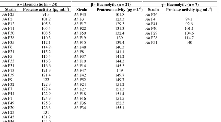
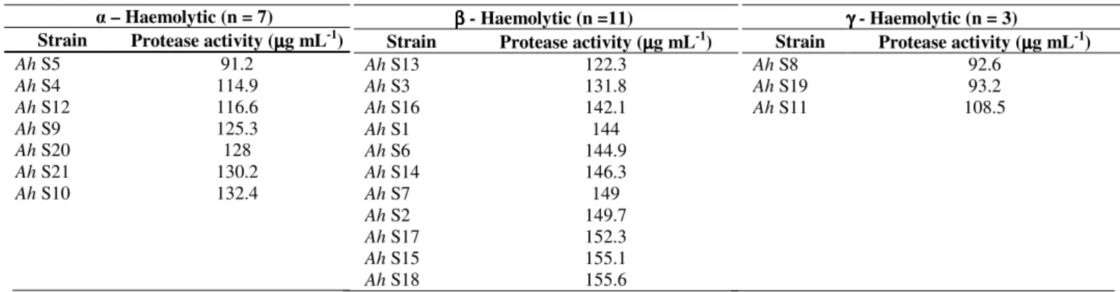
Proteolytic activity was determined for all the 73 strains
of A. hydrophila. The maximum and minimum level of
protease activity observed in fish isolates was 155.1 µg mL-1
and 91.3 µg mL-1 respectively, likewise the level of protease
activity observed in shrimp isolates was between 155.6 µg
mL-1 and 91.2 µg mL-1. Notably, the strain Ah F26 showed
no protease activity. Among the 32 β - haemolytic isolates, 31
of them showed proteolytic activity ≥ 122.3 µg mL-1 and of the
41 and γ- haemolytic isolates, 29 (26 and 3 γ - haemolytic) of
them showed proteolytic activity ≥ 108.5 µg mL-1 (Tables 3 and
4).
All the 73 A. hydrophila isolates were subjected to
cytotoxicity analysis on Vero cells. Cell shrinkage, detachment
and rounding were observed as cytotoxicity changes.
Vacuolating effects on Vero cells started within 6 hr of
bacterial supernatant addition. Complete cell death was
observed within 72 hr, with total detachment and destruction of
the monolayer. The cytotoxicity on Vero cells was observed till
1:16 dilution of the isolates’ cell free filtrate. The cytotoxic
effect was not observed in 1:32 and further dilutions. The cell
free filtrate of strain Ah F26 did not show cytotoxic effects on
Vero cells.
Table 3. Proteolytic activity of A. hydrophila isolates from fish samples (n = 52)
– Haemolytic (n = 24) Strain Protease activity (µµµµg mL-1)
Ah F25 91.3
Ah F2 101.2
Ah F12 105.3
Ah F11 105.4
Ah F30 108.5
Ah F38 110.3
Ah F35 112.1
Ah F6 114.2
Ah F21 115.2
Ah F5 115.4
Ah F33 116.3
Ah F31 116.6
Ah F13 121.3
Ah F39 121.4
Ah F9 122
Ah F32 122.3
Ah F7 122.4
Ah F44 122.9
Ah F1 124.3
Ah F49 125.3
Ah F20 126.3
Ah F23 131
β β β
β - Haemolytic (n = 21) Strain Protease activity (µµµµg mL-1)
Ah F43 101.8
Ah F3 123.3
Ah F17 129.3
Ah F22 131.3
Ah F50 132.4
Ah F19 139
Ah F15 139.4
Ah F48 140.3
Ah F8 141.1
Ah F37 141.2
Ah F10 144.3
Ah F14 145.3
Ah F47 149
Ah F42 149.7
Ah F52 149.7
Ah F24 151.2
Ah F27 151.3
Ah F18 151.4
Ah F16 151.5
Ah F36 152.3
Ah F34 155.1
γγγγ - Haemolytic (n = 7) Strain Protease activity (µµµµg mL-1)
Ah F26 -
Ah F4 94.1
Ah F41 92.6
Ah F40 101.1
Ah F29 104.6
Ah F28 114.7
981
Table 4. Proteolytic activity of A. hydrophila isolates from shrimp samples (n = 21)DISCUSSION
When analyzing the previous investigations (8, 13, 20, 24,
26,), a considerable increase in prevalence of A. hydrophila in
seafood was observed over the years. Moreover, 98.63% of the
73 isolates were observed to be multiple antibiotic resistant
against 20 commercially available standard antibiotics (20).
The occurrences of haemolytic factors in aeromonads are
widespread (10). Of the 52 A. hydrophila isolates from fish,
46.1, 40.3 and 13.4 % of them exhibited α, β and γ -
haemolytic activity respectively. Of the 21 A. hydrophila
isolates from shrimp, 33.3, 52.3 and 14.2 % showed α, β and γ
- haemolytic pattern respectively. In the present study, equal
distribution of α and β haemolytic activity was observed
(Table 1) which is in contrary to the results of Palumbo et al.
(18) who reported that all strains isolated from retail foods of
animal origin were β - haemolytic.
Slime is another type of virulence factor, which is a viscous
glycoconjugate material, produced by most of the Gram negative
bacteria. It is also helpful in the formation of biofilm. The slime is
highly significant to the pathogenesis; it appears to inhibit the
neutrophil, chemotaxis, phagocytosis and antimicrobial drugs (17).
Among the 73 isolates of A. hydrophila, 76.9 % in fish and 80.9
% in shrimp isolates were positive for slime production.
Proteases are thought to contribute to virulence of
aeromonads in fish and other hosts (9). Considerable
differences between the number, types and quantities of
protease produced by Aeromonas have been reported and
attributed to the strain variation, origin, incubation temperature
or culture media (16). All the 73 isolates of A. hydrophila
except Ah F26 have produced protease enzyme after 24 hr, but
the amount of protease production varied depending on the
strains and species diversity, which is in agreement with the
findings of Abdullah et al. (1). Howard and Buckley (11)
reported that both the proteolytic and haemolytic activity are
interrelated. The results of the present study supported the work of
Gonzalez-Serrano et al. (9) who documented that there is no
relation between proteolytic and haemolytic activities.
Aeromonads produce a wide range of extracellular toxins
and enzymes. The multiplicity of extracellular products has led
to difficulty in characterizing these factors and to disagreement
in the enteropathogenicity. Generally, cytotoxins damage the
cell and may produce severe illness in humans. Among the 73
A. hydrophila isolates, 98.61 % exhibited cytotoxicity on Vero
cells. Cytotoxic effects on Vero cells started within 6 hr of
bacterial supernatant addition and were monitored till 72 hr of
incubation time. In this study, Cytotoxicity was produced in
98.07 % and 100% of cell free culture filtrate of A. hydrophila
isolates from fish and shrimp samples respectively. Other
studies reported that 73 % of the food isolates (15) and 100 %
of the clinical isolates produced cytotoxicity on Vero cells (23).
In the present study, the cell free filtrate of A. hydrophila
had an impact on the integrity of Vero cells and might facilitate
a more rapid translocation of bacteria through the cells in vivo. – Haemolytic (n = 7)
Strain Protease activity (µµµµg mL-1)
Ah S5 91.2
Ah S4 114.9
Ah S12 116.6
Ah S9 125.3
Ah S20 128
Ah S21 130.2
Ah S10 132.4
β ββ
β - Haemolytic (n =11)
Strain Protease activity (µµµµg mL-1)
Ah S13 122.3
Ah S3 131.8
Ah S16 142.1
Ah S1 144
Ah S6 144.9
Ah S14 146.3
Ah S7 149
Ah S2 149.7
Ah S17 152.3
Ah S15 155.1
Ah S18 155.6
γγγγ - Haemolytic (n = 3)
Strain Protease activity (µµµµg mL-1)
Ah S8 92.6
Ah S19 93.2
Snowden et al. (22) observed similar type of impact on
integrity of the monolayers of Caco-2 cell lines. However, the
increased ability of A. hydrophila isolates to adhere, vacuolate
and destruct the Vero cells was in contrast to the previous
studies (19, 27). This may be due to the reflection of
geographic variation among the cytopathic ability of the
isolates.
Interestingly a notable relation was found between the
virulence factors and the cytotoxicity of the A. hydrophila
isolates on Vero cells. Among the 31 isolates (β - haemolytic
and showing proteolytic activity ≥ 122.3 µg mL-1), 27 strains’
cell free filtrate had significant impact on Vero cells; complete
cell death was observed within 12 hr of bacterial supernatant
addition. About 20 strains of the 26 ( – haemolytic and
showing proteolytic activity ≥ 108.5 µg mL-1) isolates’ cell free
filtrate exhibited complete death of Vero cells within 15 hr of
bacterial supernatant addition. All the 3 (γ – haemolytic and
showing proteolytic activity ≥ 108.5 µg mL-1) isolates’ cell free
filtrate showed complete death of Vero cells within 15 hr of
bacterial supernatant addition. Rest of the strains showed
variations in the time to produce complete cytotoxic effects on
Vero cells. Notably, the isolate Ah F26 did not showed
haemolytic activity, protease activity, slime production and
also showed no cytotoxic effects on Vero cells. This isolate
may be regarded as non-pathogenic.
The haemolytic pattern of the strains had no direct relation
to the cytotoxic activities as observed in vitro but might play an
important role in the pathogenesis of the organism in vivo.
Proteolytic activity had direct relation with the cytotoxic
activity irrespective of haemolytic activity of the strains. Even
though the results showed some relationship between
proteolysis and cytotoxic effects, there are other extracellular
virulence factors that must be studied in relation to
pathogenicity of A. hydrophila. These extracellular factors
might have different expression or subject to the same genetic
control. It is not unlikely that different virulence factors are
important under different conditions. Thus we must not only
characterize the different protein toxins, but also address the
genetic regulation of transcription.
CONCLUSION
These results demonstrated the presence of wide spread,
pathogenically characterized, multiple antibiotic resistant,
cytotoxic seafood borne A. hydrophila in Chennai. The relation
found between the haemolysis; proteolysis and cytotoxic
activities must be further evaluated in relation to genetic
control of other extracellular virulence factors, molecular
mechanisms and network of these virulence factors in
exhibiting the pathogenesis of A. hydrophila.
REFERENCES
1. Abdullah, A.I.; Hart, C.A.; Winstanley, C. (2003). Molecular characterization and distribution of virulence - associated genes amongst Aeromonas isolates from Libya. J Appl Microbiol. 95, 1001-1007. 2. Breneder, R.; Janda, J.M. (1987). Detection, quantification and stability
of the β - haemolysin of Aeromonas spp. J Med Microbiol 24, 247-251. 3. Castro-Escarpulli G.; Figueras, M.J.; Aguilera-Arreola, G.; Soler, L.;
Fernandez- Rendon, E.; Aparicio, G.O.; Guarro, J.; Chacon, M.R. (2003). Characterization of Aeromonas species isolated from frozen fish intended for human consumption in Mexico. Int J Food Microbiol. 84 (1), 41-49. 4. Charney, J.; Tomarelli, R.M. (1947). A colorimetric method for the
determination of the proteolytic activity of duodenal juice. J Biol Chem. 171, 501-505.
5. Darlington, L.G.; Stone, T.W. (2001). Antioxidants and fatty acids in the amelioration of rheumatoid arthritis and related disorders. Br J Nutr. 85, 251-269.
6. Freeman, D.J.; Falkiner, F.R.; Keane, C.T. (1989). New method for detection slime production by coagulase negative staphylococci. J Clin Pathol. 42, 872–874.
7. Gillespie, I.A.; Adak, G.K.; O’Brien, S.J.; Brett, M.M.; Bolton, F.J. (2001). General outbreaks of infectious intestinal disease associated with fish and shellfish, England and Wales, 1992-1999. Commun Dis Public Health. Vol 4 (2), 117-123.
8. Gonazalez, G.J.; Santos, J.A.; Gareia-Lopez, H.L.; Gonazalez, N.; Otero, A. (2001). Mesophilic Aeromonas in wild and aquacultured fresh water Fish. J Food Prot. 64, 687-691.
9. Gonzalez-Serrrano, C.J.; Santos, J.A.; Garcia-Lopez, M.L.; Otero, A. (2002). Virulence markers in Aeromonas hydrophila and Aeromonas veronii biovar sobria isolates obtained from fresh water fish and from a diarrhoea case. J Appl Microbiol. 93 (3), 414-419.
983
11. Howard, S.P.; Buckley, J.T. (1985). Protein export by a gram-negativebacterium: production of aerolysin by Aeromonas hydrophila. J Bacteriol. 161, 1118-1124.
12. Howard, S.P.; Mac Intyre, S.; Buckley, J.T. (1996). Toxins. In: Austin, B., Altwegg, M., Gosling, P.J., Joseph, S. (Eds.). The Genus Aeromonas. Wiley and Sons, Chrichester, UK, p. 267–286.
13. Hudson, J.A.; De Lacy, K.M. (1991). Incidence of motile aeromonads in New Zealand retail foods. J Food Prot. 54, 696-699.
14. Janda, J.M.; Abbot, S.L. (1996). Human pathogens. In: Austin, B., Altwegg, M., Gosling, P.J., Joseph, S. (Eds.), The Genus Aeromonas. Wiley and Sons, Chrichester, UK, p. 151-170.
15. Martins, L.M.; Marquez, R.F.; Yano, T. (2002). Incidence of toxic Aeromonas isolated from food and human infection. FEMS Immunol Med Microbiol. 32, 237-242.
16. Mateos, D.; Anguita, J.; Naharro, G.; Panlagua, C. (1993). Influence of growth temperature on the production of extracellular virulence factors and pathogenicity of environmental and human strains of Aeromonas hydrophila. J Appl Bateriol. 74, 111-118.
17. Merino, S.; Rubires, X.; Knochel, S.; Tomas, J.M. (1995). Emerging pathogens: Aeromonas spp. Int J Food Microbiol. 28, 157-168.
18. Palumbo, S.A.; Bencivengo, M.M.; Corral, F.D.; Williams, A.G.; Buchanan, R.L. (1989). Characterization of the Aeromonas hydrophila group isolated from retail foods of animal origin. J Clin Microbiol. 27, 854-859.
19. Sechi, L.A.; Deriu, A.; Falchi, M.P.; Fadda, G.; Zanetti, S. (2002). Distribution of virulence genes in Aeromonas spp. isolated from Sardinian waters and from patients with diarrhoea. J Appl Bateriol. 92, 221–227.
20. Seethalakshmi, I.; Jayalakshmi, S.; Subashkumar, R.; Swaminathan, P.;
Rajagopal, K. (2006). Occurrence of multiple antibiotic resistant Aeromonas hydrophila isolated from marketed fish and shrimp samples. Indian J Appl Microbiol. 6 (1), 79-87.
21. Snieszko, S.F.; Bullock, G.L. (1976). Disease of freshwater fishes caused by bacteria of the genera Aeromonas, Pseudomonas and Vibrio. FDL 40, USKI, FWS, p.10.
22. Snowden, L.; Wernbacher, L.; Stenzel, D.; Tucker, J.; McKay, D.; O'Brien, M.; Katouli, M. (2006). Prevalence of environmental Aeromonas in South East Queensland, Australia: a study of their interactions with human monolayer Caco-2 cells. J Appl Microbiol. 101 (4), 964–975.
23. Sridharan, G.; Balaji, V.; Jesudason, M.V. (2004). Cytotoxin testing of environmental Aeromonas spp. in Vero cell culture. Ind J Med Res. 119, 1-3.
24. Thayumanavan, T.; Vivekanandhan, G.; Savithamani, K.; Subashkumar, R.; Lakshmanaperumalsamy, P. (2003). Incidence of haemolysin-positive and drug-resistant Aeromonas hydrophila in freshly caught finfish and prawns collected from major commercial fishes of Costal South India. FEMS Immunol Med Microbiol. 36, 41–45.
25. Trust, M.B.; Currie, B.R.; Buckley, J.T. (1974). Obligate anaerobic bacteria in the gastrointestinal microflora of the grass carp (Ctenopharyngodon idella), goldfish (Carassius auratus) and rainbow trout (Salmo gairdneri). J Fish Res Board Can. 36, 1174-1179.
26. Vivekanandhan, G.; Hatha, A.A.M.; Lakhmanaperumalsamy, P. (2005). Prevalence of Aeromonas hydrophila in fish and prawns from the seafood market of Coimbatore, South India. Food Microbiol. 22, 133-137. 27. Yadav, M.; Indira, G.; Ansary, A. (1992). Cytotoxin elaboration by