Dendritic Spine, Neuroinflammation and Memory
Impairment at Rats after Surgery
Yuan Le1, Shuli Liu1, Mingchao Peng1, Chang Tan1, Qin Liao1, Kaiming Duan1, Wen Ouyang1*, Jianbin Tong1,2*
1Department of Anesthesiology, the Third Xiangya Hospital, Central South University, Changsha, Hunan, P.R. China,2Center for Experimental Medicine, the Third Xiangya Hospital, Central South University, Changsha, Hunan, P.R. China
Abstract
It is known that age is an important factor for postoperative cognitive dysfunction (POCD) and the patients with POCD suffer from the impairment of multiple brain regions and multiple brain functions. However currently animal studies of POCD mainly focus on hippocampus region, therefore in this study we performed partial hepatectomy in young adult and aged rats to test the questions (1) whether POCD in animals involves other brain areas besides hippocampus; (2) how age influences POCD of young adult and aged animals. We found that (1) in young adult rats, the memory was not significantly affected (P.0.05) 1d, 3d and 7d after partial hepatectomy, but was significantly impaired (p,0.001) in aged rats 1d and 3d post-surgery; (2) in young adult rats, the surgery did not significantly affect the densities of dendritic spines of neurons at CA1, dentate gyrus (DG) and cingulate cortex (P.0.05, respectively) 1d and 3d post-surgery, but the spine densities at CA1 and DG of aged rats were significant reduced 1d and 3d post-surgery (p,0.001, respectively), however this didn’t happen at cingulate cortex (P.0.05); (3) In young adult rats, surgery didn’t affect the activation of microglia and levels of TNF-aand IL-1bat hippocampus (P.0.05), but significantly activated microglia and increased levels of TNF-aand IL-1bat hippocampus of aged rats (P,0.05). Our data suggest that (1) partial hepatectomy-induced POCD mainly involves hippocampus impairments, and (2) differential loss of neuronal dendritic spines and neuroinflammation at hippocampus are most likely the mechanism for the formation of POCD in aged rats.
Citation:Le Y, Liu S, Peng M, Tan C, Liao Q, et al. (2014) Aging Differentially Affects the Loss of Neuronal Dendritic Spine, Neuroinflammation and Memory Impairment at Rats after Surgery. PLoS ONE 9(9): e106837. doi:10.1371/journal.pone.0106837
Editor:Zhongcong Xie, Massachusetts General Hospital, United States of America
ReceivedJune 16, 2014;AcceptedAugust 1, 2014;PublishedSeptember 8, 2014
Copyright:ß2014 Le et al. This is an open-access article distributed under the terms of the Creative Commons Attribution License, which permits unrestricted use, distribution, and reproduction in any medium, provided the original author and source are credited.
Data Availability:The authors confirm that all data underlying the findings are fully available without restriction. All relevant data are within the paper and its Supporting Information files.
Funding:This work was supported by grants from the National Natural Science Foundation of China (No.81371216). The funders had no role in study design, data collection and analysis, decision to publish, or preparation of the manuscript.
Competing Interests:The authors have declared that no competing interests exist. * Email: [email protected] (WO); [email protected] (JT)
Introduction
Postoperative cognitive dysfunction (POCD) is usually detected among aged patients after surgery, especially after critical illness [1–3]. It characterizes with impaired memory, information processing, concentration and mental flexibility [1,2,4]. Occur-rence of POCD is closely associated with increased incidence of postoperative complications, longer hospitalization, and higher mortality of 6 months [5]. The profound socioeconomic signifi-cance of POCD makes it the subject of many investigations.
Clinical researches have shown that brain areas involved in POCD include frontal, parietal, temporal, occipital, hippocampal, insular, cingulated, thalamic and cerebellar regions [4]. Risk factors of POCD include preoperative factors (such as age, cognitive function level, mental illness), perioperative factors (such as surgery type, duration of surgery, anesthesia type, intraoperative hypotension), and postoperative factors (such as postoperative infection, respiratory complications) [6–9]. Among these risk factors, age is the only risk factor for long-term POCD [6–9]. Further animal studies have shown that anaesthetics neurotoxicity,
systemic inflammation induced by surgery trauma, and acceler-ation of ongoing endogenous neurodegenerative processes all contribute much to POCD [10–13]. For example, Terrando et al found that blocking the signals of TNF-a and IL-1 effectively decreased the impairment of cognitive function of adult mice after surgery [8]. Li et al found that minocycline, a drug of anti-inflammation, mitigated isoflurane-induced cognitive impairment in aged rats [14]. However, there is an obvious translational gap between clinical studies and animal studies of POCD [4]. Animal studies of POCD mainly focused on the changes of structures and functions of hippocampus induced by surgery, neglecting the fact of POCD that multiple brain regions and multiple brain functions are affected in patients. It remains unclear (1) whether POCD in animals involves other brain areas besides hippocampus; (2) how age influences POCD of young adult and aged animals.
after surgery [2]. Cingulate cortex is associated with working memory, long-term memory, mental flexibility, and selective attention, which are totally impaired in POCD [4]. In addition, synapse plasticity is the structure base of cognitive functions and is usually evaluated by changes of dendritic spines of neurons [18– 20]. For these reasons we answered above-mentioned two questions in this study by the detection of learning and memory, the measurement of the dendritic spine density of neurons at hippocampus and cingulate cortex, and the level of neuroin-flammation of hippocampus at young adult and aged rats before and after surgery. We found that surgery induced obvious loss of dendritic spines of neurons at CA1 and dentate gyrus (DG) of aged rats, corresponding to their memory impairment after surgery. But surgery didn’t significantly affect dendritic spine density and memory of young adult rats. In addition, surgery also induced strong inflammation response at hippocampus of aged rats, but not at young adult rats. Our data suggest that postoperative memory dysfunction in aged rats is closely associated with the loss of neuronal dendritic spines and strong inflammation at hippocam-pus.
Materials and Methods
Animals and Grouping
The animal care and the experimental protocol were approved by the Institutional Review Board of the Third Xiangya Hospital of Central South University. Forty-six young adult (aged 2 months, 200–250 g) and fifty-two aged (aged 18 months, 500–610 g) female Sprague-Dawley rats were purchased from Central South University (P.R. China). All rats were raised under controlled environmental conditions on a 12 h light/dark cycle with ad libitum access to food and water.
Young adult (n = 22 for behavior test, n = 24 for Golgi staining and other staining) and aged rats (n = 28 for behavior test, n = 24 for Golgi staining and other staining) were randomly divided into normal control (n = 19 for young adult, n = 22 for aged), and surgery groups (n = 27 for young adult, n = 30 for aged). Rats in surgery group received partial hepatectomy plus general anethesia. Rats in nomal control received neither anesthesia nor surgery.
Anesthesia and partial hepatectomy
Anesthesia was prepared with the procedure described by He et al [12]. Rats were exposed to sevoflurane (4.5% sevoflurane in 100% oxygen for induction followed by 2.5% for maintenance) for 2 h with endotracheal intubation with a 14-gauge catheter. Gas concentrations and respiratory rate were continuously monitored with a multi-function monitor (datex-ohmeda, Helsinki, Finland). Partial hepatectomy was performed under aseptic conditions following Wuri’s method [21]. Briefly, a small incision about 2 cm was made in the upper quadrant through skin and muscles. The left liver was visualized, isolated, and removed. After checking, the muscles and skins were sutured, respectively.
Morris Water maze test
The Morris water maze test was used for assessing learning and memory of rats. We used a computerized video track system (Logitech, Suzhou, China) to record the rat’s movement in water maze by following our previous method [12]. Briefly, a transparent round platform was placed below the water surface of southeast quadrant in a circular pool. During the training, rats were first placed on the platform for 30 seconds, and then were released into the water facing the tank wall. The maximum trial time was 60 seconds for a trial, following a relaxation of 15 seconds on the platform. If a rat couldn’t locate the platform within 60 seconds, it
was guided to the platform and remained for 30 seconds. All rats were trained for 6 days with three trials per day. After training, the memory of rats, which was evaluated by the latency for the first entrance of targeted area and the percents of searching time and distance in the targeted area, was detected 1d before surgery and 1d, 3d, and 7d after surgery.
Golgi staining and analysis
Golgi staining was performed with FD Rapid Golgi-stain Kit (FD Neurotechnologies, MD) according to the manufactures instructions [18–20]. Briefly, fresh brain was impregnated in the mixed solution A and B for two weeks at room temperature, and then at solution C for 48 hours at 4uC. The brains were cut with 150mm thickness and mounted on the gelatin-coated slides. After drying, the sections were stained with solution D and E, dehydrated in graded ethanol, cleared in xylene, and finally covered by coverslip. Neurons (5 CA1 neurons/rat, 5 neurons at the 2nd layer of cingulate cortex/rat, 8 DG neurons/rat) were analyzed following our previous methods [18–20]. The selected neurons have to be relatively isolated from neighboring neurons. Three to five tertiary apical dendrites and basal dendrites with at least one branch point were selected for counting for each neuron [18–20]. The visible spines along the branch segment (.10mm long) were counted and data were expressed as number/10mm [18–20].
Tissue Preparation
For immunofluorescence assay, under deep anesthesia, rats were firstly infused with 0.9% saline at 37uC, and then with 4% paraformaldehyde. The brains were taken out, and then post-fixed in 4% paraformaldehyde overnight at 4uC. After dehydrating with sucrose, cross sections with hippocampus were cut at a thickness of 16mm by a cryostat machine.
For western blot assay, under deep anesthesia, rats were killed. The hippocampus was removed and homogenized in cold lysis buffer containing protease inhibitors. After centrifuging, the supernatant of hippocampal homogenates were collected and stored at280uC.
Immunofluorescence
After washing with 0.01 M phosphate buffered saline (PBS) for 10 min, sections were incubated in blocking solution (5% BSA and 0.3% Triton X-100 in 0.01 M PBS) for 1 hr at room temperature. Then the sections were incubated in primary antibodies (rabbit anti-Iba-1, lot 019–19741, 1:1000, Wako Chemical) overnight at 4uC. On the second day, these sections were washed with 0.01 M PBS for three times and then incubated in the secondary antibodies labeled with fluorescent dyes (1:200, Jackson Immunor-esearch) for 2 hours at room temperature. Through three washes of PBS, these sections were covered with mounting medium with DAPI (vector). As negative controls, an adjacent series of sections were processed using the same procedures without the primary antibodies.
Western blot
Cam-bridge, UK; rabbit anti-IL-1b polyclonal antibody, 1:1000, Abzoom, Shanghai, China;rabbit anti-GAPDH, 1:4000, Protein-tech Group, Shanghai, China) overnight at 4uC. After three washes, membranes were incubated with the secondary antibodies (1:2000) at room temperature for 2 h. Finally, visualization of the proteins was accomplished by enhanced chemiluminescence detection kit (Pierce; Thermo Scientific, Shanghai, China), and the intensity of each band was quantified by densitometry. Relative expression levels of protein were normalized by the ratio of target protein (TNF-a, IL-1b) to GAPDH.
Statistical analysis
Water maze data were presented as mean 6 standard error (mean 6 SEM). The water maze data were analyzed using repeated measures ANOVA with the factor surgery and the factor measure time, complemented by repeated measures ANOVA at data of control group and surgery group, respectively. Golgi data and western data were presented as mean6 standard deviation (mean6SD) and were analyzed using two-way ANOVA followed by LSD test. P,0.05 was considered statistically significant.
Results
Hepatectomy differentially impaired the memory of young adult and aged rats
The effects of surgery on the memory of rats were detected by Morris water maze. During the training of 6 days, there was no significant difference of the latency to platform between control and surgery groups in aged (P = 0.202) and young adult rats (P = 0.057), suggesting same learning ability in control and surgery groups (Fig. 1A). During the probe test, the latency for the first entrance of targeted area and the percents of time and distance in the targeted area were measured to evaluate the memory of rats. Percent of distance in targeted area (P = 0.767), percent of time in targeted area (P = 0.769) and latency for the first entrance of targeted area (P = 0.903) in the surgery group weren’t different from that of control group at aged rats 1d before surgery (Fig. 1 B– D), suggesting that the same memory ability of aged rats at control group and surgery group before surgery. However, further repeated measures ANOVA showed that percents of searching time and distance in the targeted area of aged rats were affected by surgery (P = 0.045 for time; P = 0.041 for distance), measure time (P = 0.013 for time; P,0.001 for distance), and the interaction of surgery6measure time (P = 0.037 for time; P = 0.013 distance)
(Fig. 1B, C). Compared to 1d before surgery, the percents of time and distance in the targeted area of aged rats obviously decreased 1d (P = 0.006 for time, P,0.001 for distance) and 3d (P = 0.011 for time, P = 0.002 for distance) after surgery (Fig. 1B, C). The latency for the first entrance of targeted area at aged rats was not affected by surgery (P = 0.495), measure time (P,0.067), the interaction of surgery6measure time (P = 0.984) (Fig. 1D). These above
suggest-ed that surgery impairsuggest-ed the memory of agsuggest-ed rats 1d and 3d after hepatectomy.
For young adult rats, there was no obvious difference at the percent of distance in the targeted area (P = 0.170), the percent of time in the targeted area (P = 0.772) and the latency for the first entrance of targeted area (P = 0.938) between control and surgery groups 1d before surgery (Fig. 1). Further repeated measures ANOVA showed that the percents of time and distance, and the latency for the first entrance in the targeted area were not affected by surgery (P = 0.639 for time; P = 0.773 for distance; P = 0.983 for latency), measure time (P = 0.327 for time; P = 0.358 for distance; P = 0.173 for latency) and the interaction of surgery6measure time
(P = 0.122 for time; P = 0.095 for distance; P = 0.436 for latency)
(Fig. 1). These data suggested that surgery didn’t impair the memory of young adult rats.
Hepatectomy differentially induced the loss of dendritic spines of neurons at young adult and aged rats
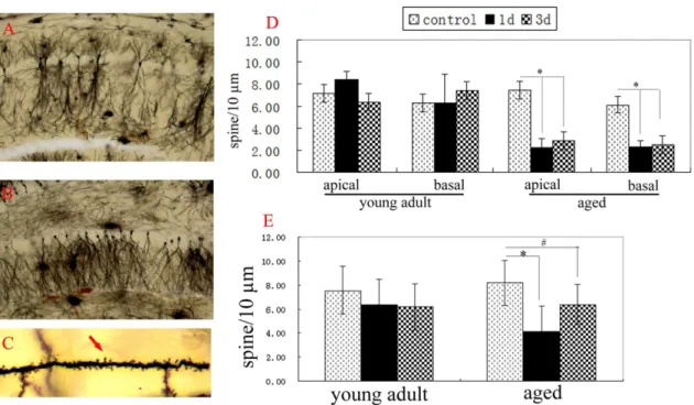
Synapse plasticity is the structure base of memory. Changes of dendritic spine density of neurons are usually used to show synapse plasticity [22,23]. In addition, dendritic spines of neurons are easily affected by the environment [24]. In order to detect the effects of surgery on different brain areas, we detected the changes of dendritic spine densities of neurons at CA1, DG and cingulate cortex of young adult and aged rats after hepatectomy. At normal aged rats, the spine densities of apical dendrites and basal dendrites of neurons at CA1 and cingulate cortex and the spine densities of basal dendrites of neurons at DG all were similar to that of normal young adult rats, respectively (p.0.05) (Fig. 2, 3). Two-way ANOVA analysis showed that spine densities of apical dendrites and basal dendrites of neurons at CA1 and DG of aged rats were affected by surgery (p,0.001 for CA1 apical, CA1 basal, and DG basal, respectively), age (p,0.001 for CA1 apical and CA1 basal; P = 0.405 for DG basal), and the interaction of surgery6age (p,0.001 for CA1 apical and CA1 basal; P = 0. 078 for DG basal). Compared to that of normal aged rats, the spine densities of apical dendrites (2. 2660.73 for 1d; 2. 8660.95 for 3d) and basal dendrites (2. 2860.60 for 1d; 2. 4960.80 for 3d) of neurons at CA1 of aged rats significantly decreased 1d and 3d after hepatectomy (P,0.001, respectively) (Fig. 2D). The spine density of basal dendrites of neurons at DG of aged rats also obviously decreased 1d and 3d after hepatectomy (P,0.001 for 1d; P = 0.004 for 3d) (Fig. 2E). In contrast, the spine densities of apical dendrites and basal dendrites of neurons at cingulate cortex of aged rats weren’t altered 1d and 3d after hepatectomy, compared to the normal aged rats (P.0.05) (Fig. 3C). These data showed that surgery decreased the spine densities of neurons at CA1 and DG, but not at cingulate cortex of aged rats.
At young adult rats, the spine densities of apical dendrites and basal dendrites of neurons at CA1 and cingulate cortex and the spine densities of basal dendrites of neurons at DG all weren’t altered 1d and 3d after hepatectomy, similar to that of normal young adult rats (p.0.05, respectively) (Fig. 2, 3).
Hepatectomy differentially induced strong
neuroinflammation at hippocampus of aged and young adult rats
Figure 1. Hepatectomy differentially impaired the memory of young adult and aged rats.Latency to platform of young adult and aged rats both at control and surgery groups decreased with the increase of training days. No significant difference was detected between control and surgery groups of young adult and aged rats by repeated measures of ANOVA (P = 0.202 for aged; P = 0.057 for young adult) (A). During probe test of aged rats, repeated measures of ANOVA showed that the latency for the first entrance of targeted area wasn’t affected by surgery (P = 0.495), measure time (P = 0.067), and the interaction of surgery6measure time (P = 0.984) (D). Yet, percents of time (B) and distance (C) in the targeted area both were significantly affected by surgery (P = 0.045 for time; P = 0.041 for distance), measure time (P = 0.013 for time; P,0.001 for distance), and the interaction of surgery6measure time (P = 0.037 for time; P = 0.013 distance). Percents of time (B) and distance (C) in the targeted area of aged rats 1d (P = 0.006 for time; P,0.001 for distance) and 3d (P = 0.011 for time, P = 0.002 for distance) after surgery all were obviously less than that of 1d before surgery, respectively. For young adult rats, there was no obvious difference at the percent of distance in the targeted area (P = 0.170), the percent of time in the targeted area (P = 0.772) and the latency for the first entrance of targeted area (P = 0.938) between control and surgery groups 1d before surgery (A). Percents of time and distance, and the latency for the first entrance in the targeted area were not affected by surgery (P = 0.639 for time; P = 0.773 for distance; P = 0.983 for latency) and measure time (P = 0.327 for time; P = 0.358 for distance; P = 0.173 for latency) (B, C, D). 1d pre: 1d before surgery; 1d aft, 3d aft and 7d aft: 1d, 3d, 7d after surgery. Data were mean6SEM. *p,0.05vs1d before surgery.
doi:10.1371/journal.pone.0106837.g001
Figure 3. Hepatectomy didn’t decrease spine densities of neuron at cingulate cortex of adult and aged rats.A, neurons of cingulate cortex. B, showing the dendritic spine of neurons at the 2ndlayer of cingulate cortex (red arrow). C, the spine densities of apical dendrites and basal
dendrites of neurons were analyzed by two-way ANOVA. Compared with the control, no difference was detected 1d and 3d after surgery (p.0.05, respectively). Data were mean6SD.
doi:10.1371/journal.pone.0106837.g003
Figure 4. Hepatectomy differentially induced strong neuroinflammation at hippocampus of aged and adult rats.A: Iba1 staining (red) of CA1 at normal young adult rat. B: the merged panel of Iba1 staining (red) and Dapi (blue) of CA1 at normal young adult rat. C: Iba1 staining (red) of CA1 at young adult rat 1d after surgery. D: the merged panel of Iba1 staining (red) and Dapi (blue) of CA1 at young adult rat 1d after surgery. E: Iba1 staining (red) of CA1 at normal aged rats. F: the merged panel of Iba1 staining (red) and Dapi (blue) of CA1 at normal aged rats. G: Iba1 staining (red) of CA1 at aged rat 1d after surgery. Activated microglia (white arrow) was observed. H: the merged panel of Iba1 staining (red) and Dapi (blue) of CA1 at aged rat 1d after surgery. I: western blot of TNF-aat hippocampus. J: western blot of IL-1bat hippocampus. Data were mean6SD. *p,0.05 vs control. Bar = 50 um.
adult rats after surgery were similar to that of control (P.0.05) (Fig. 4I, J).
Discussion
The aim of the study was to detect (1) whether POCD in animals involves other brain areas besides hippocampus; (2) how age influences POCD of young adult and aged animals. We found that corresponding to memory impairment of aged rats after surgery, surgery induced obvious loss of dendritic spines of neurons at CA1 and DG of aged rats, but not affected the density of dendritic spine of neurons at cingulate cortex. Surgery didn’t significantly decrease spine density of neuronal dendrites and memory of young adult rats. In addition, surgery induced strong neuroinflammation at hippocampus of aged rats, but not at the young adult rats. These data suggested that surgery differentially induced the loss of neuronal dendritic spine and neuroinflamma-tion at hippocampus of young adult and aged rats, which was the possible basis for postoperative memory impairment of aged rats. These data also suggested that hippocampus was the main target involved in POCD.
Many brain areas including frontal, parietal, temporal, occip-ital, hippocampal, insular, cingulated, thalamic and cerebellar regions are affected in aged patients with POCD [4]. These surgery-affected brain areas are closely related with memory, information processing, concentration and mental flexibility [4]. However, in the animal studies of POCD, most of them focused on the structure and function of hippocampus [4]. To date, it remained unclear whether surgery affected other brain areas besides hippocampus in animals with POCD. Dendritic spines of neurons were easily affected by the environment [24], and were closely associated with memory [22,23]. Thus, in the study, we used the changes of dendritic spines of neurons to evaluate the effects of surgery on neurons of hippocampus and cingulate cortex, two very important brain areas involved in POCD of patients. Our data showed that surgery induced obvious memory impairment of aged rats 1d and 3d after hepatectomy, but not at young adult rats. Corresponding to the postoperative memory impairment of aged rats, surgery also obviously reduced the spine densities of dendrites of neurons at CA1 and DG of aged rats 1d and 3d after hepatectomy, but not at cingulate cortex neurons of aged rats and at the neurons of CA1, DG and cingulate cortex of young adult rats. These data showed that (1) the loss of spines of neurons at CA1 and DG was closely associated with postoperative memory impairment of aged rats; (2) hippocampus was the main target impaired by surgery. This was consistent with our previous research of aged patents with surgery [2]. In aged patients, small hippocampal volume could be an independent risk predictor of POCD [2]. In addition, Bloss et al found that dendritic spines of prefrontal cortex neurons of aged rat were remarkably stable to the stress [22]. Thus it was possible that surgery didn’t affect the dendritic spine density of neurons at cingulate cortex of aged rats. The difference of frontal cortex involved POCD between human and rats reported in this study might be due to the species difference. In addition, anesthesia or surgery, like other stress factors, may provide dual effects on neuroprotection and neurotoxicity [4,11]. Minor stresses (e.g. general anesthetics at
low concentration for short time) may provide neuroprotection [4,11]. Detrimental stresses (e.g. general anesthesia at high concentration for long duration) may provide neurotoxicity [4,11]. The stress size of anesthesia or surgery was also the possible reason for the difference of frontal cortex involved POCD between human and rats reported in this study.
Age is the main risk factor of POCD [5]. POCD is usually detected in aged patients with surgery, but not in young adult patients [5]. Low cognitive reservation is thought to be the reason for occurrence of POCD at aged patients [2]. However the question is how age influences POCD of young adult and aged animals. Thus we first detected the spine density of neurons at young adult and aged rats after surgery. Our data showed that in contrast to the significant loss of dendritic spines of neurons at CA1 and DG of aged rats after surgery, there were no obvious changes at spine densities of dendrites of neurons at CA1, DG and cingulate cortex of young adult rats. These suggested that surgery induced more obvious impairment of neurons at aged rats than that of young adult rats. Neuroinflammation was closely associated with POCD [8,14,27,28]. So we also detected the activation of microglia and the expressions of TNF-aand IL-1bafter surgery. Corresponding to the loss of dendritic spines of hippocampal neurons, microglia was activated and levels of TNF-aand IL-1b
were up-regulated at hippocampus of aged rats after surgery. In contrast, activation of microglia and increase of TNF-aand IL-1b
were not detected in young adult rats after surgery. These data showed that surgery induced strong neuroinflammation at the hippocampus of aged rats, but not at young adult rats. Similar results were also reported by Cao [26]. They found that surgery induced more durable and stronger inflammation response in aged rats, compared with adult rats [26]. Previous studies showed that intra-hippocampal administration of IL-1b impaired contextual fear memory of rats [29]. Sustained elevation of hippocampal IL-1b levels also produced marked impairments in spatial memory [30]. In addition, over-expressing TNF-a in the brain (TNF-a
transgenic mice) impaired leaning of adult mice [31]. Intra-hippocampal administration of TNF-a impaired hippocampal-dependent working memory [32]. Blocking the signals of TNF-a
and IL-1beffectively decreased the cognitive function impairment induced by surgery [8,14,27]. These information showed that increase of TNF-a and IL-1b was detrimental in learning and memory. Based on the above information, we thought that strong neuroinflammation was possible mechanism for significant loss of dendritic spines of hippocampal neurons of aged rats, finally led to POCD.
Briefly, our data show that partial hepatectomy-induced POCD mainly involves hippocampus impairment and differential loss of neuronal dendritic spines and neuroinflammation at hippocampus are most likely the mechanism for the formation of POCD in aged rats.
Author Contributions
Conceived and designed the experiments: JBT WOY. Performed the experiments: YL SLL MCP CT. Analyzed the data: YL JBT. Contributed reagents/materials/analysis tools: QL KMD. Contributed to the writing of the manuscript: YL JBT.
References
1. Ramaiah R, Lam AM (2009) Postoperative cognitive dysfunction in the elderly. Anesthesiol Clin 27: 485–496.
2. Chen MH, Liao Y, Rong PF, Hu R, Lin GX, et al. (2013) Hippocampal volume reduction in elderly patients at risk for postoperative cognitive dysfunction. J Anesth 27: 487–492.
3. Pandharipande PP, Girard TD, Jackson JC, Morandi A, Thompson JL, et al. (2013) Long-term cognitive impairment after critical illness. N Engl J Med 369: 1306–1316.
gap between clinical and pre-clinical perspectives. Brain Behav Immun 26: 1169–1179.
5. Monk TG, Price CC (2011) Postoperative cognitive disorders. Curr Opin Crit Care 17: 376–381.
6. Ballard C, Jones E, Gauge N, Aarsland D, Nilsen OB, et al. (2012) Optimised anaesthesia to reduce post operative cognitive decline (POCD) in older patients undergoing elective surgery, a randomised controlled trial. PLoS One 7: e37410. 7. Monk TG, Weldon BC, Garvan CW, Dede DE, van der Aa MT, et al. (2008) Predictors of cognitive dysfunction after major noncardiac surgery. Anesthesi-ology 108: 18–30.
8. Terrando N, Brzezinski M, Degos V, Eriksson LI, Kramer JH, et al. (2011) Perioperative cognitive decline in the aging population. Mayo Clin Proc 86: 885–893.
9. Wang J, Su T, Liu Y, Yue Y, He R (2012) Postoperative cognitive dysfunction is correlated with urine formaldehyde in elderly noncardiac surgical patients. Neurochem Res 37: 2125–2134.
10. Hudson AE, Hemmings HC Jr (2011) Are anaesthetics toxic to the brain? Br J Anaesth 107: 30–37.
11. Yan XB, Ouyang W, Li G, Duan KM (2012) Involvement of neuronal nitric oxide synthase in cognitive impairment in isoflurane-treated rats. Neurosci Lett 506: 240–244.
12. He HJ, Wang Y, Le Y, Duan KM, Yan XB, et al. (2012) Surgery upregulates high mobility group box-1 and disrupts the blood-brain barrier causing cognitive dysfunction in aged rats. CNS Neurosci Ther 18: 994–1002.
13. Wang Y, He H, Li D, Zhu W, Duan K, et al. (2013) The role of the TLR4 signaling pathway in cognitive deficits following surgery in aged rats. Mol Med Rep 7: 1137–1142.
14. Li SY, Xia LX, Zhao YL, Yang L, Chen YL, et al. (2013) Minocycline mitigates isoflurane-induced cognitive impairment in aged rats. Brain Res 1496: 84–93. 15. Harvey J (2013) Leptin regulation of neuronal morphology and hippocampal
synaptic function. Front Synaptic Neurosci 5: 3.
16. Morrison JH, Baxter MG (2012) The ageing cortical synapse: hallmarks and implications for cognitive decline. Nat Rev Neurosci 13: 240–250.
17. Devinsky O, Morrell MJ, Vogt BA (1995) Contributions of anterior cingulate cortex to behaviour. Brain 118: 279–306.
18. Tong J, Huang C, Bi F, Wu Q, Huang B, et al. (2012) XBP1 Depletion Precedes Ubiquitin Aggregation and Golgi Fragmentation in TDP-43 Transgenic Rats. J Neurochem 123: 406–416.
19. Huang C, Tong JB, Bi Ff, Wu Qx, Huang B, et al. (2012) Entorhinal cortical neurons are the primary targets of FUS mislocalization and ubiquitin aggregation in FUS transgenic rats. Hum Mol Genet 21: 4602–4614.
20. Yau SY, Lau BWM, Tong JB, Wong R, Ching YP, et al. (2011) Hippocampal Neurogenesis and Dendritic Plasticity Support Runnin-Improved Spatial Learning and Depression-Like Behaviour in Stressed Rats. PLoS ONE 6: e24263.
21. Wuri G, Wang DX, Zhou Y, Zhu SN (2011) Effects of surgical stress on long-term memory function in mice of different ages. Acta Anaesthesiol Scand 55: 474–485.
22. Bloss EB, Janssen WG, Ohm DT, Yuk FJ, Wadsworth S, et al. (2011) Evidence for reduced experience-dependent dendritic spine plasticity in the aging prefrontal cortex. J Neurosci 31: 7831–7839.
23. Sala C, Segal M (2014) Dendritic spines: the locus of structural and functional plasticity. Physiol Rev 94: 141–188.
24. Petralia RS, Mattson MP, Yao PJ (2014) Communication breakdown: The impact of ageing on synapse structure. Ageing Res Rev 14C: 31–42. 25. Yirmiya R, Goshen I (2011) Immune modulation of learning, memory, neural
plasticity and neurogenesis. Brain Behav Immun 25: 181–213.
26. Cao XZ, Ma H, Wang JK, Liu F, Wu BY, et al. (2010) Postoperative cognitive deficits and neuroinflammation in the hippocampus triggered by surgical trauma are exacerbated in aged rats. Prog Neuropsychopharmacol Biol Psychiatry 34: 1426–1432.
27. Cibelli M, Fidalgo AR, Terrando N, Ma D, Monaco C, et al. (2010) Role of interleukin-1beta in postoperative cognitive dysfunction. Ann Neurol 68: 360– 368.
28. Kapila AK, Watts HR, Wang T, Ma D (2014) The Impact of Surgery and Anesthesia on Post-Operative Cognitive Decline and Alzheimer’s Disease Development: Biomarkers and Preventive Strategies. J Alzheimers Dis 41: 1–13. 29. Barrientos RM, Sprunger DB, Campeau S, Watkins LR, Rudy JW, et al. (2004) BDNF mRNA expression in rat hippocampus following contextual learning is blocked by intrahippocampal IL-1beta administration. J Neuroimmunol 155: 119–126.
30. Hein AM, Stasko MR, Matousek SB, Scott-McKean JJ, Maier SF, et al. (2010) Sustained hippocampal IL-1beta overexpression impairs contextual and spatial memory in transgenic mice. Brain Behav Immun 24: 243–253.
31. Fiore M, Probert L, Kollias G, Akassoglou K, Alleva E, et al. (1996) Neurobehavioral alterations in developing transgenic mice expressing TNF-alpha in the brain. Brain Behav Immun 10: 126–138.