Quim. Nova, Vol. 33, No. 10, 2083-2087, 2010
Artigo
*e-mail: [email protected]
#This paper is dedicated to Prof. Hans Viertler
PREPARATION OF PVP HYDROGEL NANOPARTICLES USING LECITHIN VESICLES#
Vânia Blasques Bueno e Luiz Henrique Catalani*
Departamento de Química Fundamental, Instituto de Química, Universidade de São Paulo, CP 26077, 05513-970 São Paulo - SP, Brasil Katia Regina Perez Daghastanli, Iolanda Midea Cuccovia e Hernan Chaimovich
Departamento de Bioquímica, Instituto de Química, Universidade de São Paulo, CP 26077, 05513-970 São Paulo - SP, Brasil
Recebido em 21/6/10; aceito em 20/8/10; publicado na web em 15/10/10
Hydrogels micro, sub-micro and nanoparticles are of great interest for drug encapsulation and delivery or as embolotherapic agents. In this work it is described the preparation of nano and sub-microparticles of pre-formed, high molecular weight and monomer free poly(N-vinyl-2-pyrrolidone) encapsulated inside the core of lecithin vesicles. The hydrogel particles are formed with a very narrow diameter distribution, of about 800 nm, and a moderate swelling ratio, of approximately 10.
Keywords: PVP hydrogel nanoparticle; lecithin vesicle; photo-Fenton reaction.
INTRODUCTION
Hydrogel nanoparticles (nanogels) are of interest due to their ability to combine the advantages of biocompatibility, inherent to most hydrogels,1 and small size.2 Potential applications of nanogels
include controlled drug delivery systems3 (oral4 and/or parenteral5
delivery) and alternative therapies, like embolotherapy.6
Nanogels can be obtained from monomer polymerization in presence of difunctional monomers, either in w/o emulsions7,8 or using the core
of reverse micelles as formatting system.9 Alternatively, nanogels can
be prepared by inclusion in reverse micelles of pre-formed polymers, followed by crosslinking.10 Vesicles also can be used to obtain hydrogels
nanoparticles, by monomer encapsulation followed by polymerization11
or gelation of encapsulated polymers, generally induced by sol-gel tem-perature transitions12 or ionic crosslinking,13 typically without removal
of the lipid bilayer.14 These gel-like vesicles work as cell models, since
they have elastic modulus comparable to that of cell cytoplasm14 and
are considered artiicial cytoskeletons.14,15 Vesicles containing poly(N
-isopropylacrylamide) are good examples. This polymer responds to temperature changes forming physical gels on a reversible process, even within vesicles.16 This temperature induced sol-gel transition within the
vesicle mimics cell stiffening.12 Hydrogel-liposome assemblies
(lipobea-ds) can also be used as drug delivery systems.17 The lipid bilayer is often
left intact and the encapsulated polymer is not crosslinking.
Vesicles have advantages compared to other systems for produ-cing nanogels, since these assemblies permit more diameter control alternatives.18 The particle diameter can be controlled varying the
vesicles diameter from a few nanometers to millimeters and can be obtained with a narrow size distribution. Vesicles can be prepared using low amounts of non-toxic solvents and this compares favorably with other methods such as emulsion polymerization. The major component of a vesicle preparation is water and a non-toxic surfac-tant can be chosen, thereby resulting in a biocompatible preparation. Poly(N-vinyl-2-pyrrolidone) hydrogels can be prepared by several straightforward methodologies.19-22 One of these methods involves the
use of photo-Fenton reaction21, using ferric ions, hydrogen peroxide
and UVA radiation.22
Here we describe the use of lecithin vesicles as formatting sys-tem to obtain poly(N-vinyl-2-pyrrolidone) hydrogel particles, using photo-Fenton or Fenton reactions for crosslinking.
EXPERIMENTAL
Materials
Egg phosphatidylcholine was puriied from egg yolks as described by Maximiano et al..23 Soy lecithin was obtained from
crude soy lecithin capsules by puriication with the same method used for egg phosphatidilcholine.23
Dioctadecyldimethylammo-nium chloride (DODAC) was obtained from the bromide salt (DODAB; Aldrich) after ion exchange with a Dowex-21K resin (Fluka) in the chloride form in methanol. 1,2-Dipalmitoyl-3-trimethylammonium-propane chloride (DPTMA) was synthesized as described previously24 as bromide salt, which was exchanged
for chloride as indicated for DODAC. Poly(N-vinyl-2-pyrrolidone) (PVP) (Plasdone K90, —Mw = 1,300,000) was kindly donated by BASF. FeCl2, FeCl3 and H2O2 30% (Aldrich), NaCl (Merck) and CH2Cl2 (Synth Brazil) were analytical grade and used as received. Water was deionized.
Methods
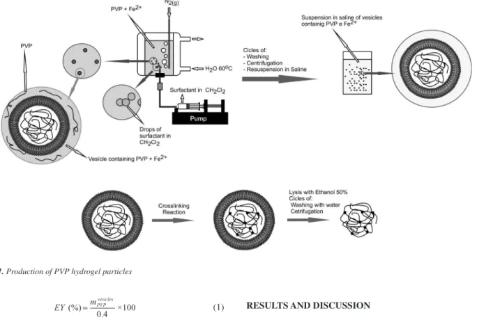
PVP encapsulation in liposome
PVP containing vesicles were obtained as follows (Figure 1): 0.5 mL of a CH2Cl2 solution containing 50 mg of egg or soy phosphatidylcholine (100 mg mL-1) were injected into 5 mL of
PVP aqueous solution (80 g L-1), containing 17 mmol L-1 FeCl 2
maintained at 50 ºC. CH2Cl2 solution was injected with a syringe adapted in a KD Scientiic Inc. Model KDS120 Push-Pull Pump, equipped with a ine-gauge needle (No 3D). During injection, a N2 stream was bubbled into PVP solution, which continued after liposome formation until removal of residual solvent. Liposome suspension was centrifuged at 22,800 × g for 30 min (Hitachi Himac CR20B2 centrifuge; Hitachi Ltd.) at 25 ºC to remove non-entrapped polymer from the external phase. The liposome-containing pellet was washed twice by centrifugation under same conditions in saline solution (51 mmol L-1). The washed pellet was
suspended on 20 mL of NaCl(aq).
PVP concentration inside vesicles was estimated using Lugol method.25 Resulting PVP complex with Lugol Reagent (PVP-I
3
-)
(1)
where is the mass of encapsulated PVP in the vesicles pellet and 0.4 is the mass of PVP in the initial solution (in g).
PVP entrapped polymerization
To crosslink PVP inside the vesicles, 4 mL of H2O2 diluted on NaCl(aq) (51 mmol L-1) was added to the PVP-containing vesicles
([H2O2]inal = 200 mmol L-1).22 As Fe2+ can be oxidized to Fe3+ inside
vesicles before H2O2 addition, to promote crosslinking by photo-Fenton reaction21 H
2O2 containing vesicles suspension was submitted
to 12 h of UV radiation (360 nm). The hydrogel particles were puriied by lysing vesicles washing 3× with ethanol 50%. The resulting pellet was resuspended in water and freeze-dried.
Dynamic light scattering measurements
The determination of particles size distribution was carried out by dynamic light scattering (DLS), using a spectrometer from Brookha-ven Instruments. The average hydrodynamic diameters were obtained from the unimodal distribution of particles analyzed by Zeta PALS Particle Sizing Software Version 2.29.
Scanning electron microscopy (SEM)
Particles were analyzed by SEM (FEG-SEM, model FEG 7401F, from Jeol). Samples were prepared by ixing the particles (freeze-dried powder) on a double-face copper tape and the specimens were analyzed without coating.
Swelling ratio determination (Q)
Swelling ratio (Q) determination was done by weighing the swollen particles pellet in an analytical balance (mswo), followed by freeze drying and weighing the dry pellet (mdry). Q was calculated directly by Equation 2.
(2)
RESULTS AND DISCUSSION
Polymer encapsulation
The eficiency of vesicle encapsulation depends on polymer size and decreases with the increase of polymer molecular weight.26,27
Szoka and Papahadjopoulos28 found that encapsulation eficiency
decreases with the increase of molecular weight of the encapsulated protein. Dominak et al.26 observed the same inverse relationship
between encapsulation eficiency and molecular weight studying polyethylene glycol and dextran encapsulation in giant vesicles. To encapsulate PVP, various surfactants were tested, including lecithin from egg yolk and soybeans and cationic surfactants. Table 1 shows the surfactants and their respective PVP encapsulation yield. As each vesicle in solution can have different encapsulation values,26
it is important to emphasize that PVP inclusion was measured by estimating the total amount that was incorporated and therefore the incorporation represents an average over all vesicles.
The best encapsulation eficiency was obtained with soy lecithin vesicles. Lecithins of different sources exhibit distinct properties.23,29
In particular the length of the apolar portion of the molecule and the insaturation of the alkyl chain will result in vesicles with different properties.30 The method used to obtain vesicle encapsulated PVP
also contributes to high encapsulation eficiency.18
Since the method used to crosslink the encapsulated polymer requires long irradiation times (photo-Fenton reaction), the addition
Table 1. PVP encapsulation in obtained vesicles
Surfactant EY (%)
Egg lecithin 0.8 ± 0.1
Soy lecithin 2.0 ± 0.3
DPPC 0.6 ± 0.1
DODAC 1% + soy lecithin 0.4 ± 0.1 DPTMA 1% + soy lecithin 2.0 ± 0.4
Preparation of PVP hydrogel nanoparticles using lecithin vesicles 2085 Vol. 33, No. 10
of cationic surfactants decreases vesicle aggregation during irradiation by increasing vesicle repulsion. Two saturated surfactants where tes-ted: DODAC and DPTMA. Encapsulation eficiency was higher with DPTMA than with DOCAC. DODAC contains two octadecyl chains linked to the tetraalkylammonium group; in DPTMA the acyl chains are attached to a propanediol moiety linked to tetraalkylammonium group (Figure 2). Bilayer packing defects are expected31,32 upon
incorporation of DODAC to lecithin causing a decrease in polymer entrapment. Measurements of polymer leak showed that entrapped PVP was retained after several washes (supernatant PVP concentra-tion was measured as <10-4 g L-1 for DPPC vesicles and as 0.02 g L-1
for DODAC/lecithin vesicles, after 5 wash cycles).
Crosslinking reaction
Two methods were used to crosslink the polymer. The Fenton reaction, an eficient method to obtain PVP hydrogels with good mechanical properties,22 has been used to obtain PVP nanogels with
superabsorbent properties.10 Photo-Fenton reaction, a photochemical
analog of Fenton reaction, also produces PVP hydrogels21 (Scheme 1).
The Fenton reaction is interesting because it allows a fast route to obtain PVP hydrogels with any desired format.33 However, this
reac-tion requires Fe2+ which autoxidizes. On the other hand, photo-Fenton
reaction uses Fe3+ ions, H
2O2 and UV radiation (λ = 365 nm). Since
the vesicles can scatter and react with H2O2 (lipoperoxidation), the UV dose and H2O2 concentrations were higher than those described.21
Fenton and photo-Fenton reactions did not change signiicantly the vesicles diameter distribution (Figures 3 and 4). This inding does not imply that the lipid bilayer remains unchanged during crosslinking reaction, since hydroxyl radical is known to react with unsaturated acyl chains.34
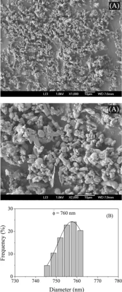
Particle characterization
When conventional Fenton reaction was used no pellet was obtained after vesicle´s lysis, evidencing absence of crosslinking, probably due Fe2+ depletion via its autoxidation. A phosphorus-free
yellow pellet was obtained after crosslinking by photo-Fenton
reac-tion and vesicles lyses.39 This pellet was resuspended in water and
analyzed by SEM and DLS. Results demonstrated the presence of a formless particulate material with dimensions comparable to vesicles (Figures 5 and 6).
The diameter of nano and sub-microgel and nanogel particles can be controlled by choosing the vesicle preparation method. Polyacrylamide nanogels (from monomers) covered by phospholipids bilayers obtained by detergent depletion35 or freeze-thaw cycles followed by extrusion36/
sonication11,17 presented diameters of 100 and 200 nm, respectively.
Larger nanogels (about 450 nm) are obtained from encapsulation of 20% dextran hydroyethylmethacrylate solution (Mn– = 19000) inside vesicles obtained by phospholipids ilm hydration, followed by
extru-Figure 2. Structures of (A) DPTMA and (B) DODAC
Scheme 1. Hydroxyl radical generation from Fenton and photo-Fenton reactions
Figure 3. Diameter distribution of egg lecithin vesicles containing PVP. (A) vesicles submitted to Fenton reaction; (B) vesicles submitted to photo-Fenton reaction and (C) vesicles prior to crosslinking reaction
sion.37 Poly(ethylenedioxythiophene)/poly(styrenesulfonate)13 and
poly(N-isopropylacrylamide)12,16 microgels, in conjunction with lipid
bilayers, are obtained by injecting a polymer solution inside GUVs, followed by freeze-thaw cycles and electroporation. The vesicle diam-eters, in these latter cases, vary from 5 to 100 µm.
The hydrogel particles obtained here showed no deined morphol-ogy, in spite of being obtained from spherical vesicles. Vesicles may be destabilized during crosslinking by H2O2 oxidation and ensuing lipoperoxidated38 (Figure 7). Fluctuation of crosslinking density, known
to inluence hydrogel particle morphology, may also account for the lack of deined shape.13 Non spherical particles have also been reported
with other nanogels prepared using vesicles.17,37
Particle swelling was estimated directly and the swelling ratio (Q) was 10, i.e., particles absorb only 10 times their mass in water. This is a strong indication that the crosslinking density is high since Q is inversely proportional to the ability of absorbing water.
Swelling of nanogels formatted from vesicles has seldom been discussed in literature. In the case of poly(N-isopropyl acrylamide) hydrogels and copolymers, coagulation of particles was observed instead of the variation in swelling. Kasakov et al. have extensively studied this systems11,17,36 mainly in regard to the
interaction between particle and liposome. Since the hydrogel used
Figure 5. SEM image of freeze-dried particles obtained from egg lecithin vesicles (A) and DLS particles diameter distribution (B)
Figure 6. SEM image of freeze-dried particles obtained from soy lecithin vesicles (A) and DLS particles diameter distribution (B)
is thermo- and pH-responsive, coagulation was observed upon heating, including coagulation of the lipid bilayer, which, after
Preparation of PVP hydrogel nanoparticles using lecithin vesicles 2087 Vol. 33, No. 10
cooling, surround the entire set of nanoparticles in a reversible process.36 The authors also observed an increase in diameter with
pH change: 100 nm at pH 6.5 to 300 nm at pH 3.0, with dimeriza-tion of particles.17
CONCLUSION
Injected lecithin vesicles can encapsulate high molecular weight PVP. The encapsulated polymer can be crosslinked by photo-Fenton reaction and hydrogel nano and sub-microparticles can be isolated. Spherical morphology was not achieved, but the produced hydrogel particles have a narrow diameter distribution compatible with the vesicles diameter distribution.
REFERENCES
1. Rosiak, J. M.; Yoshii, F.; Nucl. Instrum. Methods Phys. Res., Sect. B
1999, 151, 56.
2. Tao, S. L.; Desai, T. A.; Adv. Drug Delivery Rev.2003, 55, 315. 3. Mathews, A.; Ha, C.-S.; Cho, W.-J.; Kim, I.; Drug Delivery2006, 13,
245.
4. Mundargi, R. C.; Patil, S. A.; Kulkarni, P. V.; Mallikarjuna, N. N.; Aminabhavi, T. M.; J. Microencapsulation2008, 25, 228.
5. Nguyen, K. T.; Shukla, K. P.; Moctezuma, M.; Braden, A. R. C.; Zhou, J.; Hu, Z.; Tang, L.; J. Biomed. Mater. Res., Part A2009, 88, 1022. 6. Rao, V. R.; Ravimandalam, K.; Jayakrishnan, A.; Thanoo, B. C.;
Radhakrishnan, V. V.; Gupta, A. K.; Kumar, S.; Joseph, S.; Unni, M.; Rao, A. S.; J. Neuroradiology1991, 18, 61.
7. Sahiner, N.; Godbey, W. T.; Mcpherson, G. L.; John, V. T.; Colloid Polym. Sci.2006, 284, 1121.
8. Imaz, A.; Forcada, J.; Macromol. Symp.2009, 281, 85.
9. Bharali, D. J.; Sahoo, S. K.; Mozumdar, S.; Maitra, A. J.; J. Colloid Interface Sci.2003, 258, 415.
10. Bueno, V. B.; Cuccovia, I. M.; Chaimovich, H.; Catalani, L. H.; Colloid Polym. Sci.2009, 287, 705.
11. Kazakov, S.; Kaholek, M.; Kudasheva, D.; Teraoka, I.; Cowman, M. K.; Levon, K.; Langmuir2003, 19, 8086.
12. Jesorka, A.; Markstroem, M.; Orwar, O.; Langmuir2005, 21, 1230. 13. Jesorka, A.; Markstroem, M.; Karlsson, M.; Orwar, O.; J. Phys. Chem.
B2005, 109, 14759.
14. Kremer, S.; Campillo, C.; Pepin-Donat, B.; Viallat, A.; Brochard-Wyart, F.; EPL2008, 82, 48002/1.
15. Campillo, C.; Pepin-Donat, B.; Viallat, A.; Soft Matter2007, 3, 1421. 16. Markstroem, M.; Gunnarsson, A.; Orwar, O.; Jesorka, A.; Soft Matter
2007, 3, 587.
17. Kazakov, S.; Levon, K.; Curr. Pharm. Des.2006, 12, 4713. 18. Lasic, D. D.; Biochem. J.1988, 256, 1.
19. Rosiak, J. M.; J. Controlled Release1994, 31, 9.
20. Kadlubowski, S.; Henke, A.; Ulanski, P.; Rosiak, J. M.; Bromberg, L.; Hatton, T. A.; Polymer2007, 48, 4974.
21. Fechine, G. J. M.; Barros, J. A. G.; Catalani, L. H.; Polymer2004, 45, 4705.
22. Barros, J. A. G.; Fechine, G. J. M.; Alcantara, M. R.; Catalani, L. H.; Polymer2006, 47, 8414.
23. Maximiano, F. A.; da Silva, M. A.; Daghastanli, K. R. P.; de Araujo, P. S.; Chaimovich, H.; Cuccovia, I. M.; Quim. Nova2008, 31, 910. 24. Moss, R. A.; Gangulu, S.; Okumura, Y.; Fujita, T.; J. Am. Chem. Soc.
1990, 112, 6391.
25. Leger, C.; Nguyen, Q. T.; Neel, J.; Streicher, C.; Macromolecules1995, 28, 143.
26. Dominak, L. M.; Keating, C. D.; Langmuir2007, 23, 7148. 27. Dominak, L. M.; Keating, C. D.; Langmuir2008, 24, 13565. 28. Szoka, F., Jr.; Papahadjopoulos, D.; Annu. Rev. Biophys. Bioeng.1980,
9, 467.
29. Palacios, L. E.; Wang, T.; J. Am. Oil Chem. Soc.2005, 82, 571. 30. Nagarajan, R.; Ruckenstein, E.; J. Colloid Interface Sci.1979, 71, 580. 31. Kumar, V. V.; Indian J. Biochem. Biophys.1993, 30, 135.
32. Batenjany, M. M.; Wang, Z.-Q.; Huang, C.-H.; Levin, I. W.; Biochim. Biophys. Acta, Biomembr.1994, 1192, 205.
33. Catalani, L. H.; Fechine, G. J. M.; Barros, J. A. G.; BR PI 0402081-2
2006. (CA 147:508389)
34. Megli, F. M.; Sabatini, K.; Chem. Phys. Lipids2003, 125, 161. 35. Schillemans, J. P.; Flesch, F. M.; Hennink, W. E.; van Nostrum, C. F.;
Macromolecules2006, 39, 5885.
36. Kazakov, S.; Kaholek, M.; Teraoka, I.; Levon, K.; Macromolecules
2002, 35, 1911.
37. van Thienen, T. G.; Lucas, B.; Flesch, F. M.; Nostrum, C. F.; Demeester, J.; De Smedt, S. C.; Macromolecules2005, 38, 8503.
38. Dobretsov, G. E.; Borschevskaya, T. A.; Petrov, V. A.; Vladimirov, Yu. A.; FEBS Lett.1977, 84, 125.