Subclavian Access for Transcatheter CoreValve
®
Aortic Prosthesis Implantation:
Data from the Brazilian Registry
Fábio Sândoli de Brito Júnior
1, Luiz Antonio Carvalho
2, Dimytri Siqueira
3, João Carlos Dias
4,
José Armando Mangione
5, Rogério Sarmento-Leite
6, Pedro Alves Lemos Neto
7, Eberhard Grube
8,
Teresa Cristina Nascimento
9, Marco Antonio Perin
10, J. Eduardo Sousa
11ABSTRACT
Background: Transfemoral access is the preferred approach
for transcatheter aortic valve implantation. However, some conditions, such as the presence of peripheral vascular disease, preclude the use of such access. In these cases, subclavian access is an alternative approach for this procedure. This study aimed at evaluating the Brazilian experience using the subclavian approach for transcatheter CoreValve® prosthesis
implantation. Methods: An aortic valve area < 1 cm2; an
aortic valve ring ≥ 20 mm and ≤ 27 mm (26 mm and 29 mm CoreValve); an ascending aorta ≤ 43 mm; and a subclavian artery with a diameter ≥ 6 mm, with no significant obstruc-tive lesions, marked tortuosity, and excess calcification were requisites for the procedure. Access through the subclavian artery was obtained by surgical dissection and, under direct vision, a subclavian artery puncture was performed. Once artery access was obtained, the standard technique was used.
Results: Between January of 2008 and April of 2012, eight
patients with peripheral vascular disease underwent CoreValve®
prosthesis implantation through the subclavian artery in four institutions. The procedure was successful in all cases, with a reduction in the mean transvalvular pressure gradient from 46.4 ± 17.5 mmHg to 9.3 ± 3.6 mmHg (P = 0.0018) and symptom improvement. At 30 days and after 275 ± 231 days of follow-up, 87.5% and 62.5% of the patients, respectively, were free from major adverse events (death, myocardial
© 2012 Elsevier Editora Ltda. and Sociedade Brasileira de Hemodinâmica e Cardiologia Intervencionista. All rights reserved. 1 PhD; Medical Coordinator of the Interventional Cardiology
Depart-ment of the Hospital Israelita Albert Einstein. São Paulo, SP, Brazil. 2 PhD; Coordinator of the Cardiovascular Intervention Laboratory of the Hospital Pró-Cardíaco. Rio de Janeiro, RJ, Brazil.
3 PhD; Interventional Cardiologist at Instituto Dante Pazzanese de Cardiologia. São Paulo, SP, Brasil.
4 Physician. Coordinator of the Interventional Cardiology Department of the Hospital Vila da Serra. Belo Horizonte, MG, Brazil.
5 PhD; Head of Hemodynamics Service of Arie Interventional Car-diology of the Hospital Beneicência Portuguesa de São Paulo. São Paulo, SP, Brazil.
6 PhD; Technical Director of the Hemodynamics and Interventional Cardiology Laboratory of the Instituto de Cardiologia do Rio Grande do Sul. Porto Alegre, RS, Brazil.
7 Associate Professor. Director of the Hemodynamics and Interventio-nal Cardiology Service of the Instituto do Coração do Hospital das Clínicas da Faculdade de Medicina da Universidade de São Paulo.
São Paulo, SP, Brazil.
8 PhD; Director of the Cardiovascular Center of the Hospital Alemão Oswaldo Cruz. São Paulo, SP, Brazil.
9 Nurse. Study monitor. Sociedade Brasileira de Hemodinâmica e Cardiologia Intervencionista. São Paulo, SP, Brazil.
10 Associate Professor. Medical Manager of the Cardiovascular Intervention Sector of the Hospital Israelita Albert Einstein. São Paulo, SP, Brasil. 11 Associate Professor. Director of the Intervention Center in Structural Heart Diseases of the Instituto Dante Pazzanese de Cardiologia. São Paulo, SP, Brazil.
Correspondence to: Fábio Sândoli de Brito Júnior. Rua Dom Armando Lombardi, 819/82-A – São Paulo, SP, Brasil – CEP 05616-011. E-mail: [email protected]
Received on: 5/21/2012 • Accepted on: 7/27/2012
Original Article
RESUMO
Acesso pela Artéria Subclávia para Implante por Cateter da Bioprótese Valvar Aórtica CoreValve®: Dados do Registro Brasileiro
Introdução: A via de acesso transfemoral é preferencial para
o implante por cateter de bioprótese valvar aórtica. Entretanto, algumas situações, como a presença de doença vascular peri-férica, impossibilitam a utilização desse acesso. Nesses casos, o acesso por dissecção da artéria subclávia é uma alternativa para a realização do procedimento. Nosso objetivo foi avaliar a experiência brasileira com a utilização da artéria subclávia como via de acesso para o implante por cateter da bioprótese CoreValve®. Métodos: Foram requisitos para o procedimento
área valvar aórtica < 1 cm², ânulo valvar aórtico > 20 mm e < 27 mm (CoreValve® de 26 mm e 29 mm), aorta ascendente
< 43 mm e artéria subclávia com diâmetro > 6 mm, isenta de lesões obstrutivas signiicativas, tortuosidade acentuada e calciicação excessiva. O acesso pela artéria subclávia foi obtido por dissecção cirúrgica e, sob visão direta, punção da artéria subclávia. Obtido o acesso arterial, empregou-se a técnica padrão. Resultados: Entre janeiro de 2008 e abril de 2012, 8 pacientes com doença vascular periférica foram submetidos a implante de prótese CoreValve® pela artéria subclávia em 4
infarction, stroke, and urgent cardiac surgery). Conclusions: In the Brazilian experience, the subclavian access was a safe and effective alternative for transcatheter CoreValve®
implantation.
DESCRIPTORS: Aortic valve stenosis. Subclavian artery. Heart
valve prosthesis.
melhora dos sintomas. Aos 30 dias e no seguimento de 275 + 231 dias, 87,5% e 62,5% dos pacientes, respectivamente, apresentavam-se livres de complicações maiores (óbito, infarto do miocárdio, acidente vascular cerebral e cirurgia cardíaca de urgência). Conclusões: Na experiência brasileira, o acesso pela artéria subclávia mostrou-se seguro e eicaz como via alternativa para o implante por cateter da bioprótese CoreValve®.
DESCRITORES: Estenose da valva aórtica. Artéria subclávia.
Próteses valvulares cardíacas.
T
ranscatheter aortic valve implantation is a safe and effective procedure for the treatment of patients with symptomatic aortic stenosis that is either inoperable or that carries a high surgical risk.1,2 For this procedure, the femoral approach is preferred, since it is less invasive. However, some conditions, such as the presence of atheromatous disease, tortuosity, and calcification in the territories of the femoral and iliac arteries, prevent the procedure from being performed through the femoral artery. In these cases, alternative access routes can be used, such as the transapical and transaortic routes.1,3,4 However, complications arising from the thoracotomy and the incision in the left ventricular apex are not uncommon with these tech-niques.5,6 Access through dissection of the subclavian artery has been described as a less invasive option and therefore more attractive for the implantation ofthe CoreValve® bioprosthesis (CoreValve® Revalving
System, Medtronic, Inc. – Minneapolis, USA).7–11 In
this study, the Brazilian experience with the use of the subclavian artery as an access route for CoreV-alve® bioprosthesis implantation is reported, with data obtained from the Brazilian Registry of Transcatheter Aortic Valve Bioprostheses Implantation.12
METHODS
Patient selection
Patients in whom the subclavian artery was used as an access for CoreValve® bioprosthesis implantation were selected for this study from the Brazilian registry. The subclavian artery was used only when there were contraindications to femoral access, and whenever pos-sible, the left subclavian access was chosen. Patients considered to be suitable for the procedure were those with an aortic valve area < 1 cm2, an annular aortic
valve ≥ 20 mm and ≤ 27 mm (CoreValve® of 26 mm
and 29 mm), an ascending aorta ≤ 43 mm, and a
subclavian artery diameter ≥ 6 mm, free of signiicant
obstructive lesions, severe tortuosity, and excessive parietal calciication.
The EuroSCORE and STS scores were used to estimate the risk of surgical mortality in this series of patients.13,14
Procedure
Preparation for the procedure consisted of antibiotic and antiplatelet therapy with aspirin and/or clopidogrel. The valve implantations were performed under general anaesthesia. Alternative access through the subclavian artery was obtained by surgical dissection. For the dissection, an infraclavicular incision was used. Under direct vision, the subclavian artery was punctured for the positioning of the 18 F sheath or, when the artery was deep, a Da-cron graft was anastomosed to the artery and used for insertion of the 18 F sheath (Figure 1). After obtaining arterial access, the standard technique (Figure 2) was used for the CoreValve® bioprosthesis implantation, which consists of three porcine pericardium lealets, mounted and sutured into an auto-expansible nitinol stent 5 cm in length. At the end of the procedure, the 18 F sheath was removed, and the subclavian artery was sutured.
Data collection and outcomes
Clinical data and information on complementary exams were collected during medical appointments or by telephone contact and entered into an electronic spreadsheet developed for the Brazilian Registry. All outcomes and complications followed the criteria es-tablished by the Valve Academic Research Consortium
Consensus on Deinition Event Event (VARC).15
Statistical analysis
Continuous variables are shown as the mean ± standard deviation, and categorical variables are shown as frequencies (number and percentage). For the comparative analysis of categorical variables, the chi-squared or Fisher’s exact tests were used. For the sequential analysis of continuous variables in the same patient, a paired t-test was used. The signiicance level
was set at 5% (P < 0.05).
RESULTS
the subclavian access route was used for the treat-ment of eight (2.9%) patients: two (25%) through the right subclavian artery and six (75%) through the left subclavian artery.
The basal demographic and clinical characteristics of the eight patients receiving the CoreValve® bioprosthesis implant through the subclavian artery are described in Table 1. The high surgical risk in this group of patients was demonstrated by the EuroSCORE and STS risk scores, which were both above 30%. The contraindication for
femoral access was the presence of peripheral vascular disease, and three (37.5%) of the patients also had an abdominal aortic aneurysm.
The mean hospital stay was 14 ± 12.9 days (one to 43 days). The mean follow-up of patients was 275 ± 231 days (one to 679 days). The month, one-year, and two-year follow-up data were available for seven (87.5%) patients, ive (62.5%) patients, and one (12.5%) patient, respectively. There was no loss of clinical follow-up in any case.
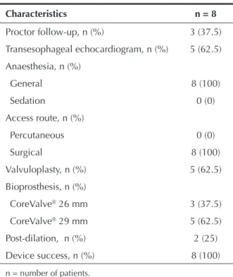
Procedure
Data from the procedure are described in Table 2. Successful CoreValve® bioprosthesis implantation was achieved in 100% of the cases. In two (25%) cases it was necessary to perform post-dilation to properly expand the bioprosthesis and to reduce the intensity of the perivalvular insuficiency. Echocardiography detected the reduction of the mean and peak aortic transvalvular pressure gradients (Table 3). At the end of the proce-dure, there was mild periprosthetic aortic regurgitation in six (75%) cases.
A B
C
A
C
B
D
Figure 1 – In A, subclavian artery dissection. In B, Dacron graft anastomosis. In C, 18 F sheath positioning in the subclavian artery through the graft.
Figure 2 – In A, aortography and placement of a guidewire inside the left ventricle. In B, introduction of the CoreValve® prosthesis for the left subclavian artery. In C, a prosthesis positioned in the aortic annulus. In D, aortography demonstrating a well-positioned and competent CoreValve® prosthesis.
TABLE 1
Basal Demographic and Clinical Data
Characteristics n = 8
Age, years (SD) 84 (7.3)
Male gender, n (%) 5 (62.5)
Logistic EUROSCORE, % (SD) 32 (16.4)
STS score, % (SD) 30.9 (23.4)
Functional Class (NYHA), n (%)
I or II 0 (0)
III or IV 8 (100)
Diabetes, n (%) 3 (37.5)
Arterial hypertension, n (%) 5 (62.5)
Renal failure*, n (%) 6 (75)
Coronary artery disease, n (%) 5 (62.5)
Previous percutaneous coronary
intervention 2 (25)
CABG 2 (25)
Cerebrovascular disease, n (%) 1 (12.5)
Peripheral vascular disease, n (%) 7 (87.5)
Chronic obstructive pulmonary
disease, n (%) 2 (25)
Deinitive pacemaker, n (%) 1 (12.5)
*Glomerular iltration rate < 60 mL/min.
Outcomes and complications
The transcatheter treatment of aortic valve stenosis was effective at alleviating the symptoms of heart failure. After 30 days and during follow-up, 85.7% of patients achieved functional class status I or II of the New York Heart Association (NYHA) criteria (P = 0.0007 vs. baseline).
Table 4 illustrates the complications that occurred within 30 days and during follow-up. During the proce-dure, one case of subclavian artery dissection occurred and was corrected with stenting, and one patient had bleeding that required a transfusion. One death from cardiovascular causes occurred one day after the proce-dure in a patient who developed refractory cardiogenic shock after valve implantation. Thus, mortality at 30 days
was 12.5%. Two other deaths from non-cardiovascular causes occurred 43 and 679 days after the CoreValve® bioprosthesis implantation.
Two patients had renal failure after the procedure, one of which was associated with refractory cardiogenic shock and death, as previously described. None of the cases had a stroke, acute myocardial infarction (AMI), or the need for urgent heart surgery; thus, throughout the 275 ± 231 days of follow-up, ive (62.5%) patients were free of major complications. In this series, excluding the patient who died one day after the procedure and one patient who was already using a pacemaker, two (33.3%) underwent implantation of a permanent pacemaker for advanced atrioventricular conduction disturbances.
DISCUSSION
The current versions of both devices available for clinical use, CoreValve® and Edwards SAPIEN (Edwards Lifesciences – Irvine, USA), with 18 F delivery systems (6 mm), allow the procedure to be performed through the femoral artery, as long as the arterial lumen diam-eter is at least 6 mm. However, in this population of patients of advanced age and with multiple comorbidi-ties, it is not uncommon to ind severe atheromatosis or excessive tortuosity, which prevent the procedure from being performed by this access route. In this case,
access through the subclavian artery for CoreValve®
bioprosthesis aortic implantation is a feasible, safe, and effective procedure, as demonstrated by several series and also by the present study with data from the Brazilian registry.9–11 In international records, subclavian access is employed in approximately 5% of cases, and the patients have, in general, a higher surgical risk than patients in whom femoral access was used.9–11,16–19 In the Brazilian Registry, only 2.9% of cases were treated using the subclavian artery, which is most likely due to the lower experience of centres with this alternative access route. As with the international series, patients from the Brazilian Registry treated through subclavian access had a very high surgical mortality risk, over 30%, demonstrating that the presence of peripheral vascular disease is also a marker of higher clinical and anatomic complexity.20
TABLE 2 Procedure Data
Characteristics n = 8
Proctor follow-up, n (%) 3 (37.5)
Transesophageal echocardiogram, n (%) 5 (62.5)
Anaesthesia, n (%)
General 8 (100)
Sedation 0 (0)
Access route, n (%)
Percutaneous 0 (0)
Surgical 8 (100)
Valvuloplasty, n (%) 5 (62.5)
Bioprosthesis, n (%)
CoreValve® 26 mm 3 (37.5)
CoreValve® 29 mm 5 (62.5)
Post-dilation, n (%) 2 (25)
Device success, n (%) 8 (100)
n = number of patients.
TABLE 3 Echocardiographic Data
Basal (n = 8)
Post-implantation
(n = 7) P
Aortic valve area, cm3 (SD) 0.7 (0.2) NA
Peak gradient, mmHg (SD) 73.6 (27.8) 17.8 (7.5) < 0.001
Mean gradient, mmHg (SD) 46.4 (17.5) 9.3 (3.6) 0.0018
LVEF, % (SD) 58.7 (9.2) 63.4 (12.4) 0.175
As a positive point regarding access through the subclavian artery, it should be emphasised that it is easier to control the stent at the time of its release in the valvular annulus when compared with femoral ac-cess, as the shorter distance and less tortuous path allow better transmission of force to the distal system portion, allowing for a more accurate positioning. Moreover, in general, the subclavian arteries are less affected by atheromatosis than the iliac and femoral arteries.
When selecting access through the subclavian ar-tery, there is a preference for the left artery because, intuitively, there would be less risk of stroke than when using the right subclavian artery, considering that the presence and manipulation of the prosthesis delivery device in the brachiocephalic trunk could cause em-bolization and limit low into the right carotid. In this case, access through the right artery is feasible only when the diameter of the brachiocephalic trunk artery is > 7 mm and free of signiicant atheromatosis.
Another point in favour of using the left subclavian artery as the access route is the more favourable orien-tation of the bioprosthesis in the valve annulus at the implantation, similar to the positioning obtained by a femoral approach. Therefore, the right subclavian artery should be considered as a good alternative only in cases where the presence of atheromatous disease or tortuosity prevent implantation through the left subclavian artery, or in cases where the diameter of the left subclavian is < 7 mm in the presence of a patent internal thoracic artery graft to a coronary artery of major anatomical importance, due to the incapacity of accommodating the prosthesis-releasing device (6 mm) and maintaining blood low to the graft. In the present study, following this recommendation, the right subclavian artery access was used in only 25% of cases.
More recently, the use of the subclavian artery for CoreValve® bioprosthesis implantation by totally per-cutaneous access and haemostasis obtained after the
procedure with ProstarTM haemostatic devices (Abbott
Vascular – Abbott Park, USA) or ProGlideTM (Abbott Vascular – Abbott Park, USA) has been reported.21 However, valida-tion of this approach also depends on the conirmavalida-tion of its safety, since severe bleeding complications may result from the failure of the device to perform haemostasis.
CONCLUSIONS
In the Brazilian experience, transcatheter CoreValve® bioprosthesis implantation through the subclavian artery route was safe and effective for use in selected patients in whom femoral access was not feasible. This alterna-tive access route allows for a larger number of patients to beneit from aortic valve bioprosthesis transcatheter implantation.
CONFLICT OF INTERESTS
See Appendix.
REFERENCES
1. Kodali SK, Williams MR, Smith CR, Svensson LG, Webb JG, Makkar RR, et al. Two-year outcomes after transcatheter or surgical aortic-valve replacement. N Engl J Med. 2012; 366(18):1686-95.
2. Makkar RR, Fontana GP, Jilaihawi H, Kapadia S, Pichard AD, Douglas PS, et al. Transcatheter aortic-valve replacement for inoperable severe aortic stenosis. N Engl J Med. 2012; 366(18):1696-704.
3. Wendler O, Walther T, Schroefel H, Lange R, Treede H, Fusari M, et al. Transapical aortic valve implantation: mid-term outcome from the SOURCE registry. Eur J Cardiothorac Surg. 2012 May 30. [Epub ahead of print]
4. Etienne PY, Papadatos S, El Khoury E, Pieters D, Price J, Glineur D. Transaortic transcatheter aortic valve implantation with the Edwards SAPIEN valve: feasibility, technical considerations, and clinical advantages. Ann Thorac Surg. 2011;92(2):746-8. 5. Lange R, Bleiziffer S, Piazza N, Mazzitelli D, Hutter A,
Tassani-Prell P, et al. Incidence and treatment of procedural cardiovascular complications associated with trans-arterial and trans-apical interventional aortic valve implantation in 412 con-secutive patients. Eur J Cardiothorac Surg. 2011;40(5):1105-13.
TABLE 4 Adverse Events
30 days
275 ± 231 days
Death, n (%) 1 (12.5) 3 (37.5)
Cardiovascular death 1 (12.5) 1 (12.5)
Stroke, n (%) 0 0
Myocardial infarction, n (%) 0 0
Acute renal failure, n (%) 2 (25) 2 (25)
Stage 1 1 (12.5) 1 (12.5)
Stage 2 0 0
Stage 3 1 (12.5) 1 (12.5)
Haemorrhagic complication,
n (%) 1 (12.5) 1 (12.5)
Life-threatening, n (%) 0 0
Major, n (%) 1 (12.5) 1 (12.5)
Minor, n (%) 0 0
Vascular complication, n (%) 1 (12.5) 0
Major, n (%) 1 (12.5) 0
Minor, n (%) 0 0
Permanent pacemaker*,
n (%) 2 (33.3) 2 (33.3)
Major-complication
free**, n (%) 7 (87.5) 5 (62.5)
Complication-free***, n (%) 5 (62.5) 4 (50)
* Six patients with pacemaker risk.
** Death, stroke, AMI, and urgency heart surgery.
6. Bleiziffer S, Piazza N, Mazzitelli D, Opitz A, Bauernschmitt R, Lange R. Apical-access-related complications associated with trans-catheter aortic valve implantation. Eur J Cardiothorac Surg. 2011;40(2):469-74.
7. Bruschi G, Fratto P, De Marco F, Oreglia J, Colombo P, Botta L, et al. The trans-subclavian retrograde approach for transcatheter aortic valve replacement: Single-center experience. J Thorac Cardiovasc Surg. 2010;140(4):911-5, 915 e1-2.
8. Fraccaro C, Napodano M, Tarantini G, Gasparetto V, Gerosa G, Bianco R, et al. Expanding the eligibility for transcatheter aortic valve implantation the trans-subclavian retrograde ap-proach using: the III generation CoreValve revalving system. JACC Cardiovasc Interv. 2009;2(9):828-33.
9. Modine T, Obadia JF, Choukroun E, Rioufoul G, Sudre A, Laborde JC, et al. Transcutaneous aortic valve implantation using the axillary/subclavian access: feasibility and early clini-cal outcomes. J Thorac Cardiovasc Surg. 2011;141(2):487-91, 491e1.
10. Moynagh AM, Scott DJ, Baumbach A, Khavandi A, Brecker SJ, Laborde JC, et al. CoreValve transcatheter aortic valve implantation via the subclavian artery: comparison with the transfemoral approach. J Am Coll Cardiol. 2011;57(5): 634-5.
11. Petronio AS, De Carlo M, Bedogni F, Marzocchi A, Klugmann S, Maisano F, et al. Safety and efficacy of the subclavian approach for transcatheter aortic valve implantation with the CoreValve revalving system. Circ Cardiovasc Interv. 2010;3(4): 359-66.
12. Brito Jr FS, Siqueira D, Sarmento-Leite R, Carvalho LA, Lemos Neto PA, Mangione JA, et al. Racional e desenho do registro brasileiro de implante de bioprótese aórtica por cateter. Rev Bras Cardiol Invasiva. 2011;19(1):145-52.
13. Hattler BG, Madia C, Johnson C, Armitage JM, Hardesty RL, Kormos RL, et al. Risk stratiication using the Society of Tho-racic Surgeons Program. Ann Thorac Surg. 1994;58(5):1348-52.
14. Roques F, Nashef SA, Michel P; EuroSCORE Study Group. Risk factors for early mortality after valve surgery in Europe in the 1990s: lessons from the EuroSCORE pilot program. J Heart Valve Dis. 2001;10(5):572-77; discussion 577-8. 15. Leon MB, Piazza N, Nikolsky E, Blackstone EH, Cutlip DE,
Kappetein AP, et al. Standardized endpoint deinitions for transcatheter aortic valve implantation clinical trials: a con-sensus report from the valve academic research consortium. J Am Coll Cardiol. 2011;57(3):253-69.
16. Abdel-Wahab M, Zahn R, Horack M, Gerckens U, Schuler G, Sievert H, et al. Aortic regurgitation after transcatheter aortic valve implantation: incidence and early outcome. Results from the German transcatheter aortic valve interventions registry. Heart. 2011;97(11):899-906.
17. Bosmans JM, Kefer J, De Bruyne B, Herijgers P, Dubois C, Legrand V, et al. Procedural, 30-day and one year outcome following CoreValve or Edwards transcatheter aortic valve implantation: results of the Belgian National Registry. Interact Cardiovasc Thorac Surg. 2011;12(5):762-7.
18. Eltchaninoff H, Prat A, Gilard M, Leguerrier A, Blanchard D, Fournial G, et al. Transcatheter aortic valve implantation: early results of the FRANCE (FRench Aortic National CoreValve and Edwards) registry. Eur Heart J. 2011;32(2):191-7.
19. Gilard M, Eltchaninoff H, Iung B, Donzeau-Gouge P, Chevreul K, Fajadet J, et al. Registry of transcatheter aortic-valve implanta-tion in high-risk patients. N Engl J Med. 2012;366(18):1705-15. 20. Sinning JM, Horack M, Grube E, Gerckens U, Erbel R,
Eggebrecht H, et al. The impact of peripheral arterial disease on early outcome after transcatheter aortic valve implantation: results from the German Transcatheter Aortic Valve Interventions Registry. Am Heart J. 2012;164(1):102-10, e1.
21. Schafer U, Ho Y, Frerker C, Schewel D, Sanchez-Quintana D, Schofer J, et al. Direct percutaneous access technique for trans-axillary transcatheter aortic valve implantation: “The Hamburg Sankt Georg approach”. JACC Cardiovasc Interv. 2012;5(5):477-86.
APPENDIX Conflict of interest
Nome
Participation
in study1 Lecturer2 Consultant3
Normative
committee4
Personal/ institutional
aid5
Sponsored scientific
texts6 Shareholder7
Dimytri Siqueira No No No No No No No
Eberhard Grube No No Yes No No No No
Fábio Sândoli de Brito Júnior No Yes No No No No No
J. Eduardo Sousa No No No No No No No
João Carlos Dias No No No No No No No
José Armando Mangione No Yes No No No No No
Luiz Antonio Carvalho No Yes No No No No No
Marco Antonio Perin No Yes Yes No No No No
Pedro Alves Lemos Neto No No No No No No No
Rogério Sarmento-Leite No Yes No No Yes No No
Teresa Cristina Nascimento No No No No No No No