Very Late Follow-up of Percutaneous Balloon Mitral
Valvotomy in Severe Mitral Stenosis
Gentil Barreira de Aguiar Filho
1, Sebastian Lluberas
2, Nisia Lyra Gomes
3, Luiz Felipe P. de Andrade
4,
Mercedes Maldonado
5, Zilda Machado Meneghelo
6, Felipe Carrha Machado
7, Mateus Veloso e Silva
8,
Cesar A. Esteves
9, Sérgio Luiz N. Braga
10, Alexandre Abizaid
11ABSTRACT
Background: Percutaneous balloon mitral valvotomy is safe and effective in patients with severe symptomatic mitral stenosis with immediate and long-term results comparable to those of surgical intervention. This study was aimed at reporting the very late follow-up results of the first percutaneous bal-loon mitral valvotomies performed at our institution and at
identifying predictive factors of restenosis. Methods: From
1987 to 1991, 200 consecutive patients were submitted to percutaneous balloon mitral valvotomy. Clinical and echocar-diographic evaluations were performed prior to the procedure,
48 hours after the procedure and annually thereafter. Results:
Mean age was 32 ± 12 years; 86.5% were female and 80.5%
were in New York Heart Association functional class III or
IV. Mean Wilkins score was 7.6 ± 1.2 and procedure success
was observed in 87.5% (175/200) of the patients. During
follow-up, 129 patients (74%) were followed up for 140 ±
79 months. Restenosis was observed after the first proce-dure in 46.5% (60/129) patients and a second percutaneous balloon mitral valvotomy was performed in 25 patients, a third one in 4 patients and a fourth one in 1 patient. The probability of being restenosis-free was 85% at 5 years, 60% at 10 years and 36% at 20 years. Left atrial diameter (P = 0.034), and preoperative (P = 0.013) and postoperative (P = 0.038) transvalvar gradient were predictors of restenosis. Conclusions: In a very late clinical follow-up, percutane-ous balloon mitral valvotomy provided long-lasting results in over one-third of the patients and showed that repeated procedures may be performed safely in selected patients. The
© 2012 Elsevier Editora Ltda. and Sociedade Brasileira de Hemodinâmica e Cardiologia Intervencionista. All rights reserved. 1 Resident Physician at the Hemodynamics Section of the Instituto
Dante Pazzanese de Cardiologia. São Paulo, SP, Brazil.
2 Resident Physician at the Valvulopathy Section of the Instituto Dante Pazzanese de Cardiologia. São Paulo, SP, Brazil.
3 Assistant Physician at the Valvulopathy Section of the Instituto Dante Pazzanese de Cardiologia. São Paulo, SP, Brazil.
4 Resident Physician at the Valvulopathy Section of the Instituto Dante Pazzanese de Cardiologia. São Paulo, SP, Brazil.
5 Assistant Physician at the Echocardiography Section of the Instituto Dante Pazzanese de Cardiologia. São Paulo, SP, Brazil.
6 Ph.D. Head of the Valvulopathy Section of the Instituto Dante Paz-zanese de Cardiologia. São Paulo, SP, Brazil.
7 Resident Physician in Cardiology of the Instituto Dante Pazzanese de Cardiologia. São Paulo, SP, Brazil.
8 Resident Physician at the Hemodynamics Section of the Instituto Dante Pazzanese de Cardiologia. São Paulo, SP, Brazil.
9 PhD; Head of the Medical Section of Acquired Valvulopathy of the Instituto Dante Pazzanese de Cardiologia. São Paulo, SP, Brazil. 10 PhD; Head of the Hemodynamics Section of the Instituto Dante Pazzanese de Cardiologia. São Paulo, SP, Brazil.
11 Associate Professor. Director of the Invasive Cardiology Service of the Instituto Dante Pazzanese de Cardiologia. São Paulo, SP, Brazil.
Correspondence to: Gentil Barreira de Aguiar Filho. Rua Borges Lagoa, 1.209 – Vila Clementino – São Paulo, SP, Brasil – CEP 04038 033. E mail: [email protected]
Received on: 6/8/2012 • Accepted on: 8/11/2012
Original Article
RESUMO
Evolução Muito Tardia da Valvotomia Percutânea por Balão na Estenose Mitral Grave
Introdução: A valvotomia mitral percutânea por balão é um procedimento seguro e eficaz em pacientes com estenose mitral grave sintomática selecionados, com resultados imediatos e a longo prazo semelhantes aos da intervenção cirúrgica. Este estudo tem o objetivo de descrever os resultados muito tardios das primeiras valvotomias mitrais percutâneas por balão realizadas em nossa instituição e identificar os fatores preditores de reestenose. Métodos: No período de 1987 a 1991, 200 pacientes consecutivos foram submetidos a valvotomia mitral percutânea por balão. Avaliações clínica e ecocardiográfica foram realizadas antes do procedimento,
48 horas após e, então, anualmente. Resultados: A média de
idade foi de 32 + 12 anos, 86,5% eram do sexo feminino e 80,5% encontravam-se em classe funcional III ou IV da New York Heart Association. A média do escore de Wilkins foi de 7,6 + 1,2 e o sucesso do procedimento ocorreu em 87,5% (175/200) dos pacientes. Durante o seguimento, foram acompanhados 129 pacientes (74%) por 140 + 79 meses. Reestenose após o primeiro procedimento ocorreu em 46,5% (60/129) dos pacientes, sendo realizada uma segunda valvotomia mitral percutânea por balão em 25 pacientes, uma terceira em 4 pacientes, e uma quarta em 1 paciente. Em cinco anos, a probabilidade livre de reeste-nose foi de 85%, em 10 anos foi de 60% e em 20 anos, de 36%. O diâmetro do átrio esquerdo (P = 0,034) e o gradiente transvalvar mitral tanto pré (P = 0,013) como pós-procedimento
identification of restenosis predictors is useful for patient selection.
DESCRIPTORS: Mitral valve stenosis. Balloon dilation. Treat-ment outcome.
seguimento clínico muito tardio, a valvotomia mitral percutânea por balão mostrou que os resultados são duradouros em mais de um terço dos pacientes e que a repetição do procedimento pode ser realizada com segurança em pacientes selecionados. A identificação dos preditores de reestenose é útil para guiar a seleção de casos para o procedimento.
DESCRITORES: Estenose da valva mitral. Dilatação com balão. Resultado de tratamento.
P
ercutaneous mitral valvotomy with balloon catheter was first described in 1984 by Inoue et al.1 as an option for treating patients with severe mitral stenosis. In 1986, Al Zaibag et al.2 started using the double balloon technique for the transseptal route, and McKay et al.3 and Palacios et al.4 simplified the technique by performing only a septal puncture and subsequent dilation of the orifice to accommodate the passage of the two balloons.Currently, percutaneous mitral valvotomy is con-sidered to be the method of choice in the treatment of mitral stenosis.5,6 When compared with surgical mitral commissurotomy, mitral balloon valvotomy presents similar or higher success rates;7,8 the restenosis rates are equivalent to that of conventional surgical treatment.8
The techniques available for mitral balloon angio-plasty include the conventional double balloon,9,10 which was most frequently used in the past; the Cribier metal commissurotome;10,11 the double balloon with single guidewire approach (MultiTrack);10,12,13 and the Inoue balloon,1,9,10 which is currently the most frequently used device. This technique has some advantages compared to the double-balloon technique, since it requires a shorter procedural time and is simpler. The Inoue bal-loon procedure may be performed by a single surgeon and provides a more stable positioning of the balloon in the mitral annulus. It has been demonstrated that the post-valvotomy mitral valve area is similar regardless of the technique used.14–16
When assessing a patient with mitral stenosis, the following factors must be considered to indicate the procedure: the New York Heart Association (NYHA) functional class; the mitral valve area; the mitral valve anatomy, which is determined by the echocardiographic score proposed by Wilkins et al.;17 and the associated valve lesions. The following factors may promote better outcomes: young patients, valve anatomy with Wilkins echocardiographic score ≤ 8 points, sinus rhythm, absence of mitral regurgitation prior to the procedure and prior surgical commissurotomy.
The aim of this study was to evaluate the immedia te and long-term results of the first 200 percutaneous mitral balloon valvotomies performed at this institution. The base-line characteristics of the patients, the restenosis predictors, and the need for new interventions were also assessed.
METHODS
This observational, longitudinal and retrospective study represents a cohort of 200 consecutive patients with mitral stenosis submitted to percutaneous mitral balloon valvotomy at this institution from August 1987 to July 1991. Early and late outcomes and predictive factors of restenosis were evaluated.
The patients who underwent percutaneous mitral valvotomy were symptomatic, with a mitral valve area ≤ 1 cm2 and favourable anatomy according to Wilkins echocardiographic score.
Contraindications to the procedure included patients with mobile thrombus or at-risk positions (septum or mitral annulus) in the left atrium, associated mitral regurgitation ≥ 2/4+ according to the classification of Sellers et al.,18 or other valve or coronary diseases or congenital heart diseases associated with surgical indication.
DEFINITIONS
Successful procedure: obtaining final mitral valve area ≥ 1.5 cm2 with no regurgitation and when present, < 2/4+.
Failure: procedural interruption due to technical difficulties, cardiac tamponade, or severe mitral regur-gitation that required surgical intervention.
Echocardiographic restenosis: mitral valve area assessed at echocardiography by planimetry and/or pressure half-time < 1.5 cm2 and/or loss ≥ 50% of the initial gain19,20 at late follow-up.
Clinical restenosis: symptom recurrence with worsen-ing of functional class confirmed by echocardiography.
Clinical follow-up
Statistical analysis
The Statistical Package for Social Sciences (SPSS) software was used for statistical analysis. Values were expressed as mean ± standard deviation. The variation of the mitral valve area mean and maximum diastolic gradients, the left atrial diameter, and the ejection fraction of the left ventricle were evaluated by Stu-dent’s t-test. The restenosis-free probabilities during the study were analysed by Kaplan-Meier curves. The Cox regression model was used to identify predictors of restenosis. P-values < 0.05 were considered to be statistically significant.
RESULTS
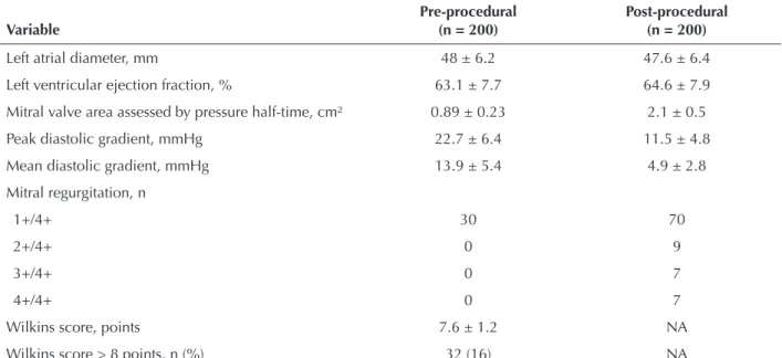
Patient characteristics are described in Table 1. The mean age of the patients was 32 ± 12 years, and 173 (86.5%) were females. A total of 39 patients (19%) were classified as NYHA functional class II, and 161 patients (80.5%) were classified as functional class III or IV. Atrial fibrillation was present in eight patients (4%), and five (2.5%) underwent previous surgical commissurotomy. The conventional double balloon technique was applied in 198 patients (99%), and the Inoue technique was used in only two patients (1%). When evaluating the pre-procedural echocardiographic characteristics (Table 2), the mean Wilkins score was 7.6 ± 1.2 points, and only 32 patients (16%) had a score > 8 points. The mean left atrial diameter was 48 ± 6.2 mm, the mean ejection fraction of the left ventricle was 63.1 ± 7.7%, the previous mitral valve area measured by pressure half-time corresponded to 0.89 ± 0.23 cm2, and the peak and mean valvular gradients were 22.7 ± 6.4 mmHg and 13.9 ± 5.4 mmHg, respectively. A total of 30 (15%) patients experienced pre-procedural mitral regurgitation.
The procedure was successful in 175 patients (87.5%). Failure occurred in 24 patients (12%); in seven (3.5%) patients it was due to severe mitral regurgitation, and in 17 (8.5%) patients it was due to technical problems related to the learning curve (12 cases of cardiac tam-ponade). It was not possible to perform the transseptal puncture in five patients (Figures 1 and 2). There was one death (0.5%) in the studied population.
The echocardiographic results obtained after the procedure showed a final mitral valve area of 2.1 ± 0.5 cm2, with post-procedural peak and mean diastolic gradients of 11.5 ± 4.8 mmHg and 4.9 ± 2.8 mmHg, respectively. The mean left atrial diameter was 47.6 ± 6.4 mm. After the procedure, 70 patients experienced mitral regurgitation 1+/4+, and 9 patients experienced mitral regurgitation 2+/4+ (Table 2).
During the follow-up, 129 patients (74%) were fol-lowed for a mean time of 140 ± 79 months. Restenosis occurred in 60 patients (46.5%), a second percutaneous balloon mitral valvotomy was performed in 25 patients, a third valvotomy was performed in four patients, and a fourth one was performed in one patient. In the other patients who developed restenosis, 27 required surgery, and eight remained in the clinical follow-up (Table 3). The mitral valve area gain declined progres-sively after the second, third, and fourth valvotomies, as shown in Figure 3.
The probability that restenosis would not occur was 85% after five years, 60% after ten years, and 36% after 20 years, as shown in the Kaplan-Meier curve (Figure 4).
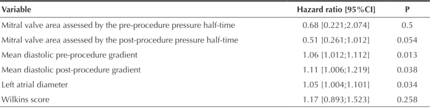
The left atrial diameter (P = 0.034) and pre- and post-procedure mitral transvalvular gradients (P = 0.013 and P = 0.038, respectively) were independent predic-tors of restenosis, but the Wilkins score (P = 0.258) and the pre- and post-procedure mitral valve areas (P = 0.5 and P = 0.054, respectively) were not independent predictors of restenosis (Table 4).
DISCUSSION
The long-term results of the treatment of mitral stenosis by balloon catheter are well established. Bal-loon catheter procedures allowed up to 90% of treated patients to remain asymptomatic or mildly symptomatic over a seven-year interval,8,21 with results similar to surgical treatment22 but with lower costs.23 Thus, bal-loon catheter procedures are considered to be the first choice in patients with favourable anatomy.
In late follow-up studies,24,25 the study populations differ regarding socioeconomic, ethnic, cultural, age, clinical and echocardiographic characteristics and the aetiology of the mitral disease.26 Therefore, it is crucial to define therapeutics, estimate risks, and evaluate the prognosis to recognize the determinants of immediate and long-term success.
TABLE 1
Patients’ Clinical Characteristics
Variable n = 200
Age, years 32 ± 12
Female gender, n (%) 173 (86.5)
Sinus rhythm, n (%) 192 (96)
NYHA FC, n (%)
II 39 (19.5)
III or IV 161 (80.5)
Pregnant patients, n (%) 14 (7)
Previous commissurotomy, n (%) 5 (2.5)
Patients in the present study had a mean age of 32 years when the procedure was performed. Patients demonstrated a low prevalence of atrial fibrillation, which was similar to the prevalence found in patients from developing countries,27 but different from that ob-served in developed countries, in which patients were older and had a higher incidence of arrhythmias.28,29
This difference can be attributed to different aetiologies of mitral stenosis: rheumatic stenosis is more common in the Brazilian population, and degenerative stenosis is more prevalent in developed countries.
Fawzy et al.22,30 demonstrated an event-free survival of 79% at ten years and 43% at 15 years in a population TABLE 2
Pre- and Post-procedural Echocardiographic Characteristics
Variable
Pre-procedural (n = 200)
Post-procedural (n = 200)
Left atrial diameter, mm 48 ± 6.2 47.6 ± 6.4
Left ventricular ejection fraction, % 63.1 ± 7.7 64.6 ± 7.9
Mitral valve area assessed by pressure half-time, cm² 0.89 ± 0.23 2.1 ± 0.5
Peak diastolic gradient, mmHg 22.7 ± 6.4 11.5 ± 4.8
Mean diastolic gradient, mmHg 13.9 ± 5.4 4.9 ± 2.8
Mitral regurgitation, n
1+/4+ 30 70
2+/4+ 0 9
3+/4+ 0 7
4+/4+ 0 7
Wilkins score, points 7.6 ± 1.2 NA
Wilkins score > 8 points, n (%) 32 (16) NA
n = number of patients; NA = not available.
Success n = 175 (87.5%)
Restenosis n = 60 (46.5%)
Redilation n = 25 (19.4%)
Surgery n = 20 (20.9%)
Clinical treatment n = 8 (6.2%) Follow-up
n = 129 (74%)
Loss at follow-up n = 46 (26%)
Severe mitral regurgitation n = 7 (3.5%)
Technical problems n = 17 (3.5%) Failure
n = 24 (12%)
Death after MPV n = 1 (0.5%) n = 200
with a mean age of 31 years; the survival was even higher in patients with favourable anatomic features (88% at ten years and 66% at 15 years). The following
factors were related to a better prognosis: age, post-procedure valve area, and previous valve anatomy. Age is a predictor of restenosis because it is associated with a longer time of disease evolution and, consequently, a higher possibility of valve degeneration.31
In Brazil, Cardoso et al.26 demonstrated an event-free survival of 61% at ten years in patients with a mean age of 35 years, with predictors of age, echocardiographic score, and immediate procedural outcome. The mag-nitude of the long-term benefit is strongly influenced by the immediate outcome, and mitral insufficiency with severe regurgitation is a factor of poor prognosis related to the need for surgery.31,32
Other factors are related to late success, such as the functional class (NYHA), the mean diastolic gradient, and the presence of atrial fibrillation.32 However, these factors characterise patients with more severe anatomic features and a lower probability of procedural success.32 In cases of restenosis, percutaneous mitral valvotomy can be performed as the first choice;32 there have been reports of patients submitted to up to four procedures with good results.33
CONCLUSION
In selected cases, percutaneous balloon mitral val-votomy demonstrates high procedural success and good late follow-up results, with over a third of the patients being restenosis-free. The identification of restenosis predictors is useful to select patients who will benefit from intervention.
CONFLICT OF INTEREST
The authors declare no conflicts of interest.
1
0
25 20 15
10 5
0 0.2 0.4 0.6 0.8
Restenosis-fr
ee su
rv
iv
al
Time (years)
TABLE 3 Late results
Variable n = 129
Follow-up, months 140 ± 79 (12 -288)
Restenosis, n (%) 60 (46.5)
Clinical treatment 8 (6.2)
Redilation 25 (19.4)
Surgery 27 (20.9)
Severe mitral regurgitation, n (%) 7 (5.4)
Clinical treatment 1 (0.8)
Surgery 6 (4.7)
Death at follow-up, n (%) 8 (6.2)
6%3.5% 2.5%
87.5%
Unsuccessful transseptal puncture (five patients) Success (175 patients)
Tamponade (12 patients)
Severe mitral regurgitation (seven patients)
Figure 2 – Procedural results.
2.5
1 PMV 2 PMV 3 PMV 4 PMV
2.5 ± 0.5
0.89 ± 0.2
1.9 ± 0.3 1.85 ± 0.3
1.5
1.05 ± 0.1
0.99 ± 0.03
0.95
2
1.5
1
0.5
1
Mitral valve area (cm2)
POST PRE
Figure 3 – Mitral valve area after the first, second, third and fourth valvotomies. POST = post-procedure; PRE = pre-procedure; PMV = percutaneous balloon mitral valvotomy.
REFERENCES
1. Inoue K, Owaki T, Nakamura T, Kitamura F, Miyamoto N. Clinical application of transvenous mitral commissurotomy by a new balloon catheter. J Thorac Cardiovasc Surg. 1984;87(3): 394-402.
2. Al Zaibag M, Kasab JA, Ribeiro PA, Fagih MR. Percutaneous double balloon mitral valvotomy for rheumatic mitral valve stenosis. Lancet. 1986;1(8484):757-61.
3. McKay RG, Lock JE, Klane JF, Safian RD, Aroesty JM, Grossman W. Percutaneous mitral valvoplasty in an adult patient with calcific rheumatic mitral stenosis. J Am Coll Cardiol. 1986; 7(6):1410-5.
4. Palacios I, Lock JE, Klane JF, Block PC. Percutaneous trans-venous balloon valvotomy in a patient with severe calcified mitral stenosis. J Am Coll Cardiol. 1986;7(6):1416-9. 5. Gomes NL, Esteves CA, Braga SL, Ramos AI, Meneghelo ZM,
Mattos LA, et al. Valvoplastia mitral com duplo cateter-balão: análise de 200 casos. Arq Bras Cardiol. 1992;58(4):269-74. 6. Gomes NL, Esteves C, Braga S, Machado L, Meneghelo Z.
Evolução tardia da valvoplastia mitral. Rev Soc Cardiol Estado de São Paulo. 2002;12(2):315-26.
7. Turi ZG, Reyes VP, Raju BS, Raju AR, Kumar DN, Rajagopal P, et al. Percutaneous balloon versus surgical closed commis-surotomy for mitral stenosis: a prospective, randomized trial. Circulation. 1991;83(4):1179-85.
8. Arora R, Nair M, Kalra GS, Nigam M, Khalilullah M. Im-mediate and long-term results of balloon and surgical closed mitral valvotomy: a randomized comparative study. Am Heart J. 1993;125(4):1091-4.
9. Esteves CA. Valvotomia mitral percutânea por cateter-balão em pacientes grávidas portadoras de estenose mitral reumática: resultados imediatos e seguimento tardio [tese doutorado]. São Paulo: Faculdade de Medicina, Universidade de São Paulo; 2002.
10. Esteves CA, Braga SLN. Aspectos técnicos atuais da valvoplas-tia mitral. Rev Soc Cardiol Estado de São Paulo. 2002;12(2): 306-14.
11. Cribier A, Eltchaninoff H, Koning R, Rath PC, Arora R, Imam A, et al. Percutaneous mechanical mitral commissurotomy with a newly designed metallic valvulotome. Immediate results of the initial experience in 153 patients. Circulation. 1999; 99(6):793-9.
12. Bonhoeffer P, Piechaud JF, Sidi D, Yonga G, Jowi C, Joshi M, et al. Mitral dilatation with the Multi-Track system: an alternative approach. Cathet Cardiovasc Diag. 1995;36(2): 189-93.
13. Bonhoeffer P, Esteves C, Casal U, Tortoledo F, Yonga G, Patel T, et al. Percutaneous mitral valve dilatation with the Multi-Track System. Catheter Cardiovasc Interv. 1999;48(2):178-83. 14. Peixoto E, Oliveira P, Salles M. Inoue balloon versus monofoil
balloon in mitral valvuloplasty: results and complications. Am J Cardiol. 1997;80 Supl. 7A:735.
15. Ben Farhat M, Betbout F, Gamra H, Maatouk F, Ayari M, Cherif A, et al. Results of percutaneous double-balloon mitral com-missurotomy in one medical center in Tunisia. Am J Cardiol. 1995;76(17):1266-70.
16. Fawzy ME. Long-term results up to 19 years of mitral balloon valvuloplasty. Asian Cardiovasc Thorac Ann. 2009;17(6):627-33. 17. Wilkins GT, Weyman AE, Abascal VM, Block PC, Palacios IF.
Percutaneous balloon dilatation of the mitral valve: an analysis of echocardiographic variables related to outcome and the mechanism of dilatation. Br Heart J. 1988;60(4):299-308. 18. Sellers RD, Levy MJ, Amplatz K, Lillehei CW. Left retrogade
cardioangiography in acquired cardiac disease: technic, indi-cation and interpretation in 700 cases. Am J Cardiol. 1964; 14:437-47.
19. Palacios IF, Block PC, Wilkins GT, Weyman AE. Follow-up of patients undergoing percutaneous mitral balloon valvot-omy: analysis of factors determining restenosis. Circulation. 1989;79(3):573-9.
20. Kang DH, Park SW, Song JK, Kim HS, Hong MK, Kim JJ, et al. Long-term clinical and echocardiographic outcome of percutaneous mitral valvuloplasty: randomized comparison of Inoue and double-balloon techniques. J Am Coll Cardiol. 2000;35(1):169-75.
21. Ben Farhat M, Ayari M, Maatouk F, Betbout F, Gamra H, Jarra M, et al. Percutaneous balloon versus surgical closed and open mitral commissurotomy: seven-year follow-up results of a randomized trial. Circulation. 1998;97(3):245-50.
22. Fawzy ME, Shoukri M, Al Buraiki J, Hassan W, El Widaal H, Kharabsheh S, et al. Seventeen years’ clinical and echocar-diographic follow up of mitral balloon valvuloplasty in 520 patients, and predictors of long-term outcome. J Heart Valve Dis. 2007;16(5):454-60.
23. Reyes VP, Raju BS, Wynne J, Stephenson LW, Raju R, Fromm BS, et al. Percutaneous balloon valvuloplasty compared with open surgical commissurotomy for mitral stenosis. N Engl J Med. 1994;331(15):961-7.
24. Borges IP, Peixoto EC, Peixoto RT, Oliveira PS, Salles Netto M, Labrunie P, et al. Percutaneous mitral balloon valvotomy: long-term outcome and assessment of risk factors for death and major events. Arq Bras Cardiol. 2005;84(5):397-404.
TABLE 4 Restenosis predictors
Variable Hazard ratio [95%CI] P
Mitral valve area assessed by the pre-procedure pressure half-time 0.68 [0.221;2.074] 0.5
Mitral valve area assessed by the post-procedure pressure half-time 0.51 [0.261;1.012] 0.054
Mean diastolic pre-procedure gradient 1.06 [1.012;1.112] 0.013
Mean diastolic post-procedure gradient 1.11 [1.006;1.219] 0.038
Left atrial diameter 1.05 [1.004;1.101] 0.034
Wilkins score 1.17 [0.893;1.523] 0.258
25. Herrmann HC, Feldman T, Isner JM, Bashore T, Holmes DR Jr, Rothbaum DA, et al. Comparison of results of percutaneous bal-loon valvuloplasty in patients with mild and mo derate mitral stenosis to those with severe mitral stenosis. The North American Inoue Balloon Investigators. Am J Cardiol. 1993;71(15):1300-3. 26. Cardoso LF, Ayres CV, Bento AM, Tarasoutchi F, Vieira ML,
Grinberg M. Immediate and late results of percutaneous mitral valve repair in patients with mitral stenosis. Arq Bras Cardiol. 2010;94(3):383-90, 406-13.
27. Ben Farhat M, Betbout F, Gamra H, Maatouk F, Ben Hamda K, Abdellaoui M, et al. Predictors of long-term event-free survival and of freedom from restenosis after percutaneous balloon mitral commissurotomy. Am Heart J. 2001;142(6):1072-9. 28. Iung B, Garbarz E, Michaud P, Helou S, Farah B, Berdah P,
et al. Late results of percutaneous mitral commissurotomy in a series of 1024 patients: analysis of late clinical deterioration: frequency, anatomic findings, and predictive factors. Circula-tion. 1999;99(25):3272-8.
29. Palacios IF, Tuzcu ME, Weyman AE, Newell JB, Block PC. Clinical follow-up of patients undergoing percutaneous mitral balloon valvotomy. Circulation. 1995;91(3):671-6.
30. Fawzy ME, Shoukri M, Hassan W, Nambiar V, Stefadouros M, Canver CC. The impact of mitral valve morphology on the long-term outcome of mitral balloon valvuloplasty. Catheter Cardiovasc Interv. 2007;69(1):40-6.
31. Bouleti C, Iung B, Laouénan C, Himbert D, Brochet E, Messika-Zeitoun D, et al. Late results of percutaneous mitral commissurotomy up to 20 years: development and validation of a risk score predicting late functional results from a series of 912 patients. Circulation. 2012;125(17):2119-27.
32. Nobuyoshi M, Arita T, Shirai S, Hamasaki N, Yokoi H, Iwabuchi M, et al. Percutaneous balloon mitral valvuloplasty: a review. Circulation. 2009;119(8):e211-9.