Chronic kidney disease — determinants of progression
and cardiovascular risk. PROGREDIR cohort study:
design and methods
Doença renal crônica — determinantes de progressão e risco cardiovascular.
Coorte PROGREDIR: desenho de estudo e métodos
Maria Alice Muniz Domingos
I, Alessandra Carvalho Goulart
II, Paulo Andrade Lotufo
III, Isabela Judith Martins Benseñor
IV,
Silvia Maria de Oliveira Titan
VClinical Research Center, University Hospital, Faculdade de Medicina da Universidade de São Paulo (FMUSP), São Paulo (SP), Brazil
ABSTRACT
CONTEXT AND OBJECTIVE: Chronic kidney disease (CKD) has become an important public health issue. The socioeconomic burden of renal replacement therapy (RRT) is very high, as is CKD-related cardiovas-cular mortality and morbidity. Preventive and therapeutic measures only have modest impact and more research is needed. Few cohort studies have been conducted on populations with CKD. Our aim was to establish a cohort that would include more advanced forms of CKD (stages 3 and 4). Data collection was focused on renal and cardiovascular parameters.
DESIGN AND SETTING: Prospective cohort study; São Paulo, Brazil.
METHODS: Recruitment took place in Hospital das Clínicas, São Paulo, from March 2012 to December 2013. Data relating to medical history, food-frequency questionnaire, anthropometry, laboratory work-up, calcium score, echocardiography, carotid intimal-medial thickness, pulse-wave velocity, retinography and heart rate variability were collected. A biobank including serum, plasma, post-oral glucose tolerance test serum and plasma, urine (morning and 24-hour urine) and DNA was established.
RESULTS: 454 participants (60% men and 50% diabetics) of mean age 68 years were enrolled. Their mean estimated glomerular iltration rate-CKD Epidemiology Collaboration was 38 ml/min/1.73 m2. Follow-up is
ongoing and the main outcomes are the start of RRT, cardiovascular events and death.
CONCLUSIONS: The PROGREDIR cohort is a promising prospective study that will allow better understanding of CKD determinants and validation of candidate biomarkers for the risks of CKD progression and mortality.
RESUMO
CONTEXTO E OBJETIVO: A doença renal crônica (DRC) tornou-se um problema de saúde pública. A carga socioeconômica da terapia renal substitutiva é muito elevada, assim como a morbimortalidade cardio-vascular associada à DRC. Medidas terapêuticas e preventivas têm impacto parcial e novos estudos são necessários. Há poucos estudos de coorte em populações com DRC. Nosso objetivo foi criar uma coorte que contemplasse formas mais avançadas de DRC (estágios 3 e 4). A coleta de dados foi centrada em parâmetros renais e cardiovasculares.
TIPO DE ESTUDO E LOCAL: Estudo de coorte prospectivo; São Paulo, Brasil.
MÉTODOS: O recrutamento ocorreu entre março de 2012 e dezembro de 2013, no Hospital das Clínicas, em São Paulo. Foram coletados dados de história médica, questionário de frequência alimentar, antropo-metria, exames laboratoriais, escore de cálcio, ecocardiograia, espessura de camada médio-intimal de carótidas, velocidade de onda de pulso, retinograia e variabilidade de frequência cardíaca. Um biobanco incluindo soro, plasma, soro e plasma pós-teste oral de tolerância à glicose, urina (manhã e 24 horas) e DNA foi estabelecido.
RESULTADOS: 454 participantes (60% homens e 50% diabéticos) com idade média de 68 anos foram recrutados. A taxa média de iltração glomerular estimada-Colaboração da Epidemiologia para DRC foi de 38,4 ml/min/1,73 m2. O seguimento está em andamento e os desfechos principais são: início de terapia
renal substitutiva, eventos cardiovasculares e óbito.
CONCLUSÃO: A coorte PROGREDIR é um estudo prospectivo promissor que permitirá melhor compre-ensão dos determinantes de DRC e a validação de biomarcadores candidatos para o risco de progressão de DRC e de mortalidade.
IMD, PhD. Nephrologist, Renal Division, Department of Clinical Medicine, Faculdade de Medicina da Universidade de São Paulo (FMUSP), São Paulo (SP), Brazil.
IIMD, PhD. Clinical Epidemiologist and Researcher, Clinical Research Center, University Hospital, Faculdade de Medicina da Universidade de São Paulo (FMUSP), São Paulo (SP), Brazil.
IIIMD, PhD. Full Professor, Clinical Research Center, University Hospital, Faculdade de Medicina da Universidade de São Paulo (FMUSP), São Paulo (SP), Brazil.
IVMD, PhD. Assistant Professor, Clinical Research Center, University Hospital, Faculdade de Medicina da Universidade de São Paulo (FMUSP), São Paulo (SP), Brazil.
VMD, PhD. Research Investigator, Renal Division, Department of Clinical Medicine, Faculdade de Medicina da Universidade de São Paulo (FMUSP), São Paulo (SP), Brazil.
KEY WORDS:
Renal insuiciency, chronic. Cardiovascular diseases. Renal replacement therapy. Biomarkers.
Risk factors.
PALAVRAS-CHAVE: Insuiciência renal crônica. Doenças cardiovasculares. Terapia de substituição renal. Biomarcadores.
INTRODUCTION
Chronic kidney disease (CKD) has become an important pub-lic health issue worldwide. Increasing prevalence of obesity and diabetes mellitus and today’s high life expectancy, particularly among patients with atherosclerosis, are all contributory factors. In addition, CKD progression is still a major challenge, with few new speciic therapeutic measures available. he socioeconomic burden on individuals who need renal replacement therapy (RRT) is very high and comes together with CKD-related high cardiovascular mortality and morbidity, with incidence that may in fact even exceed the igures for RRT.1-7
In the United States, according to the Annual Report of the United States Renal Data System (USRDS),8 the prevalence of CKD
stages 1-4 was around 14% in the general population and the inci-dence of end-stage renal disease (ESRD) was 353 cases per mil-lion/year in 2012. he prevalence of cardiovascular disease reached 69.8% among CKD patients versus 34.8% among individuals with-out CKD and the adjusted mortality rates for CKD patients was 76 deaths per 1000 patients, compared with 52 deaths per 1,000 individuals without CKD in 2012. Medicare expenses relating to CKD reach US$ 1700, 3500 and 12,700 per person-year for CKD patients with stages 2, 3 and 4, respectively.9 Overall, CKD accounts
for 6.7% of total Medicare costs.9
In Brazil, there were 100,397 patients on dialysis at the end of 2013, with incidence of 170 cases per million/year and an estimated mortality rate of 17.9% per year.10 In 2013, 5,433 kidney
transplan-tations were performed in Brazil, mostly using public resources. Preventive measures are highly necessary, and the search for new biomarkers and new therapeutic strategies is intense. While several studies on general populations and cardiovascu-lar cohorts have yielded important contributions towards CKD knowledge, more specific cohorts focusing on CKD progres-sion instead of CKD incidence are necessary within nephrology. In response to this need, several countries like the United States (CRIC study), Germany (GCKD), Canada (CanPREDDICT), Japan, Australia and Uruguay, among others, have ongoing CKD cohort studies.11
Along the same lines, the PROGREDIR cohort study was designed to enable better understanding of the determinants of CKD progression and CKD-related mortality, with particular emphasis on mineral metabolism as a cardiovascular risk fac-tor. he cohort comprises people with CKD stages 3 and 4 in São Paulo, Brazil. he cohort was established and baseline data were collected in 2012-2013. Prospective data on hard outcomes such as the start of renal replacement therapy, cardiovascular events and death are currently being gathered. he PROGREDIR cohort is funded by the Research Support Foundation of the State of São Paulo (Fundação de Amparo à Pesquisa do Estado de São Paulo, FAPESP; 2011-17341-0), São Paulo, Brazil.
OBJECTIVE
he aim of this study was to establish a CKD cohort that would include participants with more advanced forms of this disease (CKD stages 3 and 4), with data collection focused on renal and cardiovascular parameters.
METHODS
Study population and recruitment
Patients attending the outpatient service of Hospital das Clínicas, São Paulo, a public university facility providing quaternary-level care for patients with chronic diseases, were invited to participate in this study. Initially, from the outpatient records, all patients aged
≥ 30 years and at least two measurements of creatinine (with a min-imum interval of 3 months) ≥ 1.6 mg/dl for men and ≥ 1.4 mg/ dl for women were considered potential candidates. Patients attend-ing oncology, psychiatry, human immunodeiciency virus/acquired immunodeiciency syndrome (HIV/AIDS), viral hepatitis and glo-merulonephritis services were excluded. he remaining candidates were then contacted by phone and were invited to participate if they did not meet any exclusion criteria. he exclusion criteria checked by the interviewer were: hospitalization within the last six months, acute myocardial infarction within the last six months, autoimmune diseases, pregnancy, psychiatric diseases, ongoing chemotherapy or immunosuppressive therapy, ongoing RRT, glomerulonephritis, HIV/AIDS infection, hepatitis B or C and any organ transplantation. Recruitment took place between March 2012 and December 2013, and 454 participants were enrolled. he study was approved by two local ethics committees and written informed consent was obtained from all participants.
Sample size estimation
he sample size was calculated using an estimate of the annual inci-dence of end-stage renal disease (ESRD) of 2% and an annual rate of cardiovascular events of 2-3.5% among diabetic nephropathy patients.12 By assuming a diference in event rate incidence of 3%
between exposed and non-exposed subjects, a sample size of 500 was estimated, with an alpha error of 0.05 and a power of 80%.
Baseline examination and data collection
he baseline assessment lasted approximately six hours and was performed on a single-day visit to our study center. Data collec-tion included all the variables depicted in Table 1. Sex and
self-declared race were also registered. Anthropometry was performed irst, with the participants wearing light clothes, following standard techniques.13 Blood pressure (BP) was measured using a validated
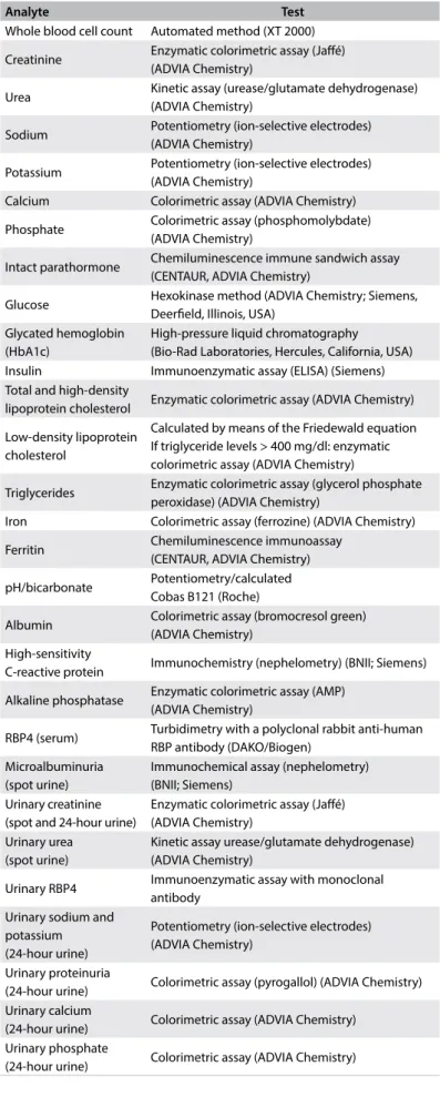
pressure. Overnight fasting blood samples and 24-hour and spot urine samples were collected. A standard 75-g oral glucose tol-erance test was administered to all participants without known diabetes. Urine and blood aliquots were prepared and stored at -180 °C in nitrogen. DNA extraction was performed and the mate-rial was stored at -80 °C. Baseline laboratory measurements were made using conventional techniques (Table 2).
Interviews were conducted by trained personal under strict quality control. Data on medical history, socioeconomic variables, family history, medication use, physical activity, smoking and alco-hol consumption were obtained. A food-frequency questionnaire was also administered. he food list was deined on the basis of the dietary intake of the Brazilian population and the reproducibility and validity of the questionnaire has been measured elsewhere.14
Conventional 12-lead electrocardiograms (ECG) were per-formed using a digital device (Atria 6100, Burdick, Cardiac Science Corporation, USA) with automated readings of heart rate; P wave, QRS complex and T wave duration, amplitude and axis; and QT, QTc and QT dispersion. All precordial electrodes were positioned ater identifying the location for the V4 electrode with a square. he Electrocardiogram Reading Center (ERC) at the Heart Institute of the University of São Paulo (INCOR) pro-vided all ECG readings.
For heart rate variability determinations, a 10-minute con-tinuous ECG was obtained from a single lead (usually D2) using a digital electrocardiograph (Micromed, Brazil) at a frequency of 250 Hz, with subjects in the supine position. Computer sotware (WinCardio) was used to generate time series of RR intervals that were sent to the central cardiovascular physiology laboratory
Anthropometry Body mass index
Waist and hip circumferences Blood pressure Mean from three seated measurements
Laboratory data
Serum fasting samples
Two-hour glucose-tolerance test (75 g oral glucose) Spot urine
24-hour urine (home collected)
Biobank (serum, plasma, post-glucose serum, urine and DNA)
Questionnaires Medical history
Food frequency questionnaire Non-alcoholic
fatty liver disease assessment
Measurement of anterior-posterior diameter of right lobe of the liver
Cardiac evaluation
Electrocardiogram
Transthoracic echocardiography Heart rate variability
Vascular evaluation
Carotid-femoral pulse-wave velocity (PWV) Carotid intima-media thickness (IMT) Coronary artery calcium score (CAC) Retinography
Table 1. Baseline assessments in PROGREDIR cohort study
Analyte Test
Whole blood cell count Automated method (XT 2000)
Creatinine Enzymatic colorimetric assay (Jafé) (ADVIA Chemistry)
Urea Kinetic assay (urease/glutamate dehydrogenase) (ADVIA Chemistry)
Sodium Potentiometry (ion-selective electrodes) (ADVIA Chemistry)
Potassium Potentiometry (ion-selective electrodes) (ADVIA Chemistry)
Calcium Colorimetric assay (ADVIA Chemistry)
Phosphate Colorimetric assay (phosphomolybdate) (ADVIA Chemistry)
Intact parathormone Chemiluminescence immune sandwich assay (CENTAUR, ADVIA Chemistry)
Glucose Hexokinase method (ADVIA Chemistry; Siemens, Deerield, Illinois, USA)
Glycated hemoglobin (HbA1c)
High-pressure liquid chromatography (Bio-Rad Laboratories, Hercules, California, USA) Insulin Immunoenzymatic assay (ELISA) (Siemens) Total and high-density
lipoprotein cholesterol Enzymatic colorimetric assay (ADVIA Chemistry)
Low-density lipoprotein cholesterol
Calculated by means of the Friedewald equation If triglyceride levels > 400 mg/dl: enzymatic colorimetric assay (ADVIA Chemistry)
Triglycerides Enzymatic colorimetric assay (glycerol phosphate peroxidase) (ADVIA Chemistry)
Iron Colorimetric assay (ferrozine) (ADVIA Chemistry)
Ferritin Chemiluminescence immunoassay (CENTAUR, ADVIA Chemistry)
pH/bicarbonate Potentiometry/calculated Cobas B121 (Roche)
Albumin Colorimetric assay (bromocresol green) (ADVIA Chemistry)
High-sensitivity
C-reactive protein Immunochemistry (nephelometry) (BNII; Siemens)
Alkaline phosphatase Enzymatic colorimetric assay (AMP) (ADVIA Chemistry)
RBP4 (serum) Turbidimetry with a polyclonal rabbit anti-human RBP antibody (DAKO/Biogen)
Microalbuminuria (spot urine)
Immunochemical assay (nephelometry) (BNII; Siemens)
Urinary creatinine (spot and 24-hour urine)
Enzymatic colorimetric assay (Jafé) (ADVIA Chemistry)
Urinary urea (spot urine)
Kinetic assay urease/glutamate dehydrogenase) (ADVIA Chemistry)
Urinary RBP4 Immunoenzymatic assay with monoclonal antibody
Urinary sodium and potassium (24-hour urine)
Potentiometry (ion-selective electrodes) (ADVIA Chemistry)
Urinary proteinuria
(24-hour urine) Colorimetric assay (pyrogallol) (ADVIA Chemistry) Urinary calcium
(24-hour urine) Colorimetric assay (ADVIA Chemistry) Urinary phosphate
(IC-ES). All readings were made through computer sotware that eliminated artifacts and selected RR intervals lasting 0.5 to 2.0 sec-onds. Temporal and spectral analyses of heart rate variability (HRV) were then performed using an autoregressive model to identify very low-frequency (VLF, 0 to 0.04 Hz), low-frequency (LF, 0.04 to 0.1 Hz) and high-frequency spectral bands (HF, 0.1 to 0.4 Hz).
Transthoracic echocardiography was performed on all par-ticipants using a device (Aplio XG; Toshiba Corporation, Tokyo, Japan) with a 2.5 MHz sector transducer. All examinations were performed by the same echocardiographer. he readings con-sisted of qualitative analysis of echocardiographic indings and measurements of quantitative parameters such as: let ventricular (LV) geometry and size, let atrial size, LV systolic and diastolic function, segmental LV dysfunction, valvular heart disease and pericardial appearance. Cardiac mass was calculated using the Devereaux formula.15
Measurement of the anterior-posterior diameter of the right lobe of the liver was performed by means of ultrasound for quan-titative assessment of nonalcoholic fatty liver disease (NAFLD). Liver images were obtained using standard equipment (Toshiba SSA-770A Aplio, Japan) and a broadband convex transducer (PVT-375BT) with a central frequency of 3.5 MHz (2.5-5.5 MHz).16
he carotid-femoral pulse-wave velocity (PWV) was measured using a validated automated device (Complior, Artech Medicale, France), with the subject in the supine position in a temperature-controlled room (20-24 °C).First, BP was measured in the right arm with the subject in the supine position using an oscillometric device (HRM Onrom 705 CP). he distance from the sternal furcula to the right femoral pulse was determined using a measuring tape regard-less of abdominal curvature. Pulse sensors were positioned in the right carotid and femoral arteries so that pulse waves were recorded and viewed on a computer screen. Computer sotware that could adequately detect and record pulse waves was used. PWV was cal-culated by dividing the distance from the sterna furcula to the femo-ral pulse by the diference between the rise delays of the carotid and femoral pulses. A subject’s PWV was the arithmetic average of read-ings obtained in ten consecutive cardiac cycles at a regular heart rate. Carotid intimal-media thickness (IMT) was assessed in all patients in a standardized manner using a device (Aplio XG, Toshiba) with a 7.5 MHz linear transducer. he technique used for IMT measurement was as previously published.17 IMT was
mea-sured in the outer wall of a predeined carotid segment of 1 cm in length from 1 cm below the carotid bifurcation, during three car-diac cycles. We considered the images acquired to be valid if they clearly showed three reference points on both sides:
1. anatomical guides for the common carotid arteries; 2. interfaces between the lumen and the far wall of the vessel; and 3. interfaces between the media and adventitia layers of the far
wall of the vessel.
We used the MIA sotware to standardize the readings and interpret the carotid scans as previously described. IMT was then deined as the mean of the right and let carotid measurements.
To determine the coronary artery calcium score, the participants underwent non-contrast computed tomography. he scans were performed using a 64-slice detector computed tomography scan-ner (Philips Brilliance, Philips, Netherlands). Ater scout images had been produced, each patient also underwent an ECG-gated prospective calcium score examination with a tube potential of 120 kV and a tube current adjusted to body habitus. Images were reconstructed at 2.5 mm slice thickness using standard iltered back projection. he coronary artery calcium score was expressed in terms of Agatston units and the percentiles were evaluated in a blinded manner by an experienced cardiologist using semi-auto-mated sotware (Calcium Scoring, Philips Workstation). Coronary calcium scores were not obtained for participants who reported that they had been itted with coronary stents, since the stent mate-rial greatly overestimates the calcium scores.
Retinography was performed using a nonmydriatic retinograph (CR-1, Canon, Japan) with a 10-megapixel digital camera (Canon EOS 40 D). he subjects underwent natural dilation of their pupils through resting in a darkened room for about four minutes, and for each eye two 45° fundus images were obtained: one centered on the optical disk and the second on the maculae. Our institu-tion’s central retinography laboratory (IC-RS) developed standard-ized image acquisition and reading protocols, and DICOM images (approximately 30 MB) and JPEG images (approximately 3 MB) were acquired. he JPEG images were recorded on CD/DVD at the study sites and were mailed to the central retinography laboratory.
Follow-up
he participants are being contacted again annually, for tele-phone interviews that include questions on hospitalizations, need for RRT and self-rated health. he main clinical endpoints inves-tigated are death, acute myocardial infarction, unstable angina pectoris, cardiac revascularization, heart failure, stroke and RRT. Any cardiovascular and renal clinical events that are reported are then investigated and classiied in line with the study protocol, by a panel of physicians that has received training in accordance with international classiication criteria.18 In the event of the
participant’s death, information regarding this event is sought. Surveillance of clinical events is also conducted through state databases such as the Mortality Registry and the São Paulo State Registry of Dialysis and Transplantation.
RESULTS
Over the two-year recruitment period, 454 participants were enrolled. Table 3 shows the main clinical and laboratory
in stages 3 and 4, with a mean estimated glomerular iltration rate-CKD Epidemiology Collaboration (eGFR-CKDEPI) of 38.4 (± 14.6) ml/min/1.73 m2. he albuminuria range was wide, with
similar frequencies of normoalbuminuria (35%), microalbu-minuria (31%) and macroalbumicroalbu-minuria (34%). he participants’ median age was 67 years; 63% were men; 60% were current or past smokers; 45% self-reported diabetes; and 32% reported hav-ing had previous myocardial infarction. Coronary artery calcii-cation scores were also high, with more than half of the cohort presenting an Agatston score above 100.
Follow-up is ongoing. Up to the present date, i.e. over the irst three years of follow-up, event rates have been high, with a 5-7% mortality rate per year and 2-3% incidence of ESRD and non-fatal cardiovascular events per year. With this event rate, from 2017 onwards, survival analysis will be started, focusing on biomark-ers for mineral metabolism.
DISCUSSION
he PROGREDIR cohort was designed speciically to address CKD progression among patients with moderate to advanced dis-ease. Over a two-year recruitment period, we were able to enroll 454 participants, and thus nearly reached the estimated sam-ple size. he baseline characteristics of these participants were in accordance with the proile expected from the inclusion and exclu-sion criteria: older age, predominance of men and high rates of diabetes and previous cardiovascular disease. In the PROGREDIR cohort, we avoided overrepresentation of glomerulonephritis and other speciic kidney diseases such as those relating to HIV, hepa-titis C and lupus. Transplantation patients (any organ) were also not included. his decision was mostly related to the fact that PROGREDIR was designed to be a cohort of general CKD cases and not to address mechanisms relating to speciic systemic or pri-mary diseases. he eligible participants were originally from a qua-ternary-level hospital, which might have yielded an excessive num-ber of glomerulonephritis cases if exclusion criteria had not been applied. Other CKD cohort studies have applied similar inclusion and exclusion criteria and have ended up with recruited popula-tions compatible with the proile observed in PROGREDIR.19-21
One important accomplishment was to have nearly equal rep-resentation of normoalbuminuria, microalbuminuria and macro-albuminuria subpopulations in the baseline proile of the cohort. he prevalence of and interest in normoalbuminuric CKD is increas-ing,22-24 since it is now known that 30-45% of diabetic patients may
in fact present CKD and normoalbuminuria. It is currently of inter-est not only to understand the determinants of CKD progression in the normoalbuminuric CKD population, but also to compare the performance of traditional and new risk factors in normoal-buminuric and alnormoal-buminuric populations, in order to test whether the results can be generalized to a broad spectrum of diseases.
All n = 454
Age (years; mean, SD) 67.5 11.9
Sex (men; n, %) 287 63.20%
Race (self-declared white; n, %) 300 66.10%
Hypertension (n, %) 409 90.10%
Diabetes (n, %) 208 45.80%
Acute myocardial infarction (n, %) 147 32.40%
Stroke (n, %) 73 16.10%
Smoking (current + former; n, %) 270 59.50% Systolic blood pressure (mmHg; mean, SD) 140 24.1 Diastolic blood pressure (mmHg; mean, SD) 76.2 12.9
Body-mass index (mean, SD) 29.4 5.4
Waist-to-hip ratio (mean, SD) 0.97 0.1
Potassium (mEq/l; mean, SD) 4.58 0.52
Urea (mg/dl; median, IQR) 69 54-89.0
Creatinine (mg/dl; median, IQR) 1.7 1.4-2.1 Albuminuria (mg/dl; median, IQR) 80 15.0-640.0 Urinary RBP (mg/g creatinine; median, IQR) 0.29 0.08-1.47 eGFR-CKDEPI (ml/min/1.73 m2; mean, SD) 38.4 14.6
Phosphorus (mg/dl; mean, SD) 3.6 0.6
Calcium (mg/dl; mean, SD) 9.6 0.6
Parathormone (pg/ml; median, IQR) 93 64.0-143.0 Serum RBP (mg/l; median, IQR) 66.1 55.2-79.8 Glycemia (mg/dl; median, IQR) 104 95.0-126.0 Glycated hemoglobin (%; median, IQR) 6.2 5.8-7.2 Glycemia after OGTT (mg/dl; mean, SD)* 155 51.8
HOMA-IR (median; IQR)* 3.4 2.3-5.8
Insulinemia (mUI/ml; IQR) 16.1 10.4-25.4
Total cholesterol (mg/l; mean, SD) 168.6 39.9 LDL-cholesterol (mg/dl; mean, SD) 91.4 32.2 HDL-cholesterol (mg/dl; mean, SD) 46 14.3 Triglycerides (mg/dl; median, IQR) 142 99.0-192.0
Serum pH (mean, SD) 7.35 0.04
Bicarbonate (mmol/l; mean, SD) 25.6 2.9
Hemoglobin (g/dl; mean, SD) 13.1 1.9
Iron (mcg/dl; mean, SD) 77.1 28.3
Ferritin (ng/ml; median, IQR) 104 52.0-197.0 High-sensitivity C-reactive protein (mg/l;
median, IQR) 2.8 1.0-6.5
Albumin (mg/dl; mean, SD) 4.3 0.3
Right hepatic lobe anteroposterior diameter
(mm; mean, SD) 104.8 12.9
Pulse wave velocity (cm/s; mean, SD) 12.8 3 Left atrium diameter (mm; mean, SD) 41.4 5.4 Left ventricular wall thickness (mm; median, IQR) 11 10.0-12.0 Ejection fraction (median, IQR) 0.66 0.6-0.7 Intima media thickness (mm; mean, SD) 0.75 0.2 Agatston score (median, IQR) 165 8.0-785.0 Table 3. Baseline clinical and laboratory proile of 454 participants in the PROGREDIR cohort
*calculated for participants without known diabetes.
Baseline data were collected in this study in accordance with the study design, covering traditional cardiovascular risk factors and biomarkers for CKD. Surrogate measurements of athero-sclerosis and hypertension such as coronary calcium score, car-diac hypertrophy, IMT, PWV and retinography were made, and these will allow understanding and stratiication of baseline CKD among the participants. he biobank is wide-ranging and kept under strict quality control, thus providing a source for reliable future measurements.
Follow-up is ongoing and a high event rate is being observed. Follow-up data collection is being centered on three major clin-ical events: death, non-fatal cardiovascular events and starting of RRT. hese events are of particular importance, since CKD is known to be a very important cardiovascular risk factor that makes a signiicant contribution to high rates of morbidity and mortality.2 Focusing data collection only on renal events would
lead to selection bias, because a signiicant proportion of the par-ticipants might experience cardiovascular events prior to renal events. hus, to fully address the impact of CKD biomarkers and measurements, it is very important to account for their impact both on renal events such as mortality and on fatal and non-fatal cardiovascular events.
Now that the cohort has been established, the PROGREDIR study can be used for research investigation in two major ways. First, it can be used to test the performance of candidate bio-markers for CKD progression. he current need to promote dis-covery and validation of biomarkers in CKD is highlighted by the recent launch of a CKD Biomarkers Consortium (BioCon)25
by the National Institute of Diabetes and Digestive and Kidney Diseases in the United States. Similar approaches are being used by European countries.26 Secondly, the cohort can be used to test
high throughput technologies, which are an innovative approach that may provide new insights on the mechanisms and pathways of complex diseases, as well as enabling identiication of novel biomarkers for diseases. As a irst step, untargeted metabolomic assessments are currently being performed on baseline serum and the data thus obtained will be analyzed in relation to renal func-tion and clinical events.
Additionally, to contribute towards improvement of scientiic knowledge on CKD, the PROGREDIR study will also serve the purpose of being a national data source in which biomarkers can be replicated and validated. Racial factors are known to have an important efect on the risk of diseases, and this has recently been very well illustrated by the discovery of the higher risk attribut-able to the APOL1 gene in the African-American population.27
In this regard, it is very important that national datasets should be available, so that the performance of candidate biomarkers can be tested on the Brazilian population, which is known to be highly admixed.
CONCLUSION
In conclusion, the PROGREDIR cohort recruitment and base-line data collection were successfully implemented. In addi-tion to being a naaddi-tional dataset, the PROGREDIR cohort pro-vides promising prospective study material that will allow better understanding of CKD determinants and validation of candidate biomarkers for CKD progression and mortality risk.
REFERENCES
1. Anavekar NS, McMurray JJ, Velazquez EJ, et al. Relation between renal
dysfunction and cardiovascular outcomes after myocardial infarction.
N Engl J Med. 2004;351(13):1285-95.
2. Astor BC, Hallan SI, Miller ER 3rd, Yeung E, Coresh J. Glomerular iltration
rate, albuminuria, and risk of cardiovascular and all-cause mortality in
the US population. Am J Epidemiol. 2008;167(10):1226-34.
3. Rahman M, Pressel S, Davis BR, et al. Cardiovascular outcomes in
high-risk hypertensive patients stratiied by baseline glomerular iltration
rate. Ann Intern Med. 2006;144(3):172-80.
4. Wen CP, Cheng TY, Tsai MK, et al. All-cause mortality attributable to
chronic kidney disease: a prospective cohort study based on 462 293
adults in Taiwan. Lancet. 2008;371(9631):2173-82.
5. Di Angelantonio E, Chowdhury R, Sarwar N, et al. Chronic kidney disease
and risk of major cardiovascular disease and non-vascular mortality:
prospective population based cohort study. BMJ. 2010;341:c4986.
6. Meisinger C, Döring A, Löwel H; KORA Study Group. Chronic kidney
disease and risk of incident myocardial infarction and all-cause and
cardiovascular disease mortality in middle-aged men and women
from the general population. Eur Heart J. 2006;27(10):1245-50.
7. Astor BC, Hallan SI, Miller ER 3rd, Yeung E, Coresh J. Glomerular iltration
rate, albuminuria, and risk of cardiovascular and all-cause mortality in
the US population. Am J Epidemiol. 2008;167(10):1226-34.
8. United States Renal Data System, 2014 Annual Data Report:
Epidemiology of Kidney Disease in the United States. Bethesda, MD:
National Institutes of Health, National Institute of Diabetes and Digestive
and Kidney Diseases; 2014.
9. Foley RN, Collins AJ. End-stage renal disease in the United States: an
update from the United States Renal Data System. J Am Soc Nephrol.
2007;18(10):2644-8.
10. Censo da Sociedade Brasileira de Nefrologia. Censo de diálise; 2013.
Available from: http://arquivos.sbn.org.br/pdf/censo_2013-14-05.pdf.
Accessed in 2016 (Dec 28).
11. Dienemann T, Fujii N, Orlandi P, et al. International Network of Chronic
Kidney Disease cohort studies (iNET-CKD): a global network of chronic
kidney disease cohorts. BMC Nephrol. 2016;17(1):121.
12. Adler AI, Stevens RJ, Manley SE, et al. Development and progression
of nephropathy in type 2 diabetes: the United Kingdom Prospective
Diabetes Study (UKPDS 64). Kidney Int. 2003;63(1):225-32.
13. Lohman TG, Roche AF, Martorell R. Anthropometric standardization
14. Mannato LW, Pereira TS, Velasquez-Melendez G, et al. Comparison
of a short version of the Food Frequency Questionnaire with
its long version--a cross-sectional analysis in the Brazilian
Longitudinal Study of Adult Health (ELSA-Brasil). Sao Paulo Med
J. 2015;133(5):414-20.
15. Devereux RB, Alonso DR, Lutas EM, et al. Echocardiographic assessment
of left ventricular hypertrophy: comparison to necropsy indings. Am
J Cardiol. 1986;57(6):450-8.
16. Goulart AC, Oliveira IR, Alencar AP, et al. Diagnostic accuracy of a
noninvasive hepatic ultrasound score for non-alcoholic fatty liver
disease (NAFLD) in the Brazilian Longitudinal Study of Adult Health
(ELSA-Brasil). Sao Paulo Med J. 2015;133(2):115-24.
17. Santos IS, Bittencourt MS, Oliveira IR, et al. Carotid intima-media thickness
value distributions in the Brazilian Longitudinal Study of Adult Health
(ELSA-Brasil). Atherosclerosis. 2014;237(1):227-35.
18. Barreto SM, Ladeira RM, Bastos MSCBO, et al. Estratégias de identiicação,
investigação e classiicação de desfechos incidentes no ELSA-Brasil
[ELSA-Brasil strategies for outcome identiication, investigation and
ascertainment]. Rev Saúde Pública. 2013;47(Supl 2):79-86.
19. Feldman HI, Appel LJ, Chertow GM, et al. The Chronic Renal Insuiciency
Cohort (CRIC) Study: Design and Methods. J Am Soc Nephrol. 2003;14(7
Suppl 2):S148-53.
20. Levin A, Rigatto C, Brendan B, et al. Cohort proile: Canadian study
of prediction of death, dialysis and interim cardiovascular events
(CanPREDDICT). BMC Nephrol. 2013;14:121.
21. Eckardt KU, Bärthlein B, Baid-Agrawal S, et al. The German Chronic Kidney
Disease (GCKD) study: design and methods. Nephrol Dial Transplant.
2012;27(4):1454-60.
22. Kramer HJ, Nguyen QD, Curhan G, Hsu CY. Renal insuiciency in the
absence of albuminuria and retinopathy among adults with type 2
diabetes mellitus. JAMA. 2003;289(24):3273-7.
23. Afkarian M, Zelnick LR, Hall YN, et al. Clinical Manifestations of
Kidney Disease among US Adults with Diabetes, 1988-2014. JAMA.
2016;316(6):602-10.
24. Garg AX, Kiberd BA, Clark WF, Haynes RB, Clase CM. Albuminuria and
renal insuiciency prevalence guides population screening: results
from the NHANES III. Kidney Int. 2002;61(6):2165-75.
25. Hsu CY, Ballard S, Batlle D, et al. Cross-Disciplinary Biomarkers Research:
Lessons Learned by the CKD Biomarkers Consortium. Clin J Am Soc
Nephrol. 2015;10(5):894-902.
26. Brück K, Stel VS, Gambaro G, et al. CKD Prevalence Varies across the
European General Population. J Am Soc Nephrol. 2016;27(7):2135-47.
27. Genovese G, Friedman DJ, Pollak MR. APOL1 variants and kidney
disease in people of recent African ancestry. Nat Rev Nephrol.
2013;9(4):240-4.
Sources of funding: Fundação de Amparo à Pesquisa do Estado de São
Paulo (FAPESP) (2011/17341-0)
Conlicts of interest: None
Date of irst submission: July 2, 2016
Last received: November 7, 2016
Accepted: November 26, 2016
Address for correspondence: Silvia Maria de Oliveira Titan
Divisão de Nefrologia, Hospital das Clínicas
Av. Dr Enéas de Carvalho Aguiar, 255
São Paulo (SP) — Brasil
CEP 05403-010