263
Lopes JR et al. Verrucous carcinoma of the hand assessed by MRI
Radiol Bras. 2011 Jul/Ago;44(4):263–264
Verrucous carcinoma of the hand: a rare presentation
evaluated by magnetic resonance imaging
*
Carcinoma verrucoso da mão: uma rara apresentação avaliada por ressonância magnética
Jailson Rodrigues Lopes1, Marcelo Bordalo Rodrigues2, Laercio Alberto Rosemberg3, Rafael
Burgomeister Lourenço4, Giovanni Guido Cerri5
Verrucous carcinoma is a variant of squamous cell carcinoma seen in mucous membranes and skin, and rarely found in the hand. The present report describes a case of two lesions on the dorsum of the hand, with no contact to each other, which underwent en-block resection and were confirmed as verrucous carcinoma.
Keywords: Verrucous; Carcinoma; Spiculated; Sea-urchin; Squamous; Hand.
O carcinoma verrucoso é uma variante do carcinoma de células escamosas, vista em mucosas e pele, raramente encontrada na mão. Nós relatamos um caso de duas lesões no dorso da mão, sem contato entre si, que foram resse-cadas em bloco e confirmadas como carcinoma verrucoso.
Unitermos: Verrucoso; Carcinoma; Espiculado; Ouriço-do-mar; Escamoso; Mão. Abstract
Resumo
* Study developed at Instituto de Radiologia do Hospital das Clínicas da Faculdade de Medicina da Universidade de São Paulo (InRad/HC-FMUSP), São Paulo, SP, Brazil.
1. MD, Radiologist, Collaborator at Unit of Musculoskeletal Imaging, Instituto de Radiologia do Hospital das Clínicas da Fa-culdade de Medicina da Universidade de São Paulo (InRad/HC-FMUSP), São Paulo, SP, Brazil.
2. MD, Radiologist, Director for Instituto de Ortopedia e Trau-matologia do Hospital das Clínicas da Faculdade de Medicina da Universidade de São Paulo (IOT/HC-FMUSP), São Paulo, SP, Brazil.
3. PhD, MD, Radiologist, Assistant, Unit of Radiology and Ins-tituto de Radiologia do Hospital das Clínicas da Faculdade de Medicina da Universidade de São Paulo (InRad/HC-FMUSP), São Paulo, SP, Brazil.
4. MD, Radiologist, Assistant, Instituto de Radiologia do Hos-pital das Clínicas da Faculdade de Medicina da Universidade de São Paulo (InRad/HC-FMUSP), São Paulo, SP, Brazil.
5. Titular Professor, Division of Radiology, Head of the Unit of Radiology, Instituto de Radiologia do Hospital das Clínicas da Faculdade de Medicina da Universidade de São Paulo (InRad/ HC-FMUSP), São Paulo, SP, Brazil.
Mailing Address: Dr. Jailson Rodrigues Lopes. Rua Cristiano Viana, 116, ap. 133, Jardim América. São Paulo, SP, Brazil, 05411-000. E-mail: [email protected]
Received January 20, 2011. Accepted after revision March 31, 2011.
Lopes JR, Bordalo-Rodrigues M, Rosemberg LA, Lourenço RB, Cerri GG. Verrucous carcinoma of the hand: a rare presentation evalu-ated by magnetic resonance imaging. Radiol Bras. 2011 Jul/Ago;44(4):263–264.
0100-3984 © Colégio Brasileiro de Radiologia e Diagnóstico por Imagem
CASE REPORT
ties. As therapeutic success had not been achieved, biopsy was requested to guide a decision-making regarding a new therapeu-tic approach. The biopsy result was com-patible with verrucous squamous cell car-cinoma.
Hand MRI was requested for loco-regional staging of the tumor. At MRI, two skin lesions were visualized, with subcu-taneous invasion, localized on the dorsum of the hand (Figure 1). The smallest lesion measured 2.0 cm, was superficial and re-stricted to the cutaneous and subcutaneous planes in the fourth metacarpal region. The largest one measured 3.0 cm and was lo-cated on the surface of the fifth metacar-pal region, without infiltrating the exten-sor tendons of the fourth and fifth fingers an enhancement pattern described at
mag-netic resonance imaging (MRI) as spicu-lated or “teased cotton wool”.
CASE REPORT
A 24-year-old, male patient, agricultur-ist, presenting two exophytic lesion on the dorsum of his hand for one year, with a more expressive growth over the last two months. The patient also presented pain and a persistent difficulty in extending the interphalangeal joints of his fourth and fifth fingers for two weeks. The lesions had been empirically treated as probably fungal skin lesions, because of their insidious growth and based on the report, by the patient, of contact with soil during his working activi-INTRODUCTION
Verrucous carcinoma is a well differen-tiated variant of squamous cell carcinoma seen in the skin and mucosae and rarely found in the hand.
We report a case of two lesions on the dorsum of the hand, with no contact to each other, which underwent en-block resection and were confirmed as verrucous carci-noma. The largest lesion presented a sea-urchin-like exophytic growth pattern and
264
Lopes JR et al. Verrucous carcinoma of the hand assessed by MRI
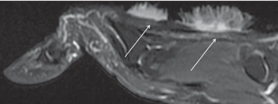
Radiol Bras. 2011 Jul/Ago;44(4):263–264 (Figure 2). Both lesions presented
interme-diate signal intensity on T1-weighted se-quences, a spiculated pattern with high sig-nal intensity on T2-weighted sequences (Figure 3A) and also a spiculated enhance-ment pattern on post-gadolinium T1-weighted sequences (Figure 3B). The ex-ternal contours of the lesions resembled the shape of a sea-urchin.
Bone invasion and distant lesions were not observed. Both lesions underwent en-block resection in the next day following the MRI study, with free surgical margins.
DISCUSSION
Verrucous carcinoma is an indolent variant of squamous cell carcinoma that is
the second most common type of skin can-cer(1). Verrucous carcinomas are not fre-quently seen on hands. As far as the authors are concerned, less than twenty cases have been reported up to the end of 2010. Much more frequently, verrucous carcinomas are seen on the feet (more than 90% of cases in the skin)(2). The “verrucous” denomina-tion is adopted because of the lesions’ simi-larity with viral warts(3). The etiology of such condition is still to be completely understood. Immunosuppression and greater Sun exposure increase the risk for disease development. Also, there are indi-cations of association with human papil-loma virus infection(4,5). Generally, verru-cous carcinomas are single lesions; and cases of multiple lesions on the hand had
not been reported yet. Rarely, such lesions produce metastases, but they may present deep tissue invasion, which is related to relapse(3). Although extensor tendons
inva-sion has not been observed in the present case, the observed perilesional edema might be implicated in the difficulty to extend the fingers and in the presence of pain.
The spiculated enhancement pattern on post-gadolinium T1-weighted sequences had already been described(6), which is also
confirmed on T2-weighted images. Was-serman et al.(7) have reported that such
image pattern resembled “teased cotton wool” and would be a result from inflam-matory infiltrate as a response to the stro-mal tissue of the tumor.
What the present report emphasizes is a particular tumor presentation, with typi-cal MRI findings and location which are useful in the development of the diagnos-tic rationale. However, the main role of the radiologist is still in the locoregional stag-ing of the disease.
REFERENCES
1. Amaral ACN, Azulay RD, Azulay DR. Neoplasias epiteliais. In: Azulay RD, Azulay DR, editores. Dermatologia. 4ª ed. Rio de Janeiro, RJ: Guana-bara Koogan; 2006. p. 510–26.
2. Kao GF, Graham JH, Helwig EB. Carcinoma cuniculatum (verrucous carcinoma of the skin): a clinicopathologic study of 46 cases with ultrastruc-tural observations. Cancer. 1982;49:2395–403.
3. Gertler R, Werber KD. Management of verrucous carcinoma of the hand: a case report. Int J Derma-tol. 2009;48:1233–5.
4. Noel JC, Peny MO, Detremmerie O, et al. Dem-onstration of human papillomavirus type 2 in a ver-rucous carcinoma of the foot. Dermatology. 1993; 187:58–61.
5. Zemtsov A, Koss W, Dixon L, et al. Anal verrucous carcinoma associated with human papilloma virus type 11: magnetic resonance imaging and flow cytometry evaluation. Arch Dermatol. 1992;128: 564–5.
6. Theodorou SJ, Theodorou DJ, Bona SJ, et al. Pri-mary squamous cell carcinoma: an incidental toe mass. AJR Am J Roentgenol. 2005;184(3 Suppl): 110–1.
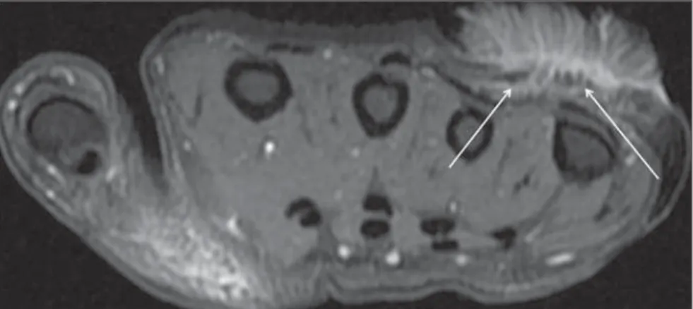
7. Wasserman PL, Taylor RC, Pinillia J, et al. Verru-cous carcinoma of the foot and enhancement assess-ment by MRI. Skeletal Radiol. 2009;38:393–5. Figure 2. Axial MRI T2-weighted image with fat saturation demonstrates the largest, noninvasive lesion
(arrows) in contact with the extensor tendons of the fourth and fifth fingers of the hand.
Figure 3. Coronal MRI T2-weighted (A) and T1-weighted, post-gadolinium fat saturated (B) images dem-onstrate a spiculated pattern (arrows) similar to a sea-urchin.