○ ○ ○ ○ ○ ○ ○ ○ ○ ○ ○ ○A BSTRA CT○ ○ ○ ○ ○ ○
Original Ar
ticle
○ ○ ○ ○ ○ ○ ○ ○ ○ ○I N TRO D U CTI O N○ ○ ○ ○ ○ ○ ○ ○ ○ ○
In the last decade, breast cancer has become the highest cause of morbidity and mortality among female malignancies, but cervical can-cer represents the leading cause of cancan-cer-re- cancer-re-lated deaths among women in developing coun-tries, including Brazil.1 Nearly 40,000 cases of cervical cancer arise among Brazilian women every year.2 Lopes et al., studying the behavior of Brazilian women in the light of cervical can-cer prevention, verified that most people had not undergone preventive examinations.3 Uter-ine cervical cancer and non-specific uterUter-ine can-cer combined are the second greatest underly-ing cause of death due to neoplasms for women aged 30-49 in the state of São Paulo.4
Evidence from laboratory and epidemio-logical studies has shown an association be-tween human papillomavirus (HPV) infection and both cervical cancer and pre-cancerous neoplasias.5,6 High-risk HPV types like HPV 16 and 18 have been strongly linked to cervi-cal carcinoma.7 In addition to HPV 16 being common in the general population, it remains among the most prevalent individual type in cervical neoplasias.8
High-risk HPVs can integrate into host cell DNA, and this is essential data regarding cancer development. Viral integration pro-motes the disruption of the HPV E2 gene leading to unregulated increases in the E6 and E7 proteins.9 These viral proteins of oncogenic HPVs inactivate the products of p53 and Rb tumor suppressor genes, respectively. The tumor suppressor gene functions include regu-lation of the cell cycle and the cellular response to DNA damage, initiation of DNA repair and
Rodrigues
• Ana Paula Terra Alvim de Salles Lopes
• André de Paula Fernandez
• Silvia Maria Baeta Cavalcanti
lesions, physical state of viral
DNA and changes in p53 gene
Department of Microbiology and Parasitology, Biomedical Institute,
Universidade Federal Fluminense, Niterói, Rio de Janeiro, Brazil
CONTEXT: Persistent infection with high risk human papillomavirus (HPV) has been linked to cervical carcinoma. Integration of viral DNA into host cell DNA is essential for this cancer development, pro-moting disruption of the HPV E2 gene, thus lead-ing to unregulated increases in E6 and E7 proteins and inactivating the products of p53 and Rb tumor suppressor genes.
OBJECTIVE: To investigate HPV 16 infection in cervi-cal lesions, physicervi-cal state of viral DNA and p53 gene alterations in a group of women attending a public health service.
DESIGN: Prospective, non-controlled, transversal study.
SETTING: Gynecological clinic of the School od Medi-cine, Universidade Federal Fluminense.
SAMPLE: 43 consective patients with cervical lesions referred to our service.
MAIN MEASUREMENTS: Cases were classified via cytology/histology as normal, HPV infection, con-dyloma, low-grade squamous intraepithelial lesion (LSIL), high-grade squamous intraepithelial lesion (HSIL) and carcinoma. HPV infection was studied via polymerase chain reaction (PCR) using two PCR primer sets, to determine DNA integration. p53 gene changes were investigated by single-strand conformation polymorphism (SSCP) analysis.
RESULTS: One normal case, 7 HPV infections, 6 con-dylomas, 7 LSIL, 14 HSIL and 8 cancers were found, with 95% positive for HPV genome when tested using both L1 and E6 primers. HPV 16 was most prevalent (73.1%). HPV 16 DNA was integrated within the host genome in 3 LSIL. One LSIL pro-gressed to HSIL by 13 months after first diagnosis. Among HPV 16-positive HSIL cases, 50% contained integrated viral DNA. HPV 16 E2 gene disruption was seen in 7 cancers (87.5%). Only smal-cell car-cinoma showed intact HPV 16 E2 gene. Abnor-mal p53 bands detected by PCR/SSCP were ob-served in 4 cases: 2 squamous carcinoma with parametrium (exon 8) and two cervical intra-epithelial neoplasia (CIN) III (exons 5 and 7). All cases presented HPV 16 E2 gene loss.
CONCLUSIONS: The sample had a high rate of high-risk HPV detected in benign and malignant lesions; high cervical cancer burden; HPV 16 DNA inte-gration in all except one case of cancer; p53 gene changes in CIN III and in invasive cancer cases associated with DNA integration.
KEY WORDS: HPV. Viral. DNA. Integration. Gene. Cervical. Lesions.
replication, induction of apoptosis and pro-motion of cell differentiation.10 Inactivation of the p53 tumor suppressor gene is correlated to a critical step in the development of many human cancers.11 It may result from a number of events including mutation of the p53 gene (with or without associated allelic deletions) and binding of the p53 to cellular or viral pro-teins. In cervical carcinoma, loss of p53 func-tion can occur by interacfunc-tion with E6 protein of high-risk HPV types.12
In this work, we conducted a study on a group of women referred to our public health service for assessment of cytological/histologi-cal abnormalities in genital lesions. We de-tected HPV type 16 infection, the physical status of viral DNA and p53 gene alterations.
○ ○ ○ ○ ○ ○ ○ ○ ○ ○ ○ ○ ○ ○M ETH O D S○ ○ ○ ○ ○ ○
We studied 43 consecutive women at-tended at Hospital Universitário Antônio Pedro of the Universidade Federal Fluminense, Rio de Janeiro, between April 2000 and June 2002. These women have litlle or no access to rou-tine, annual Papanicolaou exams in local serv-ices and were referred to our hospital for inves-tigation of different kinds of cervical lesions. Colpocytology test screening was performed at the first or subsequent visit to our clinic. The age range was 18-68 years with an average of 37.8 years, with a standard deviation of 11.9.
Pa-panicolaou exam, by three examiners per slide. For women with abnormal cervical cytology, biopsies were performed.
The cases were then classified by our Serv-ice as normal, HPV infection, low-grade squa-mous intraepithelial lesions (LSIL - CIN I, cervical intraepithelial neoplasia), high-grade squamous intraepithelial lesions (HSIL - CIN II and III), and carcinoma (in situ carcinoma, squamous invasive carcinoma, adenocarci-noma, small-cell carcinoma).
Samples were incubated for 4 hours at 50° C in 200 ml of digestion buffer (10 mM Tris-hydrochloric acid pH 8.3, 1 mM EDTApH 8.0, 0.5% Tween 20, proteinase K; final con-centration of 400 µg/ml). Later, they were extracted with phenol-chloroform-isoamyl alcohol (25:24:1). DNA was precipitated with one-tenth volume of 0.3 M sodium acetate and three volumes of 100% ice-cold ethanol, washed with 70% ethanol, air-dried and sus-pended in 50 µl of sterile water.
MY09/11 consensus primers, which am-plify 450-bp (base pair) DNA sequences within the L1 region of HPV, were used to detect generic HPV DNA. Amplification was carried out in 50 µl of reaction mixture (1 X
polymerase chain reaction [PCR] buffer, 200 mM dNTPs, 1.5 mM MgCl2, 50 pmol of each primer, 0.25 U unit of Taq polymerase, and 5 µl of sample) with 35 cycles of amplification. Each cycle included a dena-turation step at 94° C for 1 minute, an annealing step at 55° C for 2 minutes, and a chain elongation step at 72° C for 2 minutes using DNA Thermal Cycler (Perkin Elmer, CETUS). The beta-actin primers (0.1 pmol each), which amplify a 330-bp region of the human DNA, were used as an internal con-trol. Polymerase chain reaction products were analyzed on 1.3% agarose gel with ethidium bromide staining for visualization of DNA under ultraviolet light and their molecular weight was determined by comparison with a 100-bp DNA ladder.
Human papillomavirus typing was done by polymerase chain reaction amplification us-ing primers from the E6 gene DNA sequences of HPVs 6, 11, 16, 18, 31, 33, and 35. These primers yielded 230, 89, 134, 119, 97, 132 and 186-bp fragments, respectively.13 The PCR in-cluded steps at 94° C for 30 seconds, 60° C for 1 minute, and 72° C for 1 minute. Negative controls for background contamination did not
add to the DNA template. The polymerase chain reaction run was completed by extension for 10 minutes at 72° C.
HPV 16 E2 type-specific primers, which amplify 1026-bp fragments, were used to de-termine DNA integration. The following primers were used: sense 2810-5’ ATGAAAATGATAGTACAGAC-2819 and antisense 3836-5’ CCAGTAGACACT-GTAAATAG-3818.14 Absence of the E2 gene was considered to be a sign of E2 region dis-ruption. The PCR was done as above.
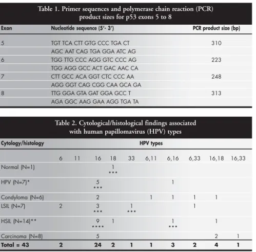
The primers used for polymerase chain reaction amplification of p53 gene exons 5 to 8 and resulting product sizes are given in Ta-ble 1. The PCR was carried out in a volume of 20 µl consisting of 1 X buffer, (Tris-HCl pH 8.0, 50 mM KCl), 50 µM dNTPs, 1.5 mM MgCl2,1 µM of each primer, 0.25 U unit of Taq polymerase, and 1 ml of sample. It was hot-started by the addition of the reaction mixture at 94° C, 35 cycles of 30 seconds at 94° C, 1 minute at 60° C, and 1 minute at 72° C. The polymerase chain reaction ampli-fication was completed by extension for 10 minutes at 72° C. The positive controls were DNA from tumor specimens, whereas nega-tive control for background contamination did not add to the DNA template. Extracted DNA from a benign wart was used as a normal con-trol. Polymerase chain reaction products were visualized by electrophoresis performed at room temperature for 3 hours at 30 mA. Sin-gle-strand DNA for sinSin-gle-strand conforma-tion polymorphism (SSCP) analysis was pro-duced by combining equal volumes of PCR product and formamide-loading buffer (95% formamide, 10 mM EDTA, 0.05% bromophenol blue, 0.05 xylene cyanol) and heating at 95°C, for 10 minutes. The reac-tion was left on ice until submitting to elec-trophoresis. Non-denaturing polyacrylamide gel (49:1 acrylamide-bisacrylamide ratio) was used. After the run, the gel was fixed in 7.5% acetic acid, washed, and silver-stained. Briefly, the gel was soaked in 10% ethanol and 1% nitric acid, and immersed in impregnation so-lution, visualized in developing soso-lution, and fixed in 10% acetic acid.
○ ○ ○ ○ ○ ○ ○ ○ ○ ○ ○ ○ ○ ○ ○RESU LTS○ ○ ○ ○ ○
In accordance with the cytological/histo-logical diagnosis, we found one normal case, seven HPV infections, six condylomas, seven lLSIL, fourteen HSIL and eight carcinomas. The average age of patients with malignant lesions was about 45 years. Among the 43 women, six (14%) were also infected with the
Table 1. Primer sequences and polymerase chain reaction (PCR) product sizes for p53 exons 5 to 8
Exon Nucleotide sequence (5’- 3’) PCR product size (bp)
5 TGT TCA CTT GTG CCC TGA CT 310
AGC AAT CAG TGA GGA ATC AG
6 TGG TTG CCC AGG GTC CCC AG 223
TGG AGG GCC ACT GAC AAC CA
7 CTT GCC ACA GGT CTC CCC AA 248
AGG GGT CAG CGG CAA GCA GA
8 TTG GGA GTA GAT GGA GCC T 313
AGA GGC AAG GAA AGG TGA TA
Table 2. Cytological/histological findings associated with human papillomavirus (HPV) types
Cytology/histology HPV types
6 11 16 18 33 6,11 6,16 6,33 16,18 16,33
Normal (N=1) 1
***
HPV (N=7)* 5 1
***
Condyloma (N=6) 2 1 1 1 1
LSIL (N=7) 2 3 1 1
*** ***
HSIL (N=14)** 9 1 1 1
**** ***
Carcinoma (N=8) 5 2 1
Total = 43 2 24 2 1 1 3 2 4 1
human immunodeficiency virus (HIV). The results are summarized in Table 2.
Ninety-five percent of the patients (41) were positive for the presence of HPV genome when they were tested using both L1 and E6 primers. Out of 43 samples, 67.4% (29) were positive for at least one virus type and 25.5% (11) presented infections by multiple types. Out of six cases of condyloma, four were associated with squamous intraepithelial lesion and all of them were positive to HPV. The study revealed that fourteen women had no complaint but had several kinds of cervical lesions including three HSIL and one in situ
carcinoma. All except one were HPV-infected. HPV 16 was the most prevalent type (60.4%) in single infections and both single and mul-tiple infections (73.1%). Thus, low-risk types were detected in 18.6%, HPV 18 in 14% and HPV type 33 in 9% of the cases. HPV 16 was found in 100% of the carcinomas alone or in association with type 18 or 33.
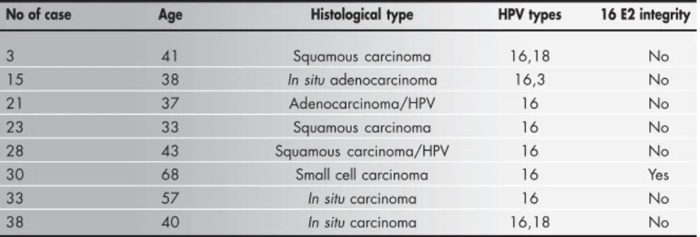
The HPV 16 DNA appeared also to be integrated within the host genome in three out of seven low-grade squamous intrae-pithelial lesions. However, one of these pa-tients had progressed to HSIL by thirteen months after the first diagnosis. Among high-grade squamous intraepithelial lesion cases positive to HPV 16, 50% (7/14) contained viral DNA in the integrated state. Disruption of the 16 E2 gene was seen in all seven cancer cases (87.5%) whereas only the small-cell carcinoma showed intact 16 E2 gene (Table 3).
Abnormal bands of p53 gene were ob-served in four cases: two squamous carcinoma with parametrium involvement (exon 8) and two CINIII (exons 5 and 7, respectively, Figu-re 1). One carcinoma had poor prognosis af-ter 24 months. All cases presented loss of the HPV 16 E2 gene.
The persistence of HPV 16 was seen in four out of five patients treated for CINIII or in situ carcinoma following conization. To verify persistent HPV infection, the PCR test was done when the patients came back for routine follow-up three months after surgery. Only one in situ carcinoma case had the vi-ral clearance demonstrated. One case (CIN III) infected with HPV 16 and 18 had their HPV 16 but not HPV 18 cleared. All pa-tients had their borderline tissue freed of ab-normal cells. After two years, three out of four HPV 16 infected patients did not present recurrence of the lesions. However, one HIV seropositive woman who presented CIN III progressed to invasive carcinoma 11 months after surgery.
○ ○ ○ ○ ○ ○ ○ ○ ○ ○ ○ ○ D I SCU SSI O N○ ○ ○ ○ ○ ○ ○ ○
The main findings of this prospective study were: HPV DNA identified in 95% of all cases; a high rate (86%) of high-risk HPV detected in benign as well as in malignant lesions; high burden of cervical cancer; HPV 16 DNA inte-gration in all except one case of cancer; p53 gene changes in CIN III; and invasive cancer cases associated with DNA integration.
Infection with high-risk human papillo-mavirus types is frequent among sexually ac-tive women, with incidence ranging from 15 to 40%.2 However, the majority of the infections are found to be transient because most individuals develop a specific immune response.15 When the infection persists, pre-cancer lesions may develop. About 1% of the general population present genital warts and 4% of all women have cervical precan-cerous lesions.16
In the population studied, human papillomavirus presence is associated with 85,7% high-grade squamous intraepithelial le-sion and 100% carcinoma. The mean age of patients with malignant lesions was about 45 years, considered low in comparison with de-veloped countries. In spite of the random fea-ture in the enrollment of the patients, the women in this sample came from lower-income classes. Moreover, the Hospital Universitário Antônio Pedro is the main general hospital in a large densely populated geographical area. The lack of public healthcare clinics for providing appropriate attendance of severe dysplasia among women and also for its adequate treat-ment oblige this hospital to receive a high number of these cases. Therefore, these are women with little or no access to annual Pa-panicolaou exams that would detect inflamma-tory or pre-neoplastic lesions (rather than neoplastic lesions) and prevent their malignant evolution. This, in association with other co-factors, may contribute to the disproportion-ately high rate of pre-cancerous and cancerous lesions observed in our sample. Lack of access
to healthcare, poor nutrition, multiple parities, human immunodeficiency virus (HIV) and other sexually transmitted diseases (STD) also increase the susceptibility to cervical carcino-genesis in our population.
Another remarkable finding was the high rate of HIV-positive women who were infected with human papillomavirus. It is an indica-tion of the misinformaindica-tion about health moni-toring among poor Brazilian people. Hetero-sexual transmission without using a condom is the main route to female human immuno-deficiency virus infection. The presence of other sexually transmitted diseases, such as human papillomavirus infection, promotes breaks in the genital tract and increases the susceptibility to acquiring the human immu-nodeficiency virus.17
In an extensive molecular epidemiological study, Villa et al.18 found that infections with non-European variants of HPV 16 and 18 had a general tendency to persist more frequently and to be more associated with pre-invasive le-sions. In their analysis, 33% (15/46) women infected with European variants were non-white
Table 3. Human papillomavirus (HPV) detection in carcinoma cases and Type 16 E2 integration
No of case Age Histological type HPV types 16 E2 integrity
3 41 Squamous carcinoma 16,18 No
15 38 In situ adenocarcinoma 16,3 No
21 37 Adenocarcinoma/HPV 16 No
23 33 Squamous carcinoma 16 No
28 43 Squamous carcinoma/HPV 16 No
30 68 Small cell carcinoma 16 Yes
33 57 In situ carcinoma 16 No
38 40 In situ carcinoma 16,18 No
Figure 1. Screening of p53 mutation on exon 5 by
polymer-ase chain reaction/single-strand conformation polymor-phism. Number 1: normal run. Number 2: abnormal
band. (CIN III).
1. Pisani P, Parkin DM, Bray F, Ferlay J. Estimates of the worlwide mortality from 25 cancers in 1990. Int J Cancer 1999;83(1):18-29. 2. Noronha V, Mello W, Villa L, et al. Papilomavírus humano associado a lesões de cérvice uterina. Rev Soc Bras Med Trop 1999; 32(3):235-40.
3. Lopes ER, Rebelo MS, Abreu E, Silva VLC, Eisenberg ALA, Lavor MF. Comportamento da população brasileira feminina em relação ao cancer cérvico- uterino. J Bras Ginecol 1995;105(11/12):505-15.
4. Haddad N, Silva MB. 2001 Mortalidade por neoplasmas em mulheres em idade reprodutiva -15 a 49 anos - no estado de São Paulo, Brasil, de 1991 a 1995. Rev Assoc Med Bras 2001;47(3):221-30. 5. Muñoz N. Human papillomavirus and cervical cancer. In:
Franco E, Monsonego J, eds. New development in cervical can-cer screening and prevention. Cambridge: Blackwell Science, 1997.p.3-13.
6. Schiffman MH, Bauer HM, Hoover RN, et al. Epidemiologic evidence showing that human papillomavirus infection causes most cervical intraepithelial neoplasia. J Natl Cancer Inst 1993;85(12):958-64.
7. Ho GY, Bierman R, Beardsley L, Chang CJ, Burk RD. Natural history of cervicovaginal papillomavirus infection in young women. N Engl J Med 1998;338(7):423-8.
○ ○ ○ ○ ○ ○ ○ ○ ○ ○ ○ ○ ○ ○ ○ ○ ○ ○ ○ ○ ○ ○ ○ ○ ○ ○ ○ ○ ○ ○ ○ ○ ○ ○ ○ ○ ○ ○ ○ ○ ○ ○ ○ ○ ○ ○ ○ ○ ○ ○ ○ ○ ○ ○ ○ ○ REFEREN CES○ ○ ○ ○ ○ ○ ○ ○
8. Bosch FX, Manos MM, Muñoz N, et al. Prevalence of human papillomavirus in cervical cancer: a worldwide perspective. In-ternational biological study on cervical cancer (IBSCC) Study Group. J Natl Cancer Inst 1995;87(11):796-802. 9. Park TW, Fugiwara H, Wright TC. Molecular biology of cervical
cancer and its precursors. Cancer 1995;76(Suppl 10):1902-13. 10. Lane DP. Cancer. p53, guardian of the genome. Nature
1992;358(6381):15-6.
11. Greenblatt MS, Bennett WP, Hollstein M, Harris CC. Muta-tions in the p53 tumour suppressor gene: clues to cancer etiology and molecular pathogenesis. Cancer Res 1994;54(18):4855-78. 12. Bremer GL, Tieboschb AT, van der Putten HW, de Haan J, Arends JW. p53 tumor suppressor gene protein expression in cervical cancer: relationship to prognosis. Eur J Obstet Gynecol Reprod Biol 1995;63(1):55-9.
13. Tinos Y, Manos M. Detection and typing of genital human papillomaviruses. In: Innis M, Gelfand D, Sninsky J, White T, eds. PCR protocols: a guide to methods and application. San Diego: Academic Press 1990.p.356-67.
14. Park JS, Hwang ES, Park SN, Ahn HK, Um SJ, Kim CJ, Kim SJ, Namkoong SE. Physical status and expression of HPV genes in cervical cancers. Gynecol Oncol 1997;65(1):121-9. 15. Hildesheim A, Schiffman M, Bromley C, et al. Human
papillomavirus type 16 variants and risk of cervical cancer. J Natl Cancer Inst 2001;93(4):315-8.
16. Mougin C, Dalstein V, Prétet JL, Gay C, Schaal JP, Riethmuller D. Epidémiologie des infections cervicales à papillomavirus. Recent Knowledge. Presse Med 2001;30(20):1017-23. 17. Trindade MP, Schiavo MR. Comportamento sexual das mulheres
em relação ao HIVA,AIDS. DST. J Bras Doenças Sex Transm 2001;13(5):17-22.
18. Villa LL, Sichero L, Rahal P, et al. Molecular variants of human papillomavirus types 16 and 18 preferentially associated with cervical neoplasia. J Gen Virol 2000;81(Pt 12):2959-68 19. Das BC, Sharma JK, Gopalakrishna V, Luthra UK. Analysis by
polymerase chain reaction of the physical state of human papillomavirus type 16 DNA in cervical preneoplastic and neoplastic lesions. J Gen Virol 1992;73(Pt 9):2327-36. 20. Klaes R, Woerner SM, Ridder R, et al. Detection of high-risk
cervical intraepithelial neoplasia and cervical cancer by amplifi-cation of transcripts derived from integrated papillomavirus oncogenes. Cancer Res 1999;59(24):6132-6.
21. Nakagawa S, Yoshikawa H, Jimbo H, et al. Elderly Japanese women with cervical carcinoma show higher proportions of both intermediate-risk human papillomavirus types and p53 muta-tions. Br J Cancer 1999;79(7-8):1139-44.
as compared to 58% (11/19) infected with non-European variants in the same ethnic category. It is worth noting that out of the eight cancer cases in our sample, seven (87.5%) were non-white women. It may be possible that, in addi-tion to the co-factors above menaddi-tioned, European variants of HPV 16 harbored in non-white patients have contributed to increasing the rate of cervical cancer in this study.
In most cervical immortalized cells, high-risk HPV DNA often integrates into the cellu-lar genome. The integration is processed throughout the E2 gene by disrupting some part of it and causing overexpression of the E6 and E7 proteins. This results in the loss of anti-oncogenic function in p53 and Rb proteins. On the basis of these events, we investigated the physical state of HPV 16 DNA using the polymerase chain reaction assay. The result was no different from other reports.9,19,20 We found loss of the E2 gene in 42.8% of low-grade sq-uamous intraepithelial lesion and in 50% of high-grade squamous intraepithelial lesions. One low-grade squamous intraepithelial lesion case had progressed to high-grade squamous intraepithelial lesion by thirteen months after
the first diagnosis. These data support the hy-pothesis that integration mainly happens at an early stage in the development of cervical neo-plasia. It is possible that the increased expres-sion of the E6 and E7 proteins may result in a selective growth advantage over cells harboring individualized HPV 16 DNA.
In a study on Korean women, 92.2% of HPV 16 cancers revealed pure or mixed forms of integrated DNA.14 We found a lack of E2 gene in 87.5% (7/8) of the carcinomas. The intact E2 gene form was only seen in small-cell carcinoma. This has also been observed by other researchers.14
We detected p53 gene changes in four cases (14% of the CINIII and 25% of the in situ carcinoma). All of them contained inte-grated HPV 16 DNA, thus showing that hu-man papillomavirus infection and p53 gene alterations are not excluding events for cancer development. In a study to determine p53 mutation by polymerase chain reaction/sin-gle-strand conformation polymorphism and sequencing techniques, point mutation was detected in 11% (5/46) of HPV-infected cer-vical carcinoma and no mutation was found
in two HPV-negative cases.21 The rapid progress of squamous invasive cancer observed in our sample may suggest a synergic effect of the two events. However, the low frequency of the p53 gene changes (19% CINIII/in situ
carcinoma) in comparison with the high fre-quency of high-risk human papillomavirus infection (85.7% CIN III/in situ carcinoma) strengthens the view that p53 inactivation by human papillomavirus proteins plays a major role in the pathogenesis of cervical cancer.
Human papillomavirus detection follow-ing conization even when CINIII and in situ
carcinoma were successfully treated is more likely to result in viral persistence than a new infection. However, the residual human papillomavirus did not promote recurrent dis-ease over a two-year period in most of the women surgically treated. The obvious im-mune failure of one HIV-seropositive patient allowed the recurrence of the disease and its progression to invasive cancer.
CONTEXTO: A infecção persistente por papilo-mavírus humano (HPV) de alto risco tem sido associada ao carcinoma cervical. Uma fase obrigatória no desenvolvimento deste câncer é a integração do DNA viral ao DNA das células hospedeiras. A integração viral promove a degradação do gene E2 do papilomavírus humano e leva a um aumento descontrolado das proteínas E6 e E7. Estas proteínas virais de HPVs oncogênicos inativam os produtos dos genes supressores de tumor p53 e Rb. Inativação da proteína p53 é o resultado de vários fatores incluindo mutação no gene para p53.
OBJETIVO: Investigar lesões cervicais associadas à infecção por papilomavírus humano, o es-tado físico do DNA viral e alterações no gene para p53 em um grupo de mulheres atendi-das em um serviço público de saúde. TIPO DE ESTUDO: prospectivo,
não-contro-lado.
LOCAL: Serviço de Patologia Cervical, Ambula-tório de Ginecologia da Faculdade de Medi-cina, Universidade Federal Fluminense. PARTICIPANTES: 43 mulheres atendidas em
consulta ambulatorial de rotina para preven-ção às lesões cervicais.
VARIÁVEIS ESTUDADAS: Pela citologia/his-tologia os casos foram classificados como nor-mal, infecção por papilomavírus humano, condiloma, lesão cervical de baixo graus, lesão cervical de alto grau e carcinoma. A presença de papilomavírus humano foi detectada por reação de polimerase em cadeia (PCR) utili-zando-se dois pares de oligonucleotídeos. Para determinar a integração do DNA viral, usa-ram-se oligonucleotídeos específicos para o gene E2 do tipo HPV 16 que amplificam um fragmento de 1.026 pares de base. Alterações no gene para p53 foram investigadas por
○ ○ ○ ○ ○ ○ ○ ○ ○ ○ ○ ○ ○ ○ ○ ○ ○ ○ ○ ○ ○ ○ ○ ○ ○ ○ ○ ○ ○ ○ ○ ○ ○ ○ ○ ○RESU M O○ ○ ○ ○ ○ ○
Ledy do Horto dos Santos Oliveira, PhD in Biological Sciences (Microbiology), Universidade Federal Fluminense, Niterói, Rio de Janeiro, Brazil.
Eliane de Vasconcelos Machado Rodrigues, MD. Gynecologist, School of Medicine, Universidade Federal Fluminense, Niterói, Rio de Janeiro, Brazil.
Ana Paula Terra Alvim de Salles Lopes, CAPES grant-holder, MSc student in Experimental Pathology, Universidade Federal Fluminense, Niterói, Rio de Janeiro, Brazil.
André de Paula Fernandez, CNPq scientific initiation grant-holder, student at the School of Medicine, Universidade Federal Fluminense, Niterói, Rio de Janeiro, Brazil. Silvia Maria Baeta Cavalcanti, PhD in Biological
Sci-ences (Microbiology), Universidade Federal Fluminense, Niterói, Rio de Janeiro, Brazil.
Sources of funding: CNPq (grant no. 470257/01-6) and FAPERJ (grant no. E-26/170.137/2001)
Conflict of interest: Not declared Date of first submission: August 27, 2002 Last received: November 8, 2002 Accepted: December 16, 2002
Address for correspondence Ledy do Horto dos Santos Oliveira
Universidade Federal Fluminense, Instituto Biomédico Departamento de Microbiologia e Parasitologia Rua Professor Ernani Melo, 101
Niterói/RJ – Brasil – CEP 24210-130 Tel. (+55 21) 2620-0623 E-mail: [email protected]
COPYRIGHT©2003, Associação Paulista de Medicina
○ ○ ○ ○ ○ ○Pu b l i sh i n g i n f o r m a t i o n○ ○ ○ ○ ○ ○ ○ ○ ○ ○ ○ ○ ○ ○
polimorfismo conformacional da fita simples. RESULTADOS: Foi detectadoum caso de cérvice normal, sete infecções por papilomavírus hu-mano, seis condilomas, sete lesões cervicais de baixo graus, 14 de alto grau e oito carcino-mas. O estudo revelou que 14 mulheres sem queixas de doença genital eram portadoras de lesões de vários tipos incluindo três lesões cervicais de alto grau e um caso de carcinoma
in situ. 95% das pacientes foram positivas para a presença do genoma de HPV quando testa-das para os genes L1 e E6, com prevalência do HPV tipo 16 (73.1%). Detectamos DNA do HPV 16 integrado ao genoma hospedeiro em três dos sete casos de lesões cervicais de baixo grau. Entretanto, um destes pacientes progre-diu a lesão cervical de alto grau 13 meses após o primeiro diagnóstico. Entre os casos de le-são cervical de alto grau positivos para HPV 16, 50% continham o DNA viral no estado integrado foi encontrado. A falta do gene E2 do HPV 16 foi observada em sete dos carci-nomas (87.5%) e somente no carcinoma de células pequenas foi detectado o gene E2. Ban-das anormais do gene p53 detectaBan-das por PCR/ SSCP foram observadas em quatro casos: dois carcinomas escamosos com envolvimento do paramétrio (exon 8) e dois casos de NIC III (exons 5 e 7, respectivamente). Todos os casos apresentaram perda do gene E2 do HPV 16. CONCLUSÕES: A amostra analisada tinha alta taxa de HPV de alto risco detectada tanto em lesões benignas como em lesões malig-nas; alto índice de câncer cervical; integração do DNA do HPV 16 em quase todos os ca-sos de câncer com uma exceção; alterações no gene para p53 em NIC III e em câncer invasivo associadas à integração do DNA.
PALAVRAS-CHAVE: HPV. Integração. DNA