SUMMARY
Objective: To detect the frequency and subtypes of HPV in the uterine cervix of HIV-infected women. Methods: Sample consisted of 288 HIV-infected women, recruited from the public health system of ive cities of Minas Gerais, Brazil. Women were seen from August 2003 to August 2008. Cervical samples were collected for cytological analysis and for HPV DNA detection, using polymerase chain reaction (PCR). HPV DNA was classi-ied according to its oncogenic potential in low risk (types 6, 11) and high risk (types 16, 18, 31, 33, 35). Colposcopy was performed, followed by cervical biopsy when necessary. Categorical variables were compared using the Chi-squared test, with a signiicance lev-el established at the 5% levlev-el. Results: HPV prevalence was 78.8%. Most frequent geno-types were HPV-6 (63.9%) and HPV-16 (48.5%). High-risk HPV were observed in 70.5% of the women; low-risk in 71.4%; both high and low-risk HPV were detected in 55.1% of the patients. Multiple HPV genotypes were detected in 64.8% of the patients; two genotypes in 23.8%, and three in 18.9%. Conclusion: HPV prevalence was high among HIV-infected women. Multiple HPV genotypes were common in samples from the uterine cervix of HIV-infected women.
Keywords: DNA probes, HPV; HIV; polymerase chain reaction; cervix uteri; papillomavirus infections, prevalence.
RESUMO
Prevalência e multiplicidade do HPV em mulheres infectadas pelo HIV em Minas Gerais
Objetivo: Detectar a frequência e os subtipos do HPV na cérvice uterina de mulheres infec-tadas pelo HIV. Métodos: A amostra era composta por 288 mulheres infectadas pelo HIV, recrutadas do sistema público de saúde de cinco cidades de Minas Gerais, Brasil. As mulheres foram avaliadas de agosto de 2003 a agosto de 2008. Amostras cervicais foram coletadas para análise citológica e para detecção do HPV DNA, usando a reação em cadeia de polimerase (PCR). O HPV DNA foi classiicado de acordo com seu potencial oncogênico em baixo risco (tipos 6,11) e alto risco (tipos 16, 18, 31, 33, 35). Foi realizada colposcopia, seguida de biópsia cervical, quando indicada. Variáveis categóricas foram comparadas usando o teste do quiqua-drado, com nível de signiicância estabelecido de 5%. Resultados: A prevalência do HPV foi 78,8%. Os genótipos mais frequentes foram HPV-6 (63,9%) e HPV-16 (48,5%). Genótipos de HPV de alto risco foram observados em 70,5% das mulheres; de baixo risco em 71,4%; HPV de alto e baixo risco foram detectados em 55,1% das pacientes. Múltiplos genótipos de HPV foram detectados em 64,8% das pacientes; dois genótipos em 23,8%, e três em 18,9%. Conclu-são: A prevalência do HPV foi alta entre mulheres infectadas pelo HIV. Múltiplos genótipos de HPV foram comuns em amostras da cérvice uterina destas mulheres.
Unitermos: Sondas DNA HPV; HIV; reação em cadeia da polimerase; colo do útero; infec- HIV; reação em cadeia da polimerase; colo do útero; infec-HIV; reação em cadeia da polimerase; colo do útero; infec-ções por papillomavirus; prevalência.
Prevalence and multiplicity of HPV in HIV women in Minas Gerais,
Brazil
CHRISTINE MIRANDA CORRÊA1, NARA CHARTUNI PEREIRA TEIXEIRA2, ANGELA CRISTINA LABANCADE ARAÚJO3, NARADE OLIVEIRA CARVALHO4, DORA MENDEZ DEL CASTILLO5, RACHEL REZENDE CAMPOS6, ISAURA VARGAS OLIVEIRA7, ANTONIO RAFAEL ALVES8, ANA FLÁVIADE MOURA FRANÇA9, VICTOR HUGO MELO10
1 M.Sc. in Women’s Health; Coordinator of the Program Atenção Integral à Saúde da Mulher de Três Rios, Três Rios, RJ, Brazil 2 M.Sc. in Woman Health; Physician at Belo Horizonte City Hall, Belo Horizonte, MG, Brazil
3 Ph.D. in Medicine; Physician at Belo Horizonte City Hall, Belo Horizonte, MG, Brazil
4 M.Sc. in Pathology; Biologist at the Genetics and Molecular Biology Laboratory NUPAD, Medical School, Universidade Federal de Minas Gerais (UFMG), Belo Horizonte, MG, Brazil 5 Specialization in Genetics; Coordinator of the Genetics and Molecular Biology Laboratory NUPAD, Medical School, UFMG, Belo Horizonte, MG, Brazil
6 M.Sc. in Women’s Health; Physician at Betim City Hall, Betim, MG, Brazil
7 Specialization in Gynecolgy and Obstetrics; Physician at Barbacena City Hall, Barbacena, MG, Brazil
8 Specialization in Gynecolgy and Obstetrics; Physician at Conselheiro Lafaiete City Hall, Conselheiro Lafaiete, MG, Brazil 9 Specialization in Gynecolgy and Obstetrics; Physician at Divinópolis City Hall, Divinópolis, MG, Brazil
10 Ph.D. in Medicine; Associate Professor, Medical School, UFMG, Belo Horizonte, MG, Brazil
Study conducted at Medical School, Hospital das Clínicas, Universidade Federal de Minas Gerais, Belo Horizonte, MG, Brazil
Submitted on: 12/18/2010 Approved on: 05/19/2011
Financial Support: Fundação de Amparo à Pesquisa do Estado de Minas Gerais (FAPEMIG)
Correspondence to: Victor Hugo Melo Av. Alfredo Balena, 190, 2o andar
Belo Horizonte, MG, Brazil CEP: 30130-100 [email protected]
Conlict of interest: None.
INTRODUCTION
he association between cervical cancer and human im-munodeiciency virus (HIV) is well established1,2. Several studies have shown that HPV infection is signiicantly more common among HIV positive women compared to HIV negative women3-10.
he association between cervical cancer and human papillomavirus (HPV) has also been demonstrated, but the potentially modifying role of HIV on the HPV/cer-vical intraepithelial neoplasia association is still under investigation11. Current data suggest that HIV infection increases HPV persistence, with increased risk for in-traepithelial cervical lesions2,4.
Published data strongly suggest that HIV-infected women are more likely to have HPV DNA, as well as mul-tiple viral types and a higher frequency of the oncogenic types6-8. Similar indings have been reported in diferent regions of Brazil9,12-14 and HPV 16 was the most frequent oncogenic type7-9.
Polimerase chain reaction (PCR) has the highest sen-sitivity for detecting HPV DNA genotypes and discrimi-nates multiples infections better than other biomolecu-lar methods. It also has good speciicity and results are quickly available2.
he aim of this study was to estimate the prevalence of HPV DNA infection in HIV-infected women from dif-ferent cities. We also aimed to investigate the prevalence of HPV genotypes, as well as the frequency of multiple types of HPV.
METHODS
PATIENTS
Women attending a Reference and Training Center on Infectious and Parasitic Diseases in Belo Horizonte (Hospital das Clínicas, Universidade Federal de Minas Gerais - UFMG) and public health units of general gyne-cological care of four other cities of Minas Gerais State (Betim, Barbacena, Conselheiro Lafaiete and Divinópolis) were randomly chosen to be enrolled in the study. his study was conducted from August 2003 to August 2008.
A standardized questionnaire was used for collecting demographic, social, and behavioral characteristics of the sample, as well as for obtaining the medical history. Gynecological exam was performed and cervical samples were collected for Pap smear, as well as for HPV-DNA, by doctors from the group study to guarantee a standard-ized collection. Blood samples were collected to obtain T CD4+ cells count and HIV viral load. Colposcopy, bi-opsy and adequate treatment of cervical lesions were per-formed as part of the routine medical care.
Inclusion criteria were: HIV infection diagnosed by ELISA and conirmed by indirect immunoluores-cence or western blot, with or without AIDS; age ≥ 18 years; consent to participate (signature of the approved
informed consent form). Exclusion criteria were: diicul-ties in obtaining information (language barrier, disori-entation); non-analyzable samples; pregnancy; history of hysterectomy.
he study protocol was approved by the UFMG Re-search Ethics Committee.
PROCEDURES
Cervical samples for DNA-HPV exams were obtained us-ing an Ayre’s spatula, and transferred to a sterile tube taining 2 mL of saline solution (0.09%). he tube was con-ditioned in a special box with cooled gel. he samples were sent to a reference laboratory (Núcleo de Ações e Pesquisas em Apoio Diagnóstico, Medical School, UFMG), within 24 hours of the procedure.
DNA was extracted using Chelex 100 chelating resin (BioRad), according to the manufacturer’s protocol15. Spe-ciic primers for HPV DNA types 6, 11, 16, 18, 31, 33 and 35 were tested.
POLYMERASECHAINREACTIONANDANALYSISCRITERIA
In order to control for DNA quality, the globin gene was ampliied in all samples16. Globin-negative samples were excluded from the study. HPV detection by PCR was car-ried out in a nested-PCR system, using MY09/1117 and GP5+/6+18 primers. Positive samples were tested with spe-ciic primers for HPV DNA types 6, 11, 16, 18, 31, 33 and 35, through independent reactions15. All products were sub-mitted to agarose gel electrophoresis 2%, treated with ethidium bromide and analyzed under UV light. DNA was classiied according to its oncogenic potential in low risk (6, 11) and high risk (16, 18, 31, 33 and 35).
STATISTICALANALYSIS
Data were transferred to a specially designed database us-ing Excel 2003. Demographic and social variables included were: age (in years) at enrollment; age of irst sexual inter-course; lifetime number of sexual partners; marital status; working status; smoking; condom use; route of transmis-sion; and drug addiction. Clinical and laboratory variables were: CDC classiication (1993)19; antiretroviral regimen use; colposcopy; CD4+ T-lymphocytes cell count; HIV-1 viral load. Frequency and type of HPV genotypes (6, 11, 16, 18, 31, 33 and 35) were also analyzed.
RESULTS
he sample consisted of 288 women from ive diferent cit-ies: A (n = 152; 52.8%); B (n = 36; 12.5%); C (n = 20; 6.9%); D (n = 44; 15.3%); and E (n = 36; 12.5%). As can be seen in Figure 1, general prevalence of HPV infection was high (78.8%) among these HIV-infected patients. here was no signiicant diference among HPV prevalence of women from these diferent cities (p = 0.19).
Median age at enrollment was 35 years; median age of irst sexual intercourse was 17 years; median number of lifetime sexual partners was three. Regarding the clinical and behavioral characteristics, most participants were in a stable union (44.1%), worked at home (51.7%) and were infected through the sexual route (92.0%). Few women smoked (31.3%), used condom (42.7%) and were addicted to drugs (0.7%). Half of them had AIDS19 and 67.7% were using antiretroviral drugs. Normal colposcopy was found in 63.2% of them. Table 1 summarizes some of the clinical and behavioral characteristics of these women.
It was detected that 151 patients presented a viral load higher than the established cut-of value of 400 copies/mL (minimum limit of detection of RNA HIV at the time of the study). Among them, in 123 (81.5%) was detected the DNA HPV. On the other hand, 119 patients had undetect-ed viral load (≤ 400 copies/mL) and 90 (75.6%) of them presented the DNA HPV. he diferences were not signii-cant (p = 0,2441).
Furthermore, a cut in the T CD4+ cell count in 200 cells/mm³ was made based on the CDC classiication of AIDS and non-AIDS19. Amongst 45 patients presenting T CD4+ cell count < 200 cells/mm³, 37 (82.2%) had the
Figure 1 – DNA-HPV detection among women from ive
different cities in Minas Gerais, Brazil.
Clinical and behavioral characteristics
City A (n = 152)
City B (n = 36)
City C (n = 20)
City D (n = 44)
City E (n = 36)
Overall
(n = 288) p*
n (%) n (%) n (%) n (%) n (%) n (%)
CDC classiication 0.46
AIDS 74 (48.7) 17 (47.2) 13 (65) 21 (47.7) 19 (52.8) 144 (50)
Not AIDS 75 (49.3) 19 (52.8) 5 (25) 20 (45.5) 17 (47.2) 136 (47.2)
Missing 3 (2) 0 (0) 2 (10) 3 (6.8) 0 (0) 8 (2.8)
Antiretroviral drugs 0.67
Use 101 (66.4) 22 (61.1) 14 (70) 32 (72.7) 26 (72.2) 195 (67.7)
Not use 51 (33.6) 14 (38.9) 5 (25) 11 (25) 10 (27.8) 91 (31.6)
Missing 0 (0) 0 (0) 1 (5) 1 (2.3) 0 (0) 2 (0.7)
Colposcopy 0.68
Normal 99 (65.1) 19 (52.8) 12 (60) 30 (68.2) 22 (61.1) 182 (63.2)
Abnormal 50 (32.9) 16 (44.4) 8 (40) 14 (31.8) 13 (36.1) 101 (35.1)
Missing 3 (2) 1 (2.8) 0 (0) 0 (0) 1 (2.8) 5 (1.7)
Condom 0.23
Use 60 (39.5) 13 (36.1) 7 (35) 22 (50) 21 (58.3) 123 (42.7)
Not use 84 (55.3) 23 (63.9) 13 (65) 22 (50) 15 (41.7) 157 (54.5)
Missing 8 (5.3) 0 (0) 0 (0) 0 (0) 0 (0) 8 (2.8)
Route of HIV acquisition 0.23
Sexual 140 (92.1) 31 (86.1) 19 (95) 39 (88.6) 36 (100) 265 (92)
Blood 0 (0) 1 (2.8) 0 (0) 1 (2.3) 0 (0) 2 (0.7)
Missing 12 (7.9) 4 (11.1) 1 (5) 4 (9.1) 0 (0) 21 (7.3)
*X² test (4 df).
Table 1 – Clinical and behavioral characteristics of women
City A (n = 152)
City B (n = 36)
City C (n = 20)
City D (n = 44)
City E (n = 36)
Total (n = 288)
Negative 23.0% 13.9% 25.0% 11.4% 30.6% 21.2%
Positive 77.0% 86.1% 75.0% 88.6% 69.4% 78.8% 0
20 40
Heterogeneity: X2 = 6.07 (4df). p = 0.19 60
80 %
DNA HPV. On the other hand, 226 patients had T CD4+ cell count ≥ 200 cells/mm³ and 177 (78.3%) of them pre-sented the DNA HPV. he diferences were not signiicant either (p = 0.5574).
High-risk HPV (patient showing at least one high-risk HPV type) was observed in 70.5% (160/288) of pa-tients; low-risk HPV (patient showing at least one low-risk HPV type) was found in 71.4% (162/288) of women; and both high and low-risk HPV were detected in 55.1% (125/288) of them. he prevalence of HPV was higher (61.7%) in patients who had three or more sexual partners. Nevertheless, this result was not signiicant (p = 0.8431).
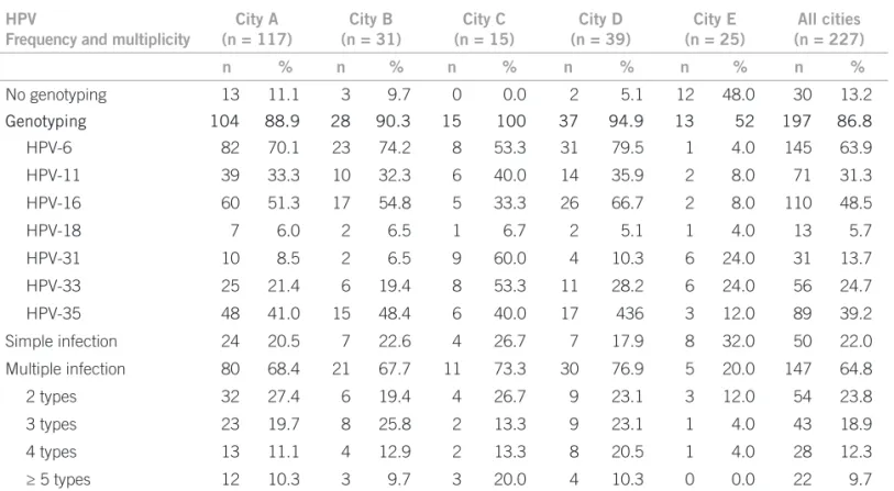
Table 2 displays the prevalence of each genotype as well as the proportion of women with multiple HPV geno-types. Most prevalent genotypes were HPV-6 (63.9%) and HPV-16 (48.5%). Diferences emerged among cities. HPV-31 and HPV-33 genotypes were the most prevalent in City C (60% and 53.3%) and City E (24% for both). Multiple genotypes were present in 64.8% of women: two (23.8%) and three (18.9%) diferent genotypes were com-mon. his table also shows that the percentage of patients with HPV which could not be typed varied from 0% to 11.1%. One of the cities (City E) presented the highest per-centage of non-identiied HPV (48%).
DISCUSSION
Prevalence of HPV infection was high (78.8%) among our sample of HIV-infected patients recruited at ive diferent cities. hese indings are consisted with other studies6,14,20.
Median age (35 years old) was similar to the age of a distinct sample in Brazil14,21. hat suggests that HPV/HIV coinfection is common among women of reproductive age.
Although the median age for the irst sexual intercourse was 17, dispersion was high and some women had irst sexual intercourse as early as at the age of 10, as previously described8.
Sexual transmission was the predominant route of HIV acquisition (92%), at higher rates than previously reported22. Condom use was not a common practice, consistently used by only 42.7% of the partners. Similar percentage has been previously reported in a sample of 75 HIV-infected patients22. Data obtained from a metanalysis from diferent coun-tries of North America, Africa, Asia, Europe and South/ Central America evaluated prevalence of HPV and geno-types among 5.578 HIV-infected patients. South and North America (Brazil and Mexico) contributed 7.8% of the sam-ple. HPV prevalence was higher among women from Brazil and Mexico (57.3%), compared to overall indings (36.5%). According to this metanalysis, HPV prevalence was asso-ciated with advanced cervical lesion. Multiple infections were frequent, and HIV-positive patients with HPV-16 were more likely to have advanced cervical lesions7.
In addition, studies conducted in Brazil9 and India8 found that HPV-16 was the most common genotype (30.9% and 33% respectively) among HIV-infected women. We found that HPV-6 was the most prevalent genotype (63.9%), followed by HPV-16 (48.5%). Other studies found percent-ages ranging from 52% 6 to 87%9.
HPV
Frequency and multiplicity
City A (n = 117)
City B (n = 31)
City C (n = 15)
City D (n = 39)
City E (n = 25)
All cities (n = 227)
n % n % n % n % n % n %
No genotyping 13 11.1 3 9.7 0 0.0 2 5.1 12 48.0 30 13.2
Genotyping 104 88.9 28 90.3 15 100 37 94.9 13 52 197 86.8
HPV-6 82 70.1 23 74.2 8 53.3 31 79.5 1 4.0 145 63.9
HPV-11 39 33.3 10 32.3 6 40.0 14 35.9 2 8.0 71 31.3
HPV-16 60 51.3 17 54.8 5 33.3 26 66.7 2 8.0 110 48.5
HPV-18 7 6.0 2 6.5 1 6.7 2 5.1 1 4.0 13 5.7
HPV-31 10 8.5 2 6.5 9 60.0 4 10.3 6 24.0 31 13.7
HPV-33 25 21.4 6 19.4 8 53.3 11 28.2 6 24.0 56 24.7
HPV-35 48 41.0 15 48.4 6 40.0 17 436 3 12.0 89 39.2
Simple infection 24 20.5 7 22.6 4 26.7 7 17.9 8 32.0 50 22.0
Multiple infection 80 68.4 21 67.7 11 73.3 30 76.9 5 20.0 147 64.8
2 types 32 27.4 6 19.4 4 26.7 9 23.1 3 12.0 54 23.8
3 types 23 19.7 8 25.8 2 13.3 9 23.1 1 4.0 43 18.9
4 types 13 11.1 4 12.9 2 13.3 8 20.5 1 4.0 28 12.3
≥ 5 types 12 10.3 3 9.7 3 20.0 4 10.3 0 0.0 22 9.7
HPV, human papillomavirus; PCR, polimerase chain reaction; HIV, human immunodeiciency virus.
he proportion of patients with non-identiied HPV was 13.2%, with the highest frequency from City E (48%). he small number of types investigated in addition to the great variety of types present in these patients may ex-plain diferences among reported prevalences23,24. We did not test all genotypes of HPV, what is one of the limita-tions of our study. Besides, comparison among cities was merely descriptive, as the sample was not calculated for this purpose and the goal was only to detect the frequen-cy and subtypes of HPV in the uterine cervix of HIV-infected women from diferent cities of Minas Gerais.
Number of sexual partners and alcohol consumption were the most signiicant risk factors for HPV infection, followed by young age and lower income in a sample of 225 Greek women attending a gynecological outpatient clinic25. On the contrary, we did not ind any association between HPV prevalence and risk factors, such as un-protected sexual intercourse, number of sexual partners, HIV viral load and T CD4+ cell count, likely relecting power limitations. Furthermore, we did not have access to longitudinal data for immunological status and HIV viral load.
According to our results the higher the HIV viral load was the more prevalent (81.5%) the HPV. How-ever, these indings were not statistically signiicant (p > 0.05). here was a trend among patients with T CD4+ cell count < 200 cells/mm³, to present a higher rate of HPV infection (82.2%). Once more the result was not signiicant (p > 0.05). Most likely the follow-up of HIV-infected women – including PCR for detecting HPV on the cervix uteri associated with T CD4+ cells count and viral load quantiication – could allow better evaluation between HPV cervical infection and low im-munity in these patients11.
An important inding was the high prevalence of mul-tiple HPV types7,9, which was found in 64.8% of women. Diferent percentages of multiple HPV genotypes were found by other authors – 36%26, 45%9 and 52%6 – likely explained by diferent methods of HPV genotyping, as well as diferent primers used for detection.
Our results difer from the indings of Cuschieri et al.26 hese authors performed PCR in 3,444 patients, and HPV infection was found in only 20% of participants. Multiple infections with at least one high risk HPV gen-otype were found in 23.3% (164/705) of cases, low risk HPV in 0.8% (6/705) and both high and low risk HPV in 19.3% (136/705), rates much lower than ours. Nonethe-less, the lack of stratiication for HIV status may explain this discrepancy.
he presence of multiple HPV genotypes can be ex-plained by the reactivation of latent HPV types with su-perimposed new infection, driven by unprotected sexual intercourse and also perhaps by the long period of data collection of our study. hey may also relect failure of
immunological response. We evaluated the T CD4+ cell counts, but did not measure local immune response. However, the mechanism of the HIV/HPV association can be only speculated at this time.
CONCLUSION
Although our study assessed women from a single state, data may relect the situation of HIV-infected women in Brazil overall. We conirmed the high prevalence of HPV and of multiple HPV infections in samples from the uter-ine cervix of HIV-infected women. Our data also dem-onstrated the importance of studying subpopulations in order to plan adequate preventive programs.
REFERENCES
1. Zur Hausen H. Papillomaviruses in the causation of human cancers - a brief historical account. Virology 2009;384:260-5.
2. Thomison III J, Thomas LK, Shroyer KR. Human papilloma-virus: molecular and citologic/histologic aspects related to cervical intraepithelial neoplasia and carcinoma. Hum Pathol 2008;39:154-66.
3. Lima MIM, Tafuri A, Araújo AC, Lima LM, Melo VH. Cervical in-Cervical in-traepithelial neoplasia recurrence ater conization in HIV-positive and HIV-negative women. Int J Gynaecol Obstet 2009;104:100-4. 4. Lehtovirta P, Paavonen J, Heikinheimo O. Risk factors, diagnosis
and prognosis of cervical intraepithelial neoplasia among HIV-infected women. Int J STD AIDS 2008;19:37-41.
5. Sanjosé S, Diaz M, Castellsagué X, Cliford G, Bruni L, Muñoz N
et al. Worldwide prevalence and genotype distribution of cervical human papillomavirus DNA in women with normal cytology: a meta-analysis. Lancet Infect Dis 2007;7:453-59.
6. Luque AE, Jabeen M, Messing S, Lane CA, Demeter LM, Rose RC
et al. Prevalence of human papillomavirus genotypes and related abnormalities of cervical cytological results among HIV-1-Infected Women in Rochester, New York. J Infect Dis 2006;194:428-34. 7. Cliford GM, Gonçalves MAG, Franceschia S. Human
papillomavi-rus types among women infected with HIV: a meta-analysis for the HPV and HIV Study Group. AIDS 2006;20:2337-44.
8. Joshi SN, Gopalkrishna V, Kumar BK, Dutta S, Nyaynirgune,
ha-kar M et al. Cervical squamous intra-epithelial changes and human
papillomavirus infection in women infected with human immuno-deiciency virus in Pune, India. J Med Virol 2005;76:470-5.
9. Levi JE, Fernandes S, Tateno AF, Motta E, Lima LP, Neto JE et al.
Presence of multiple human papillomavirus types in cervical sam-ples from HIV-infected women. Gynecol Oncol 2004;92:225-31. 10. Averbach SH, Gravitt PE, Nowak RG, Celentano DD, Dunbar MS,
Morrison CS et al. he association between cervical human
papil-lomavirus infection and HIV acquisition among women in Zimba-bwe. AIDS 2010;24:1035-42.
11. Cejtin HE. Gynecologic issues in the HIV-infected woman. Infect Dis Clin North Am 2008;22:709-39.
12. Tozetti IA, Scapulatempo IDL, Kawski VL, Ferreira W, Levi JE. Multiple types of Human Papillomavirus in cervical sam-Multiple types of Human Papillomavirus in cervical sam-ples in women in Campo Grande, Brazil. Braz J Infect Dis 2006;10:309-10.
13. Campos A, Amaral E, Levi JE, Portugal P, Villarroel M, Bezerra KC
et al. HIV vaginal viral load in brazilian hiv-infected women. Rev Assoc Med Bras 2008;54:67-71.
14. Levi JE, Kleter B, Quint WG, Fink MC, Canto CL, Matsubara R
et al. High prevalence of human papillomavirus (HPV) infections and high frequency of multiple HPV genotypes in human im-munodeiciency virus-infected women in Brazil. J Clin Microbiol 2002;40:3341-5.
16. Duggan MA, Inoue M, McGregor SE, Stuart GC, Morris S,
Chang-Poon V et al. A paired comparison of dot blot hybridization and PCR
ampliication for HPV testing for cervical scrapes interpreted as CIN 1. Eur J Gynaecol Oncol 1994;15:178-87.
17. Manos MM, Ting Y, Wright DK, Lewis AJ, Broker TR, Wolinsky SM
et al. he use of polymerase chain reaction ampliication for the detec-tion of genital human papillomavirus. Cancer Cells 1989;88:209-14. 18. Roda Husman A-M, Walboomers JMM, Van den Brule AJC,
Mei-jer CJLM, Snijders PJF. he use of general primers GP5 and GP6 elongated at their 3’ ends with adjacent highly conserved sequenc-es improvsequenc-es human papillomavirus detection by PCR J Gen Virol. 1995;76:1057-62.
19. CDC. Centers for Disease Control and Prevention. Revised classii-cation system for HIV infection and expanded surveillance case dei-nition for AIDS among adolescents and adults. MMWR Recomm Rep 1993;41(RR-17):1-19.
20. Levi G, Feldman J, Holman S, Salarieh A, Strickler HD, Alter S et
al. Relationship between HIV viral load and Langerhans cells of the
cervical epithelium. J Obstet Gynaecol Res 2005;31:178-84. 21. Valadares ALR, Mendes A, Neto P, Abdo C, Melo VH. HIV in
middle-aged women: associated factors. Rev Assoc Med Bras 2010;56:112-5.
22. Nappi L, Carriero C, Bettocchi S, Herrero J, Vimercati A, Putig-nano G. Cervical squamous intraepithelial lesions of low-grade in HIV-infected women: recurrence, persistence, and progression, in treated and untreated women. Eur J Obstet Gynecol Reprod Biol 2005;121:226-32.
23. Carvalho NO, Castillo DM, Perone C, Januário JN, Filho B, Melo VH. Comparison of HPV genotyping by type-specii c PCR and se-Comparison of HPV genotyping by type-speciic PCR and se-quencing. Mem Inst Oswaldo Cruz 2010;105:73-8.
24. Freitas TP, Carmo BB, Paula FDF, Rodrigues LF, Fernandes AP, Fernandes PA. Molecular detection of HPV 16 and 18 in cervical samples of patients from Belo Horizonte, Minas Gerais, Brazil. Rev Inst Med Trop São Paulo 2007;49:297-301.
25. Stamataki P, Papazairopoulou A, Elefsiniotis L, Giannakopoulou M, Brokalaki H, Apostolopoulou E et al. Prevalence of HPV in-Prevalence of HPV in-fection among Greek women attending a gynecological outpatient clinic. BMC Infect Dis 2010;10:27.
26. Cuschieri KS, Cubie HA, Whitley MW, Seagar AL, Arends MJ,
Moore C et al. Multiple high risk HPV infections are common in