230
Revista da Sociedade Brasileira de Medicina Tropical 40(2):230-233, mar-abr, 2007
RELATO DE CASO/CASE REPORT
Histoplasmosis is an infectious disease caused by the dimorphic fungus Histoplasma capsulatum1 26. The endemic
areas include the central and eastern states of the United States, South America, Africa and Asia1 26. In nature, it exists as a mycelium
in soil contaminated with excrement of birds and bats1 26. When
inhaled by humans or animals, it produces the yeast phase, which has an affinity for the macrophages that comprise the reticuloendothelial system1 26.
Inhalation of H. capsulatum produces a lung infection with widely varying symptoms1 26. In most people, it produces
asymptomatic lung impairment that resolves without treatment. On the other hand, in some predisposed individuals it may cause chronic pulmonary disease or disseminated disease1 26.
Cases of disseminated and extrapulmonary histoplasmosis are uncommun. Nevertheless, they have been reported from endemic areas, particularly among immunocompromised individuals and in those at the age extremities4 6 10 12 26. There is high incidence of liver,
spleen, lymph node, bone marrow and adrenal involvement26 27.
The clinical presentation of the disseminated disease includes
1. Serviço de Clínica Médica, Hospital Heliópolis, São Paulo, SP. 2. Serviço de Patologia, Hospital Heliópolis, São Paulo, SP.Serviço de Clínica Médica, Hospital Heliópolis, São Paulo, SP. 2. Serviço de Patologia, Hospital Heliópolis, São Paulo, SP.2. Serviço de Patologia, Hospital Heliópolis, São Paulo, SP.Serviço de Patologia, Hospital Heliópolis, São Paulo, SP.
Address to: Dra. Janaína Luz Narciso-Schiavon. R. Cônego Xavier 276/4º andar, Vila Heliópolis, 04231-902 São Paulo, SP. Telefax: 55 11 2274-7600 ramal 103
e-mail: [email protected] Recebido para publicação em 31/5/2006 Aceito em 17/1/2007
Bilateral adrenal histoplasmosis in an immunocompetent man
Histoplasmose adrenal bilateral em um homem imunocompetente
Carlos Frederico Lopes Benevides
1, Ronilson Oliveira Durães
1, Bianca Aquino
2,
Leonardo de Lucca Schiavon
1, Janaína Luz Narciso-Schiavon
1and Fernando da Costa Buzzoleti
1ABSTRACT
Histoplasmosis is a fungal disease that is endemic in Brazil. It may present as chronic pulmonary infection or in disseminated form. Disseminated histoplasmosis frequently affects the adrenal gland; however, unilateral involvement in immunosuppressed patients is the usual presentation. We report a case of an elderly immunocompetent male with history of weight loss, fever and bilateral adrenal mass who was successfully treated with itraconazole.
Key-words: Histoplasma capsulatum. Disseminated histoplasmosis. Adrenal tumor. Adrenal insufficiency.Adrenal tumor. Adrenal insufficiency.
RESUMO
Histoplasmose é uma doença fúngica endêmica no Brasil que pode se apresentar como infecção pulmonar crônica ou na forma disseminada. A histoplasmose disseminada freqüentemente acomete a glândula adrenal; entretanto, ocorre mais em pacientes imunossuprimidos e de forma unilateral. Relatamos um caso de um homem idoso imunocompetente com história de perda de peso, febre e massa adrenal bilateral que foi tratada com itraconazol, com sucesso.
Palavras-chave: Histoplasma capsulatum. Histoplasmose disseminada. Tumor adrenal. Insuficiência adrenal.Tumor adrenal. Insuficiência adrenal.
pyrexia, anorexia, nausea, vomiting, weight loss and fatigue. These symptoms are nonspecific and resemble other chronic infections and malignancies10 21 26.
CASE REPORT
A 74-year-old Caucasian male was admitted with a six-month history of daily fever, malaise, anorexia and weight loss of 7kg. He lived in São Paulo, a large urban area, but used to go camping in rural areas, to go fishing. He had a 10-pack-year history of smoking and a 20-year history of alcohol abuse but had quit both habits over the past thirty years. He had been recently diagnosed with benign prostatic hyperplasia and was taking doxazosin. On physical examination, the patient appeared chronically ill, normotensive and feverish (axillary temperature of 39°C). Laboratory investigations showed normocytic anemia (hematocrit 28.1%, hemoglobin 9.4g/dl, mean corpuscular volume 82.6 fl); white blood cells 3,410/mm3 (neutrophils 60.7%,
231
Benevides CFL et al
110mg/dl; albumin 2.7g/dl; sodium 138 mEq/l; potassium 4.1mEq/l; and abnormal renal function with plasma urea of 57mg/dl and creatinine of 2.2mg/dl. Other blood tests were unremarkable and HIV serology was negative. Chest X-ray and urine microscopy were normal. Upper digestive tract endoscopy exposed slight antral gastritis. PSA, CEA and CA 19-9 assays were normal, as were the serum immunoglobulin levels. Several blood cultures were obtained, but they did not yield any bacterial or fungal growth.
Radiological investigations were started. Abdominal ultrasound revealed bilateral heterogeneous hypoechoic adrenal masses, measuring 5.4 x 5.0 x 4.5 cm on the right and 1.7 x 2.5 x 1.8 cm on the left side. Abdominal computed tomography (CT) showed, as the only finding, bilateral adrenal masses with central hypodense areas and minimal peripheral enhancement on contrast, and chest tomography revealed no abnormalities (Figures 1 and 2).
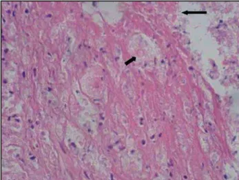
An ultrasound-guided fine-needle aspiration biopsy of the right adrenal gland yielded necrotic tissue with numerous organisms compatible with Histoplasma capsulatum (Figures 3 and 4). A diagnosis of disseminated histoplasmosis was made.
During hospitalization, the patient developed muscle weakness, arterial hypotension (blood pressure 90/60mmHg), diarrhea, hyponatremia (sodium 120mEq/l) and hyperkalemia (potassium 5.97mEq/l). A diagnosis of adrenal insufficiency was presumed (cortisol level of 6mcg/dl) and, because of the critical symptoms, treatment with hydrocortisone was started (100mg IV TID).
Figure 1 - Axial computed tomography image shows bilateral adrenal mass.
Figure 2 - Sagital computed tomography image shows bilateral adrenal mass.
Figure 3 - Micrograph of adrenal lesion, showing necrotic tissue (thin arrow) and fungic structures (large arrow). HE 400X. Hematoxylin-eosine.
Figure 4 - Arrow showing Histoplasma sp in grape cluster arrangement. 200X. Silver stain (Grocott)
The patient was initially treated with amphotericin-B 1mg/kg/d, although on the second day of therapy the laboratory findings indicated renal function impairment (creatinine 3.6mg/dl). Therefore, therapy with itraconazole (400mg per day) was started. This led to an enormous improvement in the clinical condition and laboratory tests, and the patient was gradually weaned off corticosteroids.
The patient was discharged in an improved condition of health, taking itraconazole 400mg per day and prednisone 10mg per day. Seven months after discharge, the patient was readmitted because itraconazole treatment had been interrupted two weeks prior to this admission. He relapsed with fever and adrenal insufficiency, but rapidly responded to reintroduction of medication.
DISCUSSION
232
Revista da Sociedade Brasileira de Medicina Tropical 40(2):230-233, mar-abr, 2007
diagnosis included benign or malignant adrenal tumors, metastatic tumors, subacute adrenal hemorrhage and disseminated infections such as histoplasmosis, paracoccidioidomycosis, tuberculosis, cryptococcosis and coccidioidomycosis12 13 14 17 18 20 22. In a Brazilian
series of 131,466 post-mortem examinations, there were 254 cases of adrenalitis, of which 43.7% were caused by tuberculosis, 33.8% by paracoccidioidomycosis and 1.2% by histoplasmosis7.
The CT features of adrenal histoplasmosis include bilateral symmetric enlargement with preservation of normal outlines, peripheral enhancement and central hypodense areas. Other infectious causes such as paracoccidioidomycosis are indistinguishable from histoplasmosis on imaging, and metastasis may mimic infection because central necrosis is common in both conditions9 10 12 14 17.
Percutaneous biopsy or fine-needle aspiration using either CT or ultrasound guidance is necessary for evaluating adrenal lesions12 15 17 20 21 22 23 24. Histopathological examination shows that the
intracellular forms are situated within the cytoplasm of histiocytes, where they appear as numerous small spherical or oval yeast forms surrounded by a clear ring of space that resembles a capsule, hence the misnomer, H. Capsulatum1 8. Despite its insensitivity,
histopathology was essential for the definitive diagnosis of the present case. Additional diagnostic tests for diagnosing histoplasmosis, such as tissue sample culturing, antigen detection, serology and molecular diagnosis by polymerase chain reaction1 26 were not available at that time.
Disseminated histoplasmosis may affect almost all systems, including the reticuloendothelial system, lungs, gastrointestinal tract, renal tract, central nervous system, bone marrow and adrenal glands4 10 26. It usually occurs in immunocompromised
individuals or at age extremities26. Histoplasmosis presenting
as bilateral adrenal enlargement has been previously described2 6 8 9 10 11 15 16 18 19 20 23 24 27. However, to our knowledge, this
is the fourth report of bilateral adrenal histoplasmosis associated with adrenal insufficiency in an immunocompetent host. Twelve cases of bilateral adrenal histoplasmosis have been reported in immunocompetent hosts6 12 16 20 24 27 and only three of them
presented with adrenal insufficiency12 20 24. Despite his age, this
patient had no evidence of immunosuppression and the discrete lymphocytopenia (797/mm3) on admission could have been
caused by the infection itself.
Treatment with corticosteroids was promptly started without first having a complete laboratory diagnosis of adrenal insufficiency, which would have been obtained from the blood cortisol level and the low ACTH stimulated cortisol resposes4 5 25. Among patients
with adrenal insufficiency, a large proportion require replacement therapy, although reversal of adrenal dysfunction has been described after prolonged antifungal treatment4 15 25 27.
The recommended treatment is amphotericin B for critically ill hospitalized patients1. Nevertheless, this had to be replaced
by itraconazole because of nephrotoxicity. Itraconazole is well tolerated and has excellent central nervous system penetration. Ketoconazole may be used in milder presentations. Recurrence has been described as long as nine years after cessation of treatment, and therefore treatment duration of one to two years reduces the risk of relapse3 4 6 8 9 10 11 15 19 20 23 24 25.
This case emphasizes the fact that adrenal histoplasmosis does occur in immunocompetent patients and has to be considered in the differential diagnosis of bilateral adrenal masses. Adrenal insufficiency has to be monitored and antifungal therapy should be maintained for at least one year.
REFERENCES
1. Adderson E. Histoplasmosis. The Pediatric Infectious Disease Journal 25: 73-74, 2006.
2. Anandi V, Walter A, Gammon K, Nair A, Koshi G. Histoplasmosis presenting as bilateral adrenal tumour. Indian Journal Pathology & Microbiology 32: 318-320, 1989.
3. Bamberger DM. Successful treatment of multiple cerebral histoplasmomas with itraconazole. Clinical Infectious Diseases 28: 915-916, 1999.
4. Chedid MF, Chedid AD, Geyer GR, Chedid MB, Severo LC. Histoplasmosis presenting as addisonian crisis in an immunocompetent host. Revista daRevista da Sociedade Brasileira de Medicina Tropical 37: 60-62, 2004.
5. Cooper MS, Stewart PM. Corticosteroid insufficiency in acutely ill patients. NewCorticosteroid insufficiency in acutely ill patients. New England Journal of Medicine 348: 727-734, 2003.
6. Desmet P, Vogelaers D, Afschrift M. Progressive disseminated histoplasmosis 10 years after return out of Africa in an immunocompetent host. Acta ClinicaActa Clinica Belgica 59: 274-278, 2004.
7. Fernandes VS, Bisi H, Longatto Filho A, Camargo RY. [Incidence of adrenalitis in necropsy material] Revista do Hospital das Clínicas 46: 219-222, 1991. 8. Giacaglia LR, Lin CJ, Lucon AM, Goldman J. Disseminated histoplasmosis
presenting as bilateral adrenal masses. Revista do Hospital das ClínicasRevista do Hospital das Clínicas 53: 254-256, 1998.
9. Gohar S, Sule A, Gaitonde S, Mittal G, Ejaz P, Mangat G, Bhaduri A, Joshi VR. Adrenal histoplasmosis. The Journal of the Association of Physicians of India 49: 916-917, 2001.
10. Grover SB, Midha N, Gupta M, Sharma U, Talib VH. Imaging spectrum inImaging spectrum in disseminated histoplasmosis: case report and brief review. Australasian Radiology 49: 175-178, 2005.
11. Johnston AW, Brown PA, Ewen SW. Histoplasmosis - a ten year follow-up. The Journal of Infection 33: 111-113, 1996
12. Kumar N, Singh S, Govil S. Adrenal histoplasmosis: clinical presentation and imaging features in nine cases. Abdominal Imaging 28: 703-708, 2003. 13. Lam KY, Lo CY. Metastatic tumours of the adrenal glands: a 30-year experience
in a teaching hospital. Clinical Endocrinology 56: 95-101, 2002.
14. Leal AMO, Bellucci AD, Muglia VF, Lucchesi FR. Unique Adrenal Gland Imaging Features in Addison’s Disease Caused By Paracoccidioidomycosis. American Journal of Roentgenology 181:1433-1434, 2003.
15. Lee J, Jones PH, Trowell JE, Whitear WP, Williams PF. Hypoadrenal crisis caused by disseminated histoplasmosis. The Journal of Infection 27: 181-183, 1993. 16. Mahajan R, Sharma U, Trivedi N, Prasad M, Kansra U, Bhandari S, Talib VH.
Histoplasma capsulatum in adrenal gland aspirate - a case report. Indian Journal Pathology & Microbiology 43: 165-168, 2000.
17. Moreira Jr SG, Pow-Sang JM. Evaluation and management of adrenal masses.Evaluation and management of adrenal masses. Cancer Control 9: 326-334, 2002.
18. Mukherjee JJ, Villa ML, Tan L, Lee KO. Bilateral adrenal masses due to histoplasmosis. The Journal of Clinical Endocrinology and Metabolism 90: 6725-6726, 2005.
19. Nine C, Maidana Paz C, Koch F, Stegmuller B, May A, Catalano HN. Incidental adrenal histoplasmosis. Medicina 62: 569-571, 2002.
20. Rozenblit AM, Kim A, Tuvia J, Wenig BM. Adrenal histoplasmosis manifested as Addison’s disease: unusual CT features with magnetic resonance imaging correlation. Clinical Radiology 56: 682-684, 2001.
233
22. Vaca MJ, Gonzalez C, Vega V, Martinez M, Paradelo M. Casuistic revision of adrenal pathology during last 23 years. Revista de la Facultad de Ciencias Medicas 58:Revista de la Facultad de Ciencias Medicas 58: 109-116, 2001.
23. Valente PT, Calafati SA. Diagnosis of disseminated histoplasmosis by fine needleDiagnosis of disseminated histoplasmosis by fine needle aspiration of the adrenal gland. Acta Cytologica 33: 341-343, 1989.
24. Van Zeben D, Zijlmans JM, Kuijpers TJ, Tinga CJ, Haak A. Disseminated histoplasmosis. Typical presentation with involvement of the adrenal glands. The Netherlands Journal of Medicine 34: 200-204, 1989.
25. Washburn RG, Bennett JE. Reversal of Adrenal Glucocorticoid Dysfunction in a Patient with Disseminated Histoplasmosis. Annals of Internal Medicine 110: 86-87, 1989.
26. Wheat LJ. Histoplasmosis: a review for clinicians from non-endemic areas. Mycoses 49:274-282, 2006.
27. Wilson DA, Muchmore HG, Tisdal RG, Fahmy A, Pitha JV. Histoplasmosis of the adrenal glands studied by CT. Radiology 150: 779-783, 1984.