361
Revista da Sociedade Brasileira de Medicina Tropical 48(3):361-363, May-Jun, 2015http://dx.doi.org/10.1590/0037-8682-0025-2015
Case Report
INTRODUCTION
Correspondenceauthor: Prof. Paulo Rogério Ferreti Bonan. Faculdade de Odontologia/UFPB. Cidade Universitária, Castelo Branco, 58051-800 João Pessoa, Paraíba, Brasil.
Phone: 55 83 8882-7842
e-mail: [email protected]; [email protected] Received 23 January 2015
Accepted 23 March 2015
Dirofi lariasis involving the oral cavity:
report of the fi rst case from South America
Laudenice Lucena Pereira
[1],
Ricardo Della Coletta
[2],
Larissa Cavalcanti Monteiro
[1],
Victor Yuri Nicolau Ferreira
[1],
Jorge Esquiche Leon
[3]and Paulo Rogério Ferreti Bonan
[1][1]. Faculdade de Odontologia, Universidade Federal da Paraíba, João Pessoa, Paraíba, Brasil. [2]. Faculdade de Odontologia, Universidade Estadual de Campinas, Piracicaba, São Paulo, Brasil. [3]. Faculdade de Odontologia, Universidade de São Paulo, Ribeirão Preto, São Paulo, Brasil.
ABSTRACT
Oral dirofi lariasis is very rare with non-specifi c clinical manifestations. Here, we report the case of a 65-year-old South American woman with a submucosal nodule on her right buccal mucosa. The nodule was slightly tender and painful. Differential diagnoses included mesenchymal (lipoma or fi brolipoma, solitary fi brous tumor, and neurofi broma) or glandular benign tumors (pleomorphic adenoma) with secondary infections. We performed excisional biopsy. A histopathological examination revealed a dense fi brous capsule and a single female fi larial worm showing double uterus appearance, neural plaque, well-developed musculature and
intestinal apparatus. Dirofi lariasis was diagnosed, and the patient was followed-up for 12 months without recurrence. Keywords: Pathology. Dirofi lariasis. Dentistry. Diagnosis.
Filariasis is a zoonosis that occurs worldwide but is more common in countries with tropical and subtropical climates(1). It is caused by nematodes belonging to the genus Dirofi laria, and is transmitted to humans via bites of Aedes, Culex, and Anopheles mosquitoes(2). Dirofi laria repens, Dirofi laria immitis, Dirofi laria ursi, and Dirofi laria tenuis are the species known to affect dogs, foxes, cats, raccoons, and bears, which serve as
the intermediate hosts(2).
Dirofi laria repens, one of the more common species, can cause submucosal nodules on the arms, legs, head, and even
the eyes of humans; these nodules develop as an infl ammatory
reaction to the presence of the parasite in the tissue(3). Intraoral
involvement is very rare, and diagnosis is confi rmed based on identifi cation of the parasite in the nodule under microscopy(4).
Defi nitive treatment includes surgical removal, and in some
cases, medications such as diethylcarbamazine, ivermectin, and albendazole(1). In this study, we describe a rare case of
dirofi lariasis affecting the oral mucosa. To the best of our knowledge, this case is the fi rst to be reported from Brazil and from South America.
CASE REPORT
A feoderma 65-year-old woman was referred to our hospital by the public health unit. She complained of swelling and pain on the right side of her buccal mucosa. She was unaware of when the nodule fi rst appeared, and hence could not tell when the swelling exacerbated. Her dentist had prescribed antibiotics, anti-infl ammatories, and analgesics. Despite these medications, her symptoms persisted and worsened. Our patient lived in a very crowded community with poor sanitary conditions and constant warm and humid climate. She had good general health with no gastrointestinal complaints and she did not consume tobacco. General physical examination revealed no relevant fi ndings or lymphadenopathy.
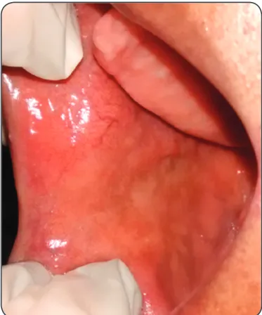
Extraoral clinical examination revealed normal structures without tumefactions. Intraoral examination revealed a movable
tender mass covered by normal mucosa on the right side of the buccal mucosa (Figure 1, left). Differential diagnosis included mesenchymal benign tumors (such as lipoma or fi brolipoma, solitary fi brous tumor, and neurofi broma) or glandular benign tumors (such as pleomorphic adenoma) with infl ammatory reactions, including secondary infections. Excisional biopsy was performed. Periodic acid-Schiff staining was carried out to
evaluate secondary fungal infections; no evidence of yeasts or
hyphae was found. Histological examination revealed a dense fi brous capsule containing an infi ltrate of mixed infl ammatory
cells-lymphocytes, macrophages, eosinophils, and neutrophils.
Within the infi ltrate, we found a single female worm with numerous larval forms. Higher magnifi cation revealed a thick
362
Pereira LL et al. - Oral dirofi lariasis
DISCUSSION
FIGURE 1 -Symptomatic well-defi ned nodule on the right side of the buccal mucosa.
intestinal tubule within the well-developed musculature,
characteristic of the Dirofi laria species (Figure 2). With these
microscopy data and considering the poor sanitary conditions,
we performed blood tests and the results were unremarkable with respect to systemic infections. Parasitological examination, too, did not reveal eggs or larvae of parasites. Thus, a defi nitive diagnosis of dirofi lariasiswas made. None additional treatment was required due to be a local manifestation. The patient was followed-up and 12 months after diagnosis she presented with
good general health and absence of recurrence (Figure 3).
A
B
Although heartworm disease occurs worldwide, it is rare in
humans. Dirofi laria species are natural parasites of mammals and can be accidentally transmitted to humans via bites of zooanthrophilic mosquitoes carrying the infectious larvae. Infectious larvae acquired during a blood meal from host
animals infected with the Dirofi laria sp. Humans are not ideal
hosts, as in the human body, Dirofi laria species develop into
worms that do not reproduce or release microfi lariae. Although
nearly 40 Dirofi laria species have been identifi ed, only a few
have been reported to cause human infections(1) (2). In our case,
the genus could be identifi ed only because we performed a morphological evaluation using microscopy, which is not the gold standard for species classifi cation(1).
It is assumed that the increase in cases of heartworm disease in humans is caused by climate change with higher temperatures favoring the survival of microfi lariae and by global tourism
increasing that can causes infection by infected carnivores in
certain regions of the world(3). Knowledge of endemic areas
with poor vector control owing to improper sanitation and
overpopulation is often helpful in diagnosis(1). In the current case, the patient did not report going abroad but she lived in a
very populated region with high temperatures and poor sanitary
conditions.
The clinical manifestations of dirofi lariasis include nodules in the subcutaneous tissues, muscles, and visceral organs. The duration between growth of the paras causing infection to the detection of the nodule is approximately 6 months. The parasite is restricted within the infl ammatory subcutaneous tissue node
or other tissues, and can survive for months or even years. Some
cases of dirofi lariasis are asymptomatic; nevertheless, cases with itching, arthritis, edema, and subcutaneous swelling have been
FIGURE 2 - (A) Mixed infl ammatory infi ltrate and female
nematoid body placed inside the tissue (HE, 100X). (B) Thick cuticle 1: developed musculature 2: double uterus 3: with eggs (arrow), intestinal apparatus 4: and neural plaque 5: revealing
363
Rev Soc Bras Med Trop 48(3):361-363, May-Jun, 2015FIGURE 3 - Buccal mucosa showing no recurrence after 12 months of follow-up.
described(4). In this case, the patient was symptomatic and an
intense infl ammatory reaction was observed. Our differential
diagnoses included mesenchymal benign tumors (such as lipoma
or fi brolipoma, solitary fi brous tumor, and neurofi broma) or glandular benign tumors (such as pleomorphic adenoma) with infl ammatory reactions, including secondary infections. Some of these lesions were also included as differential diagnoses in a previous case of dirofi lariasis affecting the oral mucosa(1).
Few studies have reported the occurrence of dirofi lariasis involving the head and neck. A study by Latifoglu et al.(5)
conducted in 2002 cited the presence of a nematode clearly
identifi ed by histopathology after excisional biopsy of a mass on the right cheek, lateral to the masseter muscle. Additional radiographic and ultrasonographic examinations of the site were performed to rule out any connection with the parotid gland. A more recent study conducted in 2014 by Friedrich et al.(6)
verifi ed the presence of Dirofi laria infection in a 40-year-old
patient without apparent symptoms. The only clinical feature was the presence of a swelling in the left temporal muscle. This patient had travelled to Sri Lanka 8 months prior to the onset of the swelling. In addition, he had travelled to the Far East and the Indian subcontinent few years ago. This information is relevant since the occurrence of dirofi lariasis in humans is
more common in those regions of the world. In 2013, Kurup
et al.(1) described the case of a 54-year-old female school teacher
who complained of intermittent swelling inside her mouth on the left side. Dirofi lariasis was diagnosed after microscopic examination, and the buccal mucosa showed features similar
to those observed in the current case.
A detailed monitoring of the patient is necessary for
diagnosis, and additional tests are needed to confirm the diagnosis(5). Blood and stool tests for parasites could be
performed. Excisional biopsy for histologic evaluation of the nodule was essential for accurate diagnosis. Aside from being a diagnostic procedure, excisional biopsy is a treatment method. The histopathology diagnosis could reveal the presence of a
double uterus, possibly containing many larval forms, neural
plaque, gut apparatus, and a thick cuticle, confi rming the diagnosis of dirofi lariasis(1). The thick cuticle and developed musculature are characteristic of Dirofi laria species(7). The treatment could involve surgical removal, and in some cases, medications such as diethylcarbamazine, ivermectin, and albendazole(1).In our case, we only observed the focal infection
without systemic involvement. After removal of the nodule, the patient was followed-up for 12 months. She was in good health
and had no recurrence.
Dirofilariasis involving the oral cavity is very rare. However, it can be found in countries with high temperatures and in populated communities with poor sanitary conditions. Health professionals need to be aware of the need for detailed examination and must consider infections caused by parasites
in their differential diagnoses. If required, ancillary tests must be performed to aid diagnosis.
REFERENCES
1. Kurup S, Veeraraghavan R, Jose R, Puthalath U. Filariasis of the buccal mucosa: a diagnostic dilemma. Contemp Clin Dent 2013; 4:254-257.
2. Orihel TC, Eberhard ML. Zoonotic fi lariasis. Clin Microbiol Rev
1998; 11:366-381.
3. Otranto D, Dantas-Torres F, Brianti E, Traversa D, Petrić D,
Genchi C, et al. Vector-borne helminths of dogs and humans in Europe. Parasit Vectors 2013; 6:16.
4. Campos JRM, Barbas CSV, Filomeno LTB, Fernandez A,
Minamoto H, B Filho JV, et al. Human pulmonary dirofi lariasis. Analysis of 24 cases from São Paulo, Brazil. Chest 1997; 112: 729-733.
5. Latifoglu O, Ozmen S, Sezer C, Yavuzer R, Altintas K¸ Uluoglu O.
Dirofi laria repens presenting as a premasseteric nodule. Oral Surg Oral Med Oral Pathol Oral Radiol Endod 2002; 94:217-220. 6. Friedrich RE, Heiland M, Burchard G, Racz P, Zustin J, Hagel C.
Human Dirofi laria repens infection of the zygomatico-temporal
region. J Craniomaxillofac Surg 2014; 42:612e615.
7. Popescu I, Tudose I, Racz P, Muntau B, Giurcaneanu C, Poppert
S. Human Dirofi laria repens infection in Romania: a case report.