HEREDITARY MOTOR AND SENSORY NEUROPATHY
WITH CONGENITAL GLAUCOMA
REPORT ON A FAMILY
WALTER O. ARRUDA*, ENIO A. COMERLATO**,
ROSANA H. SCOLA***, CARLOS E.S. SILVADO*, LINEU C. WERNECK****
ABSTRACT - We report three siblings of a family with hereditary motor and sensory polyneuropathy (HMSN) and buphthalmos. Electrophysiological studies showed a demyelinating neuropathy and pathological findings showed severe loss of myelinated fibers (MF), thin myelin sheaths and myelin infoldings in a few remaining MF. The presumed mode of inheritance is autosomal recessive. This family probably represents an unique form of CMT4 that may be related to one of the congenital glaucoma genic locus, particularly GLC3A and GLC3B, described in Turkish families.
KEY WORDS: hereditary motor and sensory neuropathy, congenital glaucoma, autosomal recessive inheritance.
Neuropatia hereditária sensitivo-motora com glaucoma congênito: descrição de uma família
RESUMO - Descrevemos três membros afetados de uma família com neuropatia hereditária sensitivo-motora tipo I (desmielinizante) e glaucoma congênito (buftalmia). O estudo eletrofisiológico dos membros afetados demonstrou polineuropatia sensitivo-motora desmielinizante, com ausência ou redução acentuada das velocidades de neurocondução sensitiva e motora. A biópsia do nervo sural revelou redução moderada a grave das fibras mielinizadas, bainhas de mielina de espessura diminuída (remielinização) com dobramentos delas nas poucas fibras mielinizadas remanescentes. Não foram observadas formações em casca de cebola, nem tampouco alterações hipertróficas. O padrão de herança desta família parece ser autossômico recessivo. Sugerimos tratar-se de uma forma singular de doença de Charcot-Marie-Tooth autossômica recessiva (CMT4), que eventualmente pode possuir locus gênico próximo a um dos locus do glaucoma congênito (GLC3A e GLC3B), localizados nos cromossomos 2p21 e 1p36.
PALAVRAS-CHAVE: neuropatia hereditária sensitivo-motora, glaucoma congênito, herança autossômica recessiva.
Hereditary motor and sensory neuropathies (HMSN) are usually classified into seven subtypes based on clinical and pathological findings1. Two main groups can be distinguished: type I primarily
presents extensive segmental demyelination in the peripheral nerves with slow nerve conduction; type II shows a primary axonal degeneration with normal nerve conduction2. Further subtypes (type V, VI,
and VII) were introduced to integrate frequently associated symptoms: spastic paraplegia, optic atrophy, and retinitis pigmentosa, though there has been an increasing number of reports on patients with HMSN plus other associated features (e.g. diaphragm/vocal cord paresis, congenital cataract/mental
Serviço de Doenças Neuromusculares, Hospital de Clínicas, Universidade Federal do Paraná: * Professor Assistente de Neurologia; ** Mestrando de Medicina Interna; *** Professora Assistente de Propedêutica Médica; **** Professor Titular de Neurologia. Aceite: 9-janeiro-1999.
retardation)1. The term “HMSN” has been employed to designate the clinical forms, whereas
“CMT”(Charcot-Marie-Tooth) has been more commonly used in the genetic literature3 (Table 1).
We describe a family with HMSN of demyelinating type associated with congenital glaucoma.
METHOD
Family description
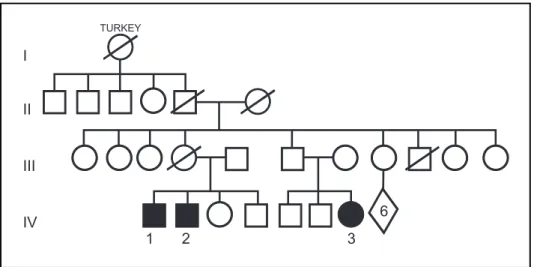
We examined three members of a family presenting a very similar clinical picture. There was no history of consanguinity. Their great-grandmother immigrated from Turkey. The parents of the two affected brothers (Patients 1 and 2) and their first degree female cousin (Patient 3) were not affected. Nerve conduction study was performed on Patients’ 1 and 2 father and was normal. The family tree is shown in Figure 1.
Patient 1. This 41-year-old male had noticed slowly progressive gait difficulty and lower limbs atrophy since 1-2 years ago; he could not precise the time his motor difficulty started. There had not been complications during pregnancy and birth, and his developmental history was normal, except for the problems caused by his visual Table 1. HMSN (CMT) classification.
Type Locus Gene
HMSN I (CMT1)
HMSN IA (CMT 1A) 17p11.2-p12 PMP22
HMSN IB (CMT 1B) 1q21.2-q23 P0
HMSN IC (CMT 1C) ?
X-linked CMT X1 Xq13.1 Cx32
X-linked CMT X2 ?
HMSN II (CMT2)
HMSN IIA (CMT 2A) 1p35-p36
HMSN IIB (CMT 2B) 3q13-q22
HMSN IIC (CMT 2C) ?
HMSN IID (CMT 2D) 7p14
HMSN III (DSD)
DSD A Mutations PMP22
DSD B Mutations P0
AD DSD 8q23-q24
AR DSD ?
HMSN IV (CMT4)
HMSN IVA (CMT4A) 8q13-21.1
HMSN IVB (CMT4B) 11q23.1
HMSN IVC (CMT4C) ?
CMT 5q23-q33
8q24
impairment. Congenital glaucoma was diagnosed during his first year of life. He was operated on at that time, but his vision acuity progressively declined. Most recently he complained of sexual impotence. Bilateral buphthalmos with corneal opacification was observed. He is presently blind. A typical “inverted bottle” configuration of his lower limbs, due to more pronounced distal muscle atrophy, could be readily observed, as well as pes cavus. There was no scoliosis or other skeletal abnormalities. His mental status was normal. There was considerable muscle atrophy and weakness (MRC 2-3) of the calves and feet and less markedly of the thighs (MRC 4). In fact, he could no longer walk, even with support. The distal muscles of the upper limbs showed a milder degree of atrophy and paresis (MRC 4). Muscle tone was decreased in all limbs and tendon jerks were absent in the upper and lower limbs. Sensory deficits affecting touch, pain, temperature, and vibration were present in the feet up to the knees and in both hands. There was no evidence of nerve hypertrophy.
Patient 2. This 29-year-old male had a diagnosis of congenital glaucoma during his first year of life, when submitted to iridocyclectomy. He was incidentally discovered to be affected, for he had neither motor nor sensory complaints. His pregnancy, delivery and developmental history were normal. Congenital glaucoma was diagnosed during his first year of life. Five years ago he suffered a left eye hemorrhage that left him blinded in this eye. His present right eye acuity is 20/200. Bilateral buphthalmos was present. There were no skeletal deformities, including pes cavus. His mental status was normal. Upper and lower limbs distal muscle atrophy (wasting of the intrinsic muscles of the hands) and a distally predominant weakness of the arms and legs (long finger extensors, intrinsic muscle of the hands, dorsiflexion and plantar flexion) (MRC 3) were present. Muscle tone was normal; he was areflexic with flexor plantar responses. There was glove and stocking sensory decrease to all modalities. Romberg’s sign was absent.
Patient 3. She was a 31-year-old woman, who complained of progressive weakness and gait difficulty since 8 years ago, with feet deformity. Her past history was unremarkable except for the diagnosis of congenital glaucoma when aged 6 months. She was operated on at that time, but she is presently blind due to bilateral leucocoria. Bilateral buphthalmos was present. Her mental status is normal. A distal upper and lower limb muscle atrophy and weakness and marked pes cavus were present. She was globally hypotonic and areflexic with flexor plantar responses. Glove and stocking sensory loss to all modalities were present. Nerve hypertrophy was absent.
RESULTS
Laboratory / Neurophysiological tests
All three affected members were submitted to the following laboratory tests, which resulted normal or negative: erythrocyte sedimentation rate, plasma glucose, serum proteins, lipidogram, liver and kidney function, CSF examination, CSF protein electrophoresis, VDRL, HTLV-1 serological tests. A head CT-scan was normal in each patient.
Standard elctromyography examination of upper and lower limbs muscles disclosed increased insertional activity, fibrillation potentials and positive sharp waves, motor unit potentials (MUP) with decreased amplitude and increased duration, long polyphasic MUP and decreased recruitment, all findings in keeping with active and chronic denervation. These findings were observed in all three patients. Nerve conduction study results are depicted in Table 2.
Neuropathological findings
Sural nerve biopsies were performed in Patients 1 and 2. Frozen sections showed normal appeareance nerve fascicles, with a moderate to severe decrease of myelinated fibers. Inflammatory changes or amyloid infiltration were not observed. Semithin sections stained with methylene blue showed a severe decrease of myelinated fibers in both patients’ nerves, without onion bulbs. Some myelinated fibers have a very thin myelin sheath, suggesting hypomyelination. Occasional myelin infoldings were seen.
DISCUSSION
Autosomal recessive inheritance has been described in HMSN I, II and III and both forms, whether demyelinating or axonal, have been labelled CMT43. Based on electrophysiological and
pathological criteria, this group has been temptatively subdivided into three groups: CMT4A, characterised by slow motor nerve conduction velocities and nerve biopsy findings of hypomyelination; CMT4B with slow motor nerve conduction velocities and focally folded myelin sheaths; and CMT4C defined by preserved motor nerve conduction velocities and absence of myelin changes in nerve biopsy. CMT4A and CMT4B forms have been linked to chromosomes 8q13-21.24 and 11q23.15,
respectivelly. Not surprising, two recent reports of CMT4 demyelinating type affected families have pointed out two other locations: chromosome 5q23-q336 and chromosome 8q247. There is no reference
about the presence of congenital glaucoma in any one of these described CMT4 families. Primary congenital or infantile glaucoma (gene symbol: GLC3), as presented in this family, is a rare inherited eye disorder that usually manifests itself within the first year of life with a typical presentation of tearing, photophobia and clouding of the cornea. Autosomal recessive inheritance has been confirmed in a significant proportion of cases and the existence of both genetic and etiological heterogeneity of the GLC3 phenotype8. So far two loci have already been identified to congenital
glaucoma: GLC3A locus, mapped to chromosome 2p21, and GLC3B locus, located on chromosome 1p368,9. Interestingly enough, these loci have been mapped in a group of Turkish families, which Table 2. Nerve conduction studies
Patient 1 Patient 2 Patient 3
Velocity Amplitude (µV) Velocity Amplitude (µV) Velocity Amplitude (µV)
Median nerve
Motor 15.3 3670 ∅ ∅
Sensory ∅ ∅ ∅
Ulnar nerve
Motor 15.9 10500 20.6 10400 ∅
Sensory 11.4 2.0 23.7 22.00 ∅
Radial nerve
Sensory21,0 21.0 ∅ ∅
Fibular nerve 21.0
Motor 15.9 2600 19.0 6000 19.1 1500
Sural nerve ∅ ∅ ∅
may give us a clue on where to pursue the CMT locus in the family hereby described. The grandparents of our patients emmigrated from Turkey, which raises the possibility of co-segregation of the CMT4 locus with one of the already identified GLC3 locus. Further genetic studies are warranted to confirm or exclude this hypothesis, and the possibility of a still unknown locus causing this particular form of CMT4 can not be excluded.
REFERENCES
1. Dyck PJ, Chance P, Lebo R, Carney JA. Hereditary motor and sensory neuropathies. In Dyck PJ, Thomas PK, Griffin JW, et al. (ed). Peripheral neuropathy, 3Ed. Philadelphia: Saunders, 1993:1094-1136.
2. Harding AE, Thomas PK. The clinical features of hereditary motor and sensory neuropathy types I and II. Brain 1980;103:259-280.
3. Reilly MM. Genetically determined neuropathies. J Neurol 1998;245:6-13.
4. Othmane KB, Hentati F, Lennon F, et al. Linkage of a locus (CMT4) for autosomal recessive Charcot-Marie-Tooth disease to chromosome 8q. Hum Mol Genet 1993;10:1625-1628.
5. Bolino A, Brancolini V, Bono F, et al. Localisation of a gene responsible for autosomal recessive demyelinating neuropathy with focally folded myelin sheaths to chromosome 11q23 by homozigosity mapping and haplotype sharing. Hum Mol Genet 1996;5:1051-1054.
6. LeGuern E, Guilbot A, Kessali M, et al. Homozygosity mapping of an autosomal recessive fdorm of demyelinating Charcot-Marie-tooth disease to chromosome 5123-q33. Hum Mol Genet 1996;5:1685-1688.
7. Kalaydjieva L, Hallmayer J, Chandler D, et al. Gene mapping of Gypsies identifies a novel demyelinating neuropathy on chromosome 8q24. Nat Genet 1996;14:214-217.
8. Akarsu AN, Turacil ME, Aktan SG, et al. A second locus (GLC3B) for primary congenital glaucoma (Buphthamos) maps to the 1p36 region. Hum Mol Genet 1996;5:1199-1203.