Simone Silvério Campos(a) Cássio Vicente Pereira(a) Márcio Gilberto Zangerônimo(b) Leandro Silva Marques(c) Luciano José Pereira(b)
(a) Curso de Odontologia, Centro Univ de Lavras - UNILAVRAS, Lavras, MG, Brasil. (b) Departamento de Medicina Veterinária,
Setor de Fisiologia e Farmacologia, Univ Federal de Lavras - UFLA, Lavras, MG, Brazil.
(c) Department of Orthodontics, School of Dentistry, Univ Federal dos Vales do Jequitinhonha e Mucuri - UFVJM, Diamantina, MG, Brazil.
Corresponding Author: Luciano José Pereira
E-mail: [email protected]
on test materials used for the
determination of masticatory
performance
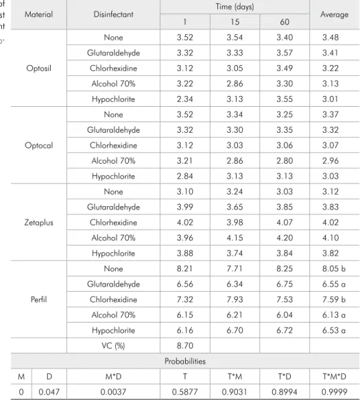
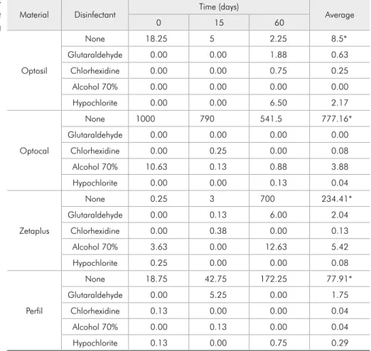
Abstract: Masticatory function can be evaluated objectively as the ca-pacity of an individual to fragment solid food after a ixed number of chewing cycles, the so-called masticatory performance (MP). The ob-jective of this study was to evaluate the reliability of four different test materials (Optosil, Optocal, Zetapuls, and Peril) and ive disinfection protocols by aspersion and immersion (no disinfection, 2% glutaralde-hyde, 2% chlorhexidine, 5.25% sodium hypochlorite, and 70% alcohol) on the MP, determined at three moments (24 hours, 15 and 60 days) after storing the fragmented blocks. MP was evaluated by calculating X50 through the sieving technique and the Rosim-Ramler equation. The weight and microbiologic count (colony forming units, CFUs) of chewed blocks were measured to identify any variations that would make MP determination unfeasible. Differences in MP were observed among the materials (p < 0.01). Peril presented the highest X50 value (worst MP determination), followed by Zetaplus (both p < 0.05), Optosil, and Opto-cal (both p > 0.05). The time and disinfection type had no inluence on MP (p > 0.05). The number of CFUs differed between the nondisinfected group and all other disinfection groups at all time points (p < 0.01). No other signiicant difference in CFU count between disinfection groups was observed. In conclusion, disinfection did not alter the reliability of the test materials for the MP calculation for up to 60 days.
Descriptors: Mastication; Disinfection; Dental Materials.
Introduction
During mastication, food is broken down into smaller particles to fa-cilitate enzymatic processing and swallowing.1,2 Although masticatory
function can be measured by various means, masticatory performance (MP) testing is the most commonly used objective method. MP is deined as the capacity of an individual to fragment a solid natural or artiicial test food during mastication.3 A decline in MP may result in changes in
the diet, as some foods become dificult to eat.4-7
The test material used to determine MP should provide an ideal bolus for the scientiic study of mastication.3,8-12 A natural food has the
advan-tage that it is normally consumed. However, the consistency of the food may vary due to seasonal and geographical inluences, and the food may
Declaration of Interests: The authors certify that they have no commercial or associative interest that represents a conflict of interest in connection with the manuscript.
Submitted: Oct 31, 2012
contamination, which can lead to the transmission of infectious diseases through the saliva or blood from the oral cavity.13 During the clinical routine
for MP determination, disinfection techniques can be applied14 to minimize the risk of contamination.
Nevertheless, the use of disinfection may result in weight alterations, creating the possibility of error in the MP determination.
The objective of this study was to evaluate the accumulation of microorganisms in four different test materials and the utility of ive disinfection pro-tocols. Additionally, the materials were appraised as to the degree of weight variation by the sieving pro-cess for the MP calculation at different times.
Methodology
Materials
This study was approved by the local ethics re-search committee (CAAE, 0041.0.189.000-09). MP blocks were made of an acrylic matrix and stainless steel15 and were manipulated according to the
man-ufacturer’s speciications. The following test materi-als were used:
1. Optosil Comfort (Heraeuz Kulzer, Hanau, Ger-many; Lot: R270493);
2. Optocal, comprised of
-
casting silicone (57%; Optosil),-
toothpaste (27%; Sorriso Super Refrescante, Colgate-Palmolive Commercial Ltda., São Pau-lo, Brazil),-
solid petroleum gel (3%, 30g; Rioquímica, São José do Rio Preto, Brazil),-
type V dental plaster (Polidental, Cotia, Bra-zil),-
irreversible hydrocolloid type 1 fast-setting al-ginate (4%; Jeltrate Plus, Dentisply, Milford, USA), and-
catalytic paste (27 mg; Optosil);16,173. Peril (Vigodent, Rio de Janeiro, Brazil; Lot: 140/08); and
4. Zetaplus (Zhermack, Badia Polesine, Italy; Lot: 78828).
Materials were disinfected by immersion and
as-• 2% glutaraldehyde solution (Glutaron, Pharma-ceutical Industry Rioquímica Ltda., São José do Rio Preto, Brazil),
• 5.25% sodium hypochlorite solution (Clorox, The Clorox Company, Oakland, USA);
• 2% chlorhexidine solution (Verde Vida Pharma-cy, Lavras, Brazil); and
• 70% alcohol solution (Start, Lima and Pergher Ind. Com. Rep. Ltda., Uberlândia, Brasil).
The accumulation of microorganisms and the weights of the materials in each disinfection group were evaluated immediately, 15 days, and 60 days after mastication.
Measurement of MP
The MP was evaluated by determining the ca-pacity of an individual to fragment the artiicial test food.18 A single subject received 17 cubes of each
material at each collection moment. Each cube was chewed for 20 masticatory cycles per collection mo-ment. The study subject had all of his permanent teeth (12 occlusal units), a mesocephalic facial pat-tern, and an Angle Class I molar relationship, was free of any systemic disease that would interfere with motor activities, and did not use any medicine that would impact saliva secretion.
The number of masticatory cycles was visually quantiied by the examiner. The triturated parti-cles were expelled from the oral cavity into plastic containers with the aid of a funnel and disinfected with one of the 5 different protocols. The particles were placed on ilter paper for drying. Dried par-ticles were removed from the ilter paper and passed through a series of 10 granulometric sieves, with the diameters of the openings ranging from 5.6 to 0.71 mm, in decreasing order. The particle distri-bution by weight was described by the cumulative function of the average size of the particles, X50.19,20
Microbiological analysis
materials and four disinfection products + 1 con-trol) with a portion divided by the time (24 hours, 15 days, and 60 days). The data were submitted to variance analysis, with the averages being compared among themselves by the Tukey test (5%). The data were subjected to the square-root transformation for normalization. For the CFU, the nonparametric statistical analysis (chi-square) was used, and the averages were compared by the Kruskal-Wallis test with the SAS statistical package (1996).
Results
No differences in the X50 values were observed between the periods studied or between the asper-sion and immerasper-sion methods. Therefore, the re-block per 2 mL of water. The mixture was agitated
in a Vortex agitator for 30 seconds, and 25 µL of the solution were inoculated into Petri dishes con-taining MITIS medium (Mitis salivarius-bacitracin; Acumedia Manufacturers, Inc., Baltimore, USA). The plates were incubated for 3 days at 37°C in mi-croaerophilic conditions. The colony forming units (CFUs) were characterized and counted (CFU/mL) on the basis of the presence of white/grey color and ground glass aspect of the colonies. This method was only used as biological marker of the presence of viable microorganisms in the samples stored over time.
Statistical analysis involved an entirely random design in a 4 × 4 + 1 factorial outline (four test
Material Disinfectant Time (days) Average
1 15 60
Optosil
None 3.52 3.54 3.40 3.48
Glutaraldehyde 3.32 3.33 3.57 3.41
Chlorhexidine 3.12 3.05 3.49 3.22
Alcohol 70% 3.22 2.86 3.30 3.13
Hypochlorite 2.34 3.13 3.55 3.01
Optocal
None 3.52 3.34 3.25 3.37
Glutaraldehyde 3.32 3.30 3.35 3.32
Chlorhexidine 3.12 3.03 3.06 3.07
Alcohol 70% 3.21 2.86 2.80 2.96
Hypochlorite 2.84 3.13 3.13 3.03
Zetaplus
None 3.10 3.24 3.03 3.12
Glutaraldehyde 3.99 3.65 3.85 3.83
Chlorhexidine 4.02 3.98 4.07 4.02
Alcohol 70% 3.96 4.15 4.20 4.10
Hypochlorite 3.88 3.74 3.84 3.82
Perfil
None 8.21 7.71 8.25 8.05 b
Glutaraldehyde 6.56 6.34 6.75 6.55 a
Chlorhexidine 7.32 7.93 7.53 7.59 b
Alcohol 70% 6.15 6.21 6.04 6.13 a
Hypochlorite 6.16 6.70 6.72 6.53 a
VC (%) 8.70
Probabilities
M D M*D T T*M T*D T*M*D
0 0.047 0.0037 0.5877 0.9031 0.8994 0.9999
VC: variance coefficient; M: material; D: disinfectant; T: time. a,b,c Different superscript letters correspond to
significant differences between lines. Table 1 - Factorial analysis of
maining analyses were conducted by combining the aspersion and immersion samples. There was inter-action (p < 0.05) between the disinfectant type and the test material; however, there was no inluence of the time (Table 1). When Peril was used as a test material, chlorhexidine presented the worst result. Peril showed the highest values for particle size (p < 0.05) in all the tested disinfectants, with Op-tosil and Optocal showing the best results (Table 2).
Data related to the CFU counts are presented in Table 3. The CFU count differed between the non-disinfected group and all other disinfection groups at all moments (p < 0.01). No other signiicant dif-ferences in the CFU counts were observed between
the disinfection groups.
The time factor and disinfection type (immersion versus aspersion) had no inluence on the MP or the number of CFUs (p > 0.05).
Discussion
The MP is determined according to an individ-ual’s capacity to fragment a test food.1,11,15,21
Proto-cols for the MP test in the literature use various test foods, and an individual’s masticatory power limits the determination of MP. Due to the dificulty of establishing precise evaluations in debilitated pa-tients (e.g., total prosthesis wearers), new methods and different test foods for the MP test have been
1 15 60
None
Optosil 3.52 3.54 3.40 3.48 a
Optocal 3.52 3.34 3.25 3.37 a
Zetaplus 3.10 3.24 3.03 3.12 a
Perfil 8.21 7.71 8.25 8.05 b
Glutaraldehyde
Optosil 3.32 3.33 3.57 3.41 a
Optocal 3.32 3.30 3.35 3.32 ab
Zetaplus 3.99 3.65 3.85 3.83 b
Perfil 6.56 6.34 6.75 6.55 c
Chlorhexidine
Optosil 3.12 3.05 3.49 3.22 a
Optocal 3.12 3.03 3.06 3.07 a
Zetaplus 4.02 3.98 4.07 4.02 b
Perfil 7.32 7.93 7.53 7.59 c
Alcohol 70%
Optosil 3.22 2.86 3.30 3.13 a
Optocal 3.21 2.86 2.80 2.96 a
Zetaplus 3.96 4.15 4.20 4.10 b
Perfil 6.15 6.21 6.04 6.13 c
Hypochlorite
Optosil 2,34 3.13 3.55 3.01 a
Optocal 2.84 3.13 3.13 3.03 a
Zetaplus 3.88 3.74 3.84 3.82 b
Perfil 6.16 6.70 6.72 6.53 c
VC (%) 8.70
Probabilities
M D M*D T T*M T*D T*M*D
0 0.0047 0.0037 0.5877 0.9031 0.8994 0.9999
VC: variance coefficient; M: material; D: disinfectant; T: time. a,b,c Different superscript letters correspond to
significant differences between lines.
employed.22 Thus, it is important to study different
test materials for MP determination, and the use of appropriate materials for different patient proiles is important from a clinical perspective.
Variations in the form, size, and hardness of foods produce differences in the tests, thereby in-luencing the inal results.23 Different test foods also
present variations in consistency, due to the incor-poration of water originating from the saliva. These variations can render the standardization and the conduction of masticatory tests dificult. Conden-sation silicone was proposed as a pioneer chewable artiicial test material for the evaluation of mastica-tory function.18 For example, the condensation
sili-cone Optosil is almost odorless and tasteless, does not incorporate water, and can be maintained for up to 7 days without undergoing any important di-mensional alterations when used for molding appli-cations. Samples of this elastomeric material can be
easily standardized and appraised after mastication, allowing weight and size control, thus reducing vari-ability during production and mastication tests.17
Among the materials employed in the present study, Peril presented signiicantly higher X50 values than the other materials. This result can be justi-ied by the fact that Peril presented a Shore A of 80 after 24 hours. As a result, it displayed greater hardness and higher resistance to mastication than the other materials and, consequently, was not eas-ily fragmented. The fragments of Peril were larger and were retained in the upper portions of the sieve sequence. In spite of being an inexpensive and easily accessible test material, use of Peril may lead to MP values that are incompatible with the international literature.
Optocal is a variation of Optosil that displays reduced consistency, for use with debilitated pa-tients or those with low masticatory capacity.17,21
Material Disinfectant Time (days) Average
0 15 60
Optosil
None 18.25 5 2.25 8.5*
Glutaraldehyde 0.00 0.00 1.88 0.63
Chlorhexidine 0.00 0.00 0.75 0.25
Alcohol 70% 0.00 0.00 0.00 0.00
Hypochlorite 0.00 0.00 6.50 2.17
Optocal
None 1000 790 541.5 777.16*
Glutaraldehyde 0.00 0.00 0.00 0.00
Chlorhexidine 0.00 0.25 0.00 0.08
Alcohol 70% 10.63 0.13 0.88 3.88
Hypochlorite 0.00 0.00 0.13 0.04
Zetaplus
None 0.25 3 700 234.41*
Glutaraldehyde 0.00 0.13 6.00 2.04
Chlorhexidine 0.00 0.38 0.00 0.13
Alcohol 70% 3.63 0.00 12.63 5.42
Hypochlorite 0.25 0.00 0.00 0.08
Perfil
None 18.75 42.75 172.25 77.91*
Glutaraldehyde 0.00 5.25 0.00 1.75
Chlorhexidine 0.13 0.00 0.00 0.04
Alcohol 70% 0.00 0.13 0.00 0.04
Hypochlorite 0.13 0.00 0.75 0.29
* Control: different from others by the Kruskal-Wallis test (p < 0.05). Table 3 - Factorial analysis of
primary advantage of Optocal is its lexible and malleable consistency.17 Nevertheless, some studies
have shown that this material is still not suficiently soft for children and for oncological patients with temporomandibular disorders.23 The mixture
capac-ity test has been considered to be more appropriate than the fragmentation test for measuring differ-ences in MP in individuals with compromised oral function.23 The Zetaplus material presented reports
of a gritty sensation.
In this study, the disinfection method did not ap-pear to alter the calculated X50. We recommend that disinfection of the MP test material be conducted, to avoid contamination of the experimenter due to contact with the patient’s saliva. After being taken into the mouth, elastomers can store viral particles and should be considered as infectocontagious dis-ease transmission vehicles. Washing removes some of the microbial lora; however, pathogenic micro-organisms can remain on the elastomer surface.24,25
Therefore, the test foods should be disinfected. The samples were evaluated at different time peri-ods to relect the fact that an experimenter may have to collect several samples and may be away from his or her laboratory/dental ofice. The time elapsed between sample collection and weight measurement could result in differences in MP determination. Be-sides, samples are often stored in the laboratory. As a result, they could present fungal growth, which
taraldehyde was previously found to have no signii-cant effect on the dimensional stability of elastomers used in moldings.26 The present indings
corrobo-rate these previous results, demonstrating that dis-infection does not lead to important alterations in the weights of samples used for MP determination. However, the use of glutaraldehyde is currently for-bidden in numerous countries. Contact of the skin and mucosa with glutaraldehyde may produce in-lammation, and handling without appropriate ex-posure to air may cause respiratory illness.27
Disinfection methods are indicated when an in-dividual has contact with saliva, because oral and respiratory passage infections can result from the transfer of pathogenic bacteria strains, such as strep-tococci, staphylococci, and pneumococcus, among others.28 Hepatitis B virus can survive in 42%
hu-midity for 7 days,29 and Staphylococcus aureus can
survive on dry surfaces for an average of 5 days.30
Conclusion
Disinfection for up to 60 days did not alter the reliability of the test foods for the MP calculation.
Acknowledgments
The authors are grateful to the Research Support Foundation of the State of Minas Gerais (FAPE-MIG) and the National Council for Scientiic and Technological Development (CNPq).
References
1. Pereira LJ, Gavião MBD, van der Bilt A. Influence of oral characteristics and food products on masticatory function. Acta Odontol Scand. 2006 Aug;64(4):193-201.
2. van der Bilt A, Engelen L, Abbink J, Pereira LJ. Effects of adding fluids to solid foods on muscle activity and number of chewing cycles. Eur J Oral Sci. 2007 Jun;115(3):198-205. 3. van der Bilt A. Assessment of mastication with
implica-tions for oral rehabilitation: a review. J Oral Rehabil. 2011 Oct;38(10):754-80.
4. Bartali B, Salvini S, Turrini A, Lauretani F, Russo CR, Corsi AM, et al. Age and disability affect dietary intake. J Nutr. 2003 Sep;133(9):2868-73.
5. Roininem K, Fillion L, Kilcast D, Lähteenmäki L. Exploring difficult textural properties of fruit and vegetables for the
elderly in Finland and the United Kingdom. Food Qual Prefer. 2004 Sep;15(6):517-30.
6. Sheiham A, Steele JG, Marcenes W, Lowe C, Finch S, Bates CJ, et al. The relationship among dental status, nutrient in-take, and nutritional status in older people. J Dent Res. 2001 Feb;80(2):408-13.
7. Takata Y, Ansai T, Awano S, Fukuhara M, Sonoki K, Waki-saka M, et al. Chewing ability and quality of life in an 80-year-old population. J Oral Rehabil. 2006 May;33(5):330-34. 8. Bates JF, Stafford GD, Harrison A. Masticatory function - a
review of the literature. III. Masticatory performance and efficiency. J Oral Rehabil. 1976 Jan;3(1):57-67.
size distributions obtained by mastication in man. Arch Oral Biol. 1993 Feb;38(2):163-7.
10. Fontijn-Tekamp FA, van der Bilt A, Abbink JH, Bosman F. Swallowing threshold and masticatory performance in dentate adults. Physiol Behav. 2004 Dec 15;83(3):431-6.
11. Matos LF, Pereira SM, Kaminagakura E, Marques LS, Pereira CV, van der Bilt A, et al. Relationships of beta-blockers and anxiolytics intake and salivary secretion, masticatory performance and taste perception. Arch Oral Biol. 2010 Feb;55(2):164-9.
12. Gambareli FR, Serra MD, Pereira LJ, Gavião MBD. Influ-ence of measurement technique, test food, teeth and muscle force interactions in masticatory performance. J Texture Stud. 2007;38(1):2-20.
13. Memarian M, Fazeli MR, Jamalifar H, Azimnejad A. Dis-infection efficiency of irreversible hydrocolloid impressions using different concentrations of sodium hypochlorite: a pilot study. J Contemp Dent Pract. 2007 May 1;8(4):27-34. 14. Kohn WG, Collins AS, Cleveland JL, Harte JA, Eklund KJ,
Malvitz DM, et al. Guidelines for infection control in den-tal health-care settings--2003. MMWR Recomm Rep. 2003 Dec;19;52(RR-17):1-61.
15. Pereira LJ, Gazolla CM, Magalhães IB, Ramos-Jorge ML, Marques LS, Gameiro GH, et al. Treatment of chronic peri-odontitis and its impact on mastication. J Periodontol. 2011 Feb;82(2):243-50.
16. Slagter AP, Bosman F, van der Bilt A. Comminution of two artificial test foods by dentate and edentulous subjects. J Oral Rehabil. 1993 Mar;20(2):159-76.
17. Pocztaruk RL, Frasca LC, Rivaldo EG, Fernandes EL, Gavião MB. Protocol for production of a chewable material for masti-catory function tests (Optocal - Brazilian version). Braz Oral Res. 2008 Oct-Dec;22(4):305-10.
18. Slagter AP, Bosman F, van der Glas HW, van der Bilt A. Human jaw-elevator muscle activity and food comminution in the dentate and edentulous state. Arch Oral Biol. 1993 Mar;38(3):195-205.
19. Slagter AP, Olthoff LW, Bosman F, Steen WH. Masticatory ability, denture quality, and oral conditions in edentulous subjects. J Prosthet Dent. 1992 Aug;68(2):299-307.
20. van der Bilt A, Olthoff LW, Bosman F, Oosterhaven SP. Chew-ing performance before and after rehabilitation of post-canine teeth in man. J Dent Res. 1994 Nov;73(11):1677-83. 21. Pereira LJ, Steenks MH, de Wijer A, Speksnijder CM, van der
Bilt A. Masticatory function in subacute TMD patients before and after treatment. J Oral Rehabil. 2009 Jun;36(6):391-402. 22. van der Bilt A, Mojet J, Tekamp FA, Abbink JH. Comparing
masticatory performance and mixing ability. J Oral Rehabil. 2010 Feb;37(2):79-84.
23. Speksnijder CM, Abbink JH, van der Glas HW, Janssen NG, van der Bilt A. Mixing ability test compared with a comminu-tion test in persons with normal and compromised masticatory performance. Eur J Oral Sci. 2009 Oct;117(5):580-6. 24. Naylor WP. Infection control in fixed prosthodontics. Dent
Clin North Am. 1992 Jul;36(3):809-31.
25. Infection control recommendations for the dental office and the dental laboratory. ADA Council on Scientific Affairs and ADA Council on Dental Practice. J Am Dent Assoc. 1996 May;127(5):672-80.
26. Can G, Ozmen G. [Effect of disinfection on linear and dimen-sional stability of impression materials]. Ankara Univ Hekim Fak Derg. 1989 May;16(1):65-70. Turkish.
27. Orsi IA, Andrade VG, Bonato PS, Raimundo LB, Herzog DS, Borie E. Glutaraldehyde release from heat-polymerized acrylic resins after disinfection and chemical and mechanical polishing. Braz Dent J. 2011;22(6):490-6.
28. Klein RS, Phelan JA, Freeman K, Schable C, Friedland GH, Trieger N, et al. Low occupational risk of human immunode-ficiency virus infection among dental professionals. N Engl J Med. 1988 Jan 14;318(2):86-90.
29. Bond WW, Favero MS, Petersen NJ, Gravelle CR, Ebert JW, Maynard JE. Survival of hepatitis B virus after drying and storage for one week. Lancet. 1981 Mar 7;1(8219):550-1. 30. Getchell-White SI, Donowitz LG, Groschel DHM. The