Arq Neuropsiquiatr 2005;63(2-B):523-526
S e rviço de Neurologia e Neuro c i ru rgia do Hospital Municipal Dr. Mário Gatti, Campinas SP, Brasil:1N e u rologista Clínica - Pre c e p t o r a , 2Residente de Neurocirurgia,3Neurocirurgião, Coordenador do Serviço
Received 6 October 2004, received in final form 6 December 2004. Accepted 9 February 2005.
Dra. Cynthia Resende Campos - Rua José Luiz Camargo Moreira 183/44 - 13087-350 Campinas SP - Brasil. E-mail: cynthiaherre r @ y a h o o . c o m
INTERNAL CAROTID ARTERY DISSECTION IN A
PATIENT WITH RECENT RESPIRATORY INFECTION
Case report of a possible link
Cynthia Resende Campos
1, Thiago Gasperini Bassi
2,
Fabiano Pinto
2, Demétrius Kasak P. Abrahão
3ABSTRACT - The pathogenesis of spontaneous cervical artery dissection remains unknown. Infection-medi-ated damage of the arterial wall may be an important triggering mechanism. We describe a 21 year-old man with re s p i r a t o ry infection (bronchial pneumonia) which was diagnosed and treated with antibiotic few days prior to the right internal carotid art e ry dissection. The patient presented ischemic retinal and cerebral stro-kes. Based on literature re v i e w, we discuss the possibility of a causal link between infection and arterial dis-section.
KEY WORDS: carotid dissection, infection, stroke.
Dissecção da artéria carótida interna em um paciente com infecção respiratória recente: re l ato de caso de uma possível associação
RESUMO - A patogênese da dissecção arterial cervical espontânea não é totalmente conhecida. O dano à p a rede arterial causado ou mediado por processos infecciosos pode ser um importante fator desencadeante. D e s c revemos o caso de um homem de 21 anos que apresentou uma infecção do trato respiratório (bro n c o p-neumonia) diagnosticada e tratada poucos dias antes da dissecção da artéria carótida interna direita. O pa-ciente apresentou lesões isquêmicas na retina e no território estriatocapsular, ambos à direita. Baseados na revisão da literatura, discutimos a possibilidade de uma relação causal entre infecção e dissecção arterial. PALAVRAS-CHAVE: dissecção de carótida, infecção, AVCI.
Non-traumatic or spontaneous cervical artery dissection (CAD) is an important cause of ischemic stroke in younger patients1. Although
increasing-ly diagnosed through modern neuroradiological t e-chniques, its pathogenesis remains unclear.
Considering that most of the patients with CAD f requently do no present clinical features of classi-cal heritable connective disorders (such as Marf a n ’s s y n d rome, Ehlers-Danlos syndrome type IV), the oc-c u rrenoc-ce of a spontaneous arterial disseoc-ction is p ro-bably the result of both an underlying arteriopa-thy and an environmental triggering factor. The l a s t includes minor precipitating events such as hyper-extension or rotation of the neck, some sportive a c-tivities, chiropractic manipulation of the neck, c o u g-hing, vomiting, sneezing and probably a recent in-fection episode1-3. The possibility of an infectious
trigger is supported by findings of a seasonal
varia-tion in the incidence of spontaneous CAD in some countries of the nort h e rn hemisphere with a peak of incidence on fall2 , 4, and it is re i n f o rced by two p ro
s-pective studies that found a higher prevalence of recent infection in patients with CAD compared w i t h patients with ischemic strokes from other cause s2 , 3.
We describe a patient with a recent respirato-ry tract infection prior to the right internal caro t i d a rt e ry dissection which caused ischemic retinal and cerebral strokes.
CASE
includ-524 Arq Neuropsiquiatr 2005;63(2-B)
ing coughing, and he was discharged. In the same night, he woke up with a sudden onset of intense right hemi-cranial and re t roorbital pain followed by visual disturban-ce and left hemiplegia. He returned to the hospital.
On physical examination he presented BP: 120/70 mmHg, HR: 64, Te m p e r a t u re: 38ºC. Neurological examintion revealed right partial oculosympathetic palsy, right a m a-u rosis with central retinal art e ryoccla-usion at fa-undoscopic examination, left-sided hemiplegia and hemisensory l o s s . The patient had no vascular risk factors, migraine or re c e n t trauma. His history was unremarkable with the excep-tion of sporadic marijuana use. There was no clinically ap-p a rent feature suggesting connective tissue disord e r.
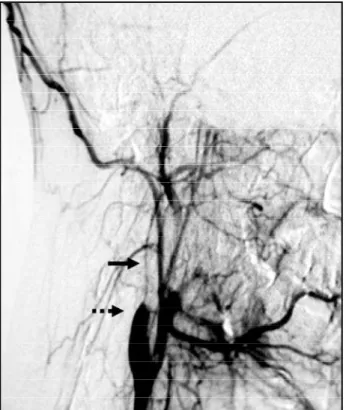
Brain CT revealed a right striatocapsular ischemic s t ro k e (Fig 2). Doppler ultrasound suggested an occlusion in the submandibular portion of the right internal carotid at e rywiath a luminal athrombus. Heparin was satarated. F o u r-vessel digital angiography showed an irregular high-gra-de stenosis at the right internal carotid art e ry (ICA) star-ting about 2 cm distal to the carotid bulb extending until an occlusion into the petrous bone. The proximal segm e n t of the right ICA had a tapered flame-like appearance. There was an accentuation of the filling of the external c a rotid art e ry branches (Fig 3). These findings supported the diagnosis of arterial dissection.
At admission, routine biochemical and hematologi-cal serum parameters were normal except by a mild leu-kocytosis (12.6/nL) and elevated ery t h rocyte sedimenta-tion rate (56mm/h). Both were normal within 1 week. F e v e r resolved within 24 hours. A new chest X-ray discharged a current pneumonia (Fig 1 B). Chest CT showed only re s i-dual pulmonary infiltrate at the site of the recent infec-tion. Echocardiography was normal, except by a slight tricuspid regurgitation. After 20 days, the patient was d i s c h a rged with anticoagulation therapy. Headache had a mild improvement and he could walk with assistance, but his right eye vision did not recover. After 3 months of follow-up and anticoagulation therapy, he was able to walk without assistance and no new ischemic event had been detected.
DISCUSSION
Vascular risk factors as smoking, hypertension, migraine, oral contraception and hyperh o m o c y s t e i-nemia have been found in many patients with ar-terial dissection. It is supposed that, chro n i c a l l y, they may weaken the arterial wall, but the exact mech-anism is unknown and athero s c l e rosis appears not to be involved1. Some studies indicate that recent
infection is among the most important risk factors for ischemic stroke in children and young adults5 -8
and is probably related to CAD2 , 3 , 9. In 1999, a pro
s-pective study comparing the prevalence of recent infection (within one week before CAD) in 43 con-secutive patients with spontaneous CAD and in younger patients with cerebral ischemia from
othFig 3. Digital angiography showing an irregular highgrade ste -nosis (solid arrow) starting about 2 cm distal to the right caro t i d bulb which has a tapered flame-like appearance (dashed arro w ) suggestive of ICA dissection.
Fig 1. Chest Xray. A: At first admission, prior to antibiotic treat -ment. B. After the treatment, at the second admission.
Arq Neuropsiquiatr 2005;63(2-B) 525
er causes found that infection in the preceding w e e k was more common in patients with CAD than in patients without it. Respiratory tract infection ( 6 8 % of infections), but not cough, sneezing and vomi-ting, was independently associated with CAD. In 3 5 % of patients with CAD the infection had been diag-nosed and treated by a physician before dissection or ischemia occurs2. In 2002, another case-control
prospective study evaluated 47 patients with CAD and found similar results. Additionally the authors o b s e rved that the association between recent i n f e c-tion (considered as within four weeks) and CAD was s t ronger in patients with multivessel dissection3. In
both studies, patients were characterized as havi n g infection on a clinical prospective basis (face-to-face i n t e rview) without knowledge of biological and se-rological tests. In our patient, the positive history w a s obtained through direct interview and the confir-mation through reviewing his medical files and re-cent laboratorial and radiological examinations.
Our patient did not present any suggestive clini-cal feature of a connective tissue disord e r. However, even when no clinically apparent connective tissue d i s o rder is detected, patients with spontaneous C A D a resuspected of harboring an underlying stru c t u r-al defect of the arterir-al wr-all, r-although the exact type of arteriopathy remains elusive in most cases1.
Considering the possibility of an underlying arte-riopathy or a connective tissue disease, the damage due to the infection could be facilitated. As a gene-ral role, CAD is not considered an inflammatory ar-teriopathy. However, it is possible that an indirect i n f l a m m a t o ry and immunological host response w i t h activation of several inflammatory biochemical s u b-stances such as cytokines, free radicals and pro t e a s-es could induce extracellular matrix degradation a n d thus weaken the vascular wall3,10,11.
Almost a century ago, a large postmortem exa-mination study1 2mentioned by Grau et al8s h o w e d
that acute infectious disease could lead to conside-rable vascular injury such as focal destruction of s m o-oth muscles and elastic fibers in the tunica media of larger arteries, which could either heal or trans-f o rm into trans-fibrous scars. Intrans-flammatory intrans-filtrates w e re r a rely present, and the intima was involved only i n the most severe cases. Although this had pointed to a possible infection-mediated vascular damage mechanism, coincidentally at the same and commo-nest site for arterial dissection (tunica media), fur-ther pathological studies to evaluate the exact me-chanism underlying the clinically observed link b e t-ween infection and arterial dissection are still
lacki-ng and the mechanism remains speculative. A l t h o u-gh an inflammatory eosinophilic infiltrate was f o u n d in the tunica media of a fatal case of spontaneous c o ro n a ry art e ry dissection1 3, there is no re p o rt of a
similar result in a CAD patient. Considering that m o s t of dissections of the cervical arteries heal spontane-ously and that the rate of death is less than 5 %1,
p e rhaps it explains why the autopsies studies a re rare and pathological and immunological studies prov i n g an infection-related mechanism remain lacking.
R e g a rding the microbial agents, although sero-epidemiological studies have shown an association between raised antibody titres against Chlamydia pneumoniae and carotid athero s c l e rosis or stro k e1 4,
there is no evidence of its relation to arterial dis-s e c t i o n2. However, it is still possible that other
bac-terial or viral agents may damage the arbac-terial wall either directly by infecting it, or indirectly by pro v o-king an autoimmune reaction against some of its components. Additionally, it is important conside-ring that mechanical stress by vomiting or coughi-ng may trigger a CAD. A case of CAD was described in a patient with pertussis and severe paroxysmal c o u-ghing15. The observation in clinical practice shows
that coughing is a far more common clinical man-ifestation than the incidence of arterial dissection. Probably the risk depends on the intensity of the coughing and the individual susceptibility to dis-section, but, as an environmental trigger, infection seems to be independently more important than coughing, as showed by Grau et al.2.
The diagnosis was made by conventional angio-graphy which has long been the gold standard d i a g-nostic test. Pathognomonic features of dissection like intimal flap or a double lumen are detected in less than 10 percent of dissected art e r i e s1. Fre q u e
n-t l y, n-the appearance of an ICA dissecn-tion is an irre g u-lar high-grade stenosis or an occlusion starting a b o u t 2 cm distal to the carotid bulb. Characteristically, at the site the ICA has a tapered flame-like appear-ance1. Actually, MRI techniques are replacing
con-ventional angiography in the diagnosis of CAD be-cause of the improvement of resolution of MRI an-giography, the capability of visualizing the intra-mural hematoma and the non-invasive approach, but, unfort u n a t e l y, it is not largely available in ma-ny hospitals in Brazil yet.
526 Arq Neuropsiquiatr 2005;63(2-B)
REFERENCES
1. Schievink WI. Spontaneous dissection of the carotid and vertebral ar-teries. N Engl J Med 2001;344:898-906.
2. Grau AJ, Brandt T, Buggle F, et al. Association of cervical artery dissec-tion with recent infecdissec-tion. Arch Neurol 1999;56:851-856.
3. Guillon B, Berthet K, Benslamia L, Bertrand M, Bousser MG, Tzourio C. Infection and the risk of spontaneous cervical artery dissection: a case-control study. Stroke 2003;34: 79-81.
4. Schievink WI, Wijdicks EFM, Kuiper JD. Seasonal pattern of sponta-neous cervical artery dissection. J Neurosurg 1998;89-101-103. 5. Syrjanen J, Valtonen VV, Iivanainen M, Kaste M, Huttunen JK. Pre c e d i n g
infection as an important risk factor for ischaemic brain infarction in young and middle aged patients. BMJ 1988;296:1156-1160.
6. Grau AJ, Buggle F, Becher H, et al. Recent bacterial and viral infection is a risk factor for cerebrovascular ischemia: clinical and biochemical studies. Neurology 1998;50:196-203.
7. Grau AJ, Buggle F, Heindl S, et al. Recent infection as a risk factor for cerebrovascular ischemia. Stroke 1995;26:373-379.
8. Grau AJ, Buggle F, Steichen-Wiehn C, et al. Clinical and biochemical analysis in infection-associated stroke. Stroke 1995;26:1520-1526. 9. Grau AJ, Brandt T, Forsting M, Winter R, Hacke W.
Infection-associat-ed cervical artery dissection: three cases. Stroke 1997;28:453-455. 10. Pober JS. Cytokine-mediated activation of vascular endothelium:
phys-iology and pathology. Am J Physiol 1988;133:426-433.
11. Smedly LA, Tonnesen MG, Sandhaus RA, et al. Neutrophil-mediated injury to endothelial cells: enhancement by endotoxin and essential ro l e of neutrophil elastase. J Clin Invest 1986;77:1233-1243.
12. Wiesel J. Die Erkrankungen arterieller Gefa beta e im Verlaufe akuter Infektionen, II: Teil. Zeitschr f Heilkunde. 1906;127:262-294. 13. Robinowitz M, Virmani R, McAllister HA. Spontaneous coronary artery
dissection and eosinophilic inflammation: a cause and effect relation-ship? Am J Med. 1982;72:923-928.