INTRODUCTION
Intraradicular posts are required for supporting a core foundation when there is insufficient clinical crown remaining (1).They have been advocated to strengthen endodontically treated teeth against intraoral forces by distributing torquing forces within the radicular dentin tissue along the roots (2). However, it has been demon-strated that the placement of a post can created stresses that lead to root fracture during function and that the strength of endodontically treated teeth was directly related to the remaining internal tooth structure (3).
Several techniques to restore endodontically treated teeth have been advocated with criteria for suc-cess dependent on variations in length, diameter, shape
Influence of Intraradicular Post and Crown
Ferrule on the Fracture Strength of
Endodontically Treated Teeth
Jefferson Ricardo PEREIRA1
Accácio Lins do VALLE2
Fábio Kenji SHIRATORI2
Janaina Salomon GHIZONI1
Murilo Pereira de MELO3
1Department of Prosthodontics, Dental School, University of Southern Santa Catarina, Tubarão, SC, Brazil 2Department of Prosthodontics, Bauru Dental School, University of São Paulo, Bauru, SP, Brazil 3Health Sciences Center, Department of Dentistry, State University of Maringá, Maringá, PR, Brazil
The aim of this study was to investigate the fracture strength of endodontically treated teeth restored with different posts and variable ferrule heights. Sixty freshly extracted human canines were treated endodontically and randomly assigned to 6 groups (n=10), being restored with custom-made cast post-and-core (CP0 and CP3 groups), prefabricated post and composite resin core (PF0 and PF3 groups), and composite resin (CR0 and CR3 groups). The CP0, PF0 and CR0 groups presented no ferrule and the CP3, PF3 and CR3 presented 3 mm of coronal structure. All teeth were restored with full metal crowns. The fracture strength was measured in a universal testing machine at 45o to the long axis of the tooth until failure. Data were analyzed statistically by 2-way ANOVA and Tukey’s test (α=0.05).
When the mean fracture strength values were compared (CP0 group - 820.20 N, CP3 group - 1179.12 N; PF0 group - 561.05 N; PF3 group - 906.79 N; CR0 group - 297.84 N; and CR3 group - 1135.15 N) there was statistically significant among the groups (p<0.05), except for the three groups with 3 mm of coronal remaining, which were similar to each other. The results of this study showed that the ferrule in crowns promoted significantly higher fracture strength in the endodontically treated teeth.
Key Words: composite resins, fracture strength, fracture stress, posts and core technique, pulpless teeth, root fracture.
and surface configuration,amount of dentinal structure, materials and techniques used in construction (4-6).
Cast post and core has been regarded as the “gold standard” in post-and-core restorations due to its superior success rate, when coronal tooth structure is missing (6,7). Alternatives to cast posts and cores have been developed. Prefabricated post systems simplify the restorative procedure because all steps can be completed chairside and clinical success can also be expected (6,7). Fraga et al. (8)showed that roots restored with cast posts exhibited significantly higher internal stresses than prefabricated posts.
Prefabricated posts and composite resin core association is a viable technique for endodontically treated teeth (6,7). Previous studies (6,8) have shown that when this association was used, fracture of the
storative material was the most common failure, while root fracture was the most common failure when a cast post and core system was used.
An important element for tooth preparation when using a post and core is the incorporation of a ferrule (9-12). The effectiveness of the ferrule has been evalu-ated by a variety of methods, including fracture testing, (10-12) impact testing, (13) fatigue testing, (14) and photoelastic analysis (15). Several authors (1,3,16)have suggested that the tooth should have a minimum amount of 2 mm of coronal structure above the cementoenamel junction to ensure proper strength. This coronal structure will provide a ferrule effect with the artificial crown to prevent root fracture and post fracture or dislodgement (3,10,11,17-20). Gegauff (10) evaluated the effect of crown ferrule on the fracture strength of endodonti-cally treated teeth and showed no significant correla-tion between amount remaining coronal structure and fracture strength. However, Pereira et al. (11) showed that increasing ferrule length significantly increased the fracture strength of endodontically treated teeth.
Although authors have different opinions about the ideal amount of remaining coronal structure, the results of fracture strength of endodontically treated teeth restored by cast posts or prefabricated posts are acceptable clinically because they are considerably higher than the maximal physiologic forces acting on the teeth in the oral cavity (20).
The aim of this study was to compare the fracture strength of endodontically treated teeth with different posts and different amounts of coronal tooth structure available for crown treatment. The tested hypothesis was that there is a significant difference in the effect of remaining coronal structures and types of posts on the fracture strength of endodontically treated teeth.
MATERIAL AND METHODS
The research project was reviewed and ap-proved by the Research Ethics Committee of Bauru Dental School, University of São Paulo. Eighty-seven human maxillary canines obtained shortly after extrac-tion for periodontal reasons were examined for the study. Twenty-seven teeth were discarded for presence of root caries, restorations, previous endodontic treatment or cracks, and the remaining teeth were cleaned and stored in distilled water at 37ºC until the end of the study. All selected teeth had root sizes between 15 mm and 16 mm
measured with a millimeter ruler from the apex to the ce-mentoenamel junction (CEJ). Instrumentation was done using a size 20 standard master apical K-file (Dentsply Maillefer, Ballaigues, Switzerland) 1 mm short of the apex and conventional step-back technique was done up to a size 35 K-file (Dentsply Maillefer) was used. The roots were irrigated copiously with 2.5% sodium hypo-chlorite solution (Asfer Industrial Química, São Paulo, SP, Brazil) throughout the preparation and dried with sterile paper points (Tanari, Tamariman Industrial Ltda., Manacapuru, AM, Brazil). The roots canals were filled by lateral condensation of gutta-percha cones (Tanari, Tamariman Industrial Ltda.) and Endomethasone (Ivory; Septodont Brasil, Barueri, SP, Brazil) sealer. The teeth were randomly assigned to 6 groups (n=10).
Post preparations were standardized using a #5 reamer (Largo; Dentsply Maillefer) to remove 9 mm of gutta-percha from each tooth. All groups named with 0 (CP0, PF0, and CR0) had the coronal portion of the teeth sectioned at the CEJ using a double-faced diamond disc (KG Sorensen Indústria e Comércio Ltda., Barueri, SP, Brazil) (Fig. 1) and the groups named with 3 (CP3, PF3, and CR3) had the coronal tooth structure reduced to a flat plane, up with 3 mm in height from the CEJ (Fig. 2). All specimens were prepared with water-cooled #3216 diamond burs (KG Sorensen Indústria e Co-mércio Ltda.) at high-speed handpiece (Super Torque 625 Autofix; Kavo do Brasil Ind. Com. Ltda, Joinville, SC, Brazil) according to a previously outlined crown preparation (1.5 mm facial reduction with a chamfer finish line and 0.5 mm chamfered lingual reduction). In all specimens and groups, the finish lines were placed at the CEJ level.
low-speed handpiece. Cement was also placed on the post and seated under a load of 5 kgf during 5 min. After this time, the load was removed and the post was held in place for 8 min until final setting of the cement. Excess cement was removed and each specimen was returned to storage in distilled water. All specimens were stored in distilled water at 37ºC until the end of the study.
The PF0 and PF3 group were restored with prefab-ricated stainless steel, parallel-sided, serrated posts with a tapered end (#5317; Screw-Post, Euro-Post Anthogyr S.A., Sallanches, France). In these groups, the teeth were cemented with the same material and technique as used in the CP0 and CP3 groups. The coronal por-tion was built with composite resin (Z250; 3M/ESPE). The root surfaces and cervical dentin were etched with 37% phosphoric acid for 30 s, rinsed and air dried. Two layers of Primer-Bond 2.1 adhesive system (Dentsply Ind. Com. Ltda., Petrópolis, RJ, Brazil) were applied to cervical dentin and coronal portion of the post, and were light-activated for 20 s with a halogen light-curing unit (Ultraled; Dabi Atlante, Ribeirão Preto, SP, Brazil) with 450 mW/cm2 light intensity. Cores were fabricated in a standardized form, using the same core-forming matrix as used in the other groups. Five increments of the composite resin were applied to complete the coronal core, each requiring 40 s of photo-activation (Ultraled; Dabi Atlante). The tip of the light-activation unit was positioned on the top of the core 1 cm from the specimens.
In the CR0 and CR3 groups, the canals were restored with Z250 composite resin (3M/ESPE) using a translucent curing post (Luminex System; Dentatus Ltd., New York, NY, USA). Root dentinal walls were etched with 37% phosphoric acid for 15 s, copiously rinsed with water, dried with paper points (Tanari; Tanari-man Industrial Ltda.,) and coated with Prime-Bond 2.1 bonding agent as recommended by the manufacturer. The translucent post (Luminex System; Dentatus Ltd.) was used in the canal during the polymerization of the bonding agent and during root filling with the compos-ite resin inserted in layers under this post. Each layer had approximately 1.0 mm thick (from the apex to the CEJ). Before the resin layer was inserted into the root canal, it was measured with a mm ruler, a thin resin layer of composite resin was inserted in the root canal that was measured again. The difference between both measurements should be 1 mm. The composite resin was light for 40 s with the translucent post in position. Core reconstruction was done in the same way as described for the PF0 and PF3 groups.
The root canals of the teeth fitted with custom cast post and core and composite resin were prepared as the root canals for the prefabricated posts.
Impressions were taken with vinyl polysiloxane impression material (Aquasil; Dentsply DeTrey GmbH, Konstanz, Germany) and Ni-Cr alloy crowns (Durabond, São Paulo, SP, Brazil) were made by the conventional casting technique. Resin modified glass ionomer
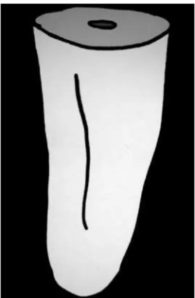
ce-Figure 1. Coronal tooth structure reduced to a flat plane at height of 0.0 mm.
ment (Rely X; 3M/ESPE) was used for cementation of the crowns.
The root surface of each tooth was coated with an approximately 60-mm-thick layer of silicone impres-sion material (Aquasil; Dentsply DeTrey) to simulate a periodontal ligament (11).Root surfaces were marked 2 mm below the CEJ and covered with a 0.6-mm-thick foil (Adapta foil; Bego). All specimens were embed-ded in acrylic resin (Artigos Odontológicos Clássico Ltda.) poured into molds (30 mm high and 22 mm in diameter) of the same material, with an internal central opening measuring 20 mm high and 10 mm in diameter. The teeth were embedded along their long axes using a surveyor (Bio-Art Equipamentos Odontológicos Ltd, São Carlos, SP, Brazil) and placed in a cool water bath during polymerization of the resin. After the first signs of polymerization, teeth were removed from the resin blocks along their long axes using the surveyor, and the spacers (Adapta) were removed from root surfaces. A silicone-base impression material (Aquasil; Dentsply DeTrey) was injected into the acrylic resin blocks, and the teeth were reinserted into the resin cylinders. A standardized silicone layer that simulated periodontal ligament was thus created.
After 48-h storage in distilled water at 37ºC, each acrylic block was placed in a custom apparatus that allowed the block of acrylic to be positioned a 45o angle with the long axis of the tooth (11,12). A universal testing machine (Kratus K2000 MP; Dinamômetros Kratos Ltda, São Paulo, SP, Brazil) was used to apply a constant load at a crosshead speed of 0.5 mm/min until failure. The force in N was applied 3 mm below the incisal edge of the palatine face of the crowns. Failure was considered as the point at which the loading force reached a maximum value by fracturing the root or core, bending or debonding the post. The failures were analyzed with a ×4 binocular microscope (Bio Art Eq-uipamentos Odontológicos Ltda.).
Two-way analysis of variance was used to deter-mine the overall differences among the mean values of the groups and the overall variability within the groups. Tukey’s multiple comparison test was used to establish
intergroup differences (α=0.05).
RESULTS
The CP3 group showed the highest mean frac-ture strength values (1179.1a ± 208.2 N) followed by
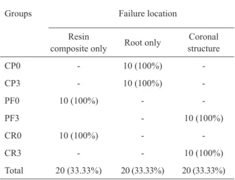
CR3(1135.3a ± 175.1 N), PF3 (906.8ab ± 270.4 N), CP0 (818.2b ± 147.2 N), , PF0 (561.1c ± 136.9 N) and CR0 (297.8d ± 78.9 N) (different letters indicate statistically significant difference at 5%). The groups differed for type of restoration (p=0.0000), corona remaining (p=0.0000) and interaction between these variables (p=0.0000). There were no significant differences (p>0.05) among the groups with 3 mm of remaining coronal structure, while the groups did not differ significantly from each other. The types of fractures occurred in all groups are shown in Table 1.
DISCUSSION
The present study tested the hypotheses that there is a significant difference in the effect of the dif-ferent posts and the amount of coronal tooth structure on the fracture strength of endodontically treated teeth.
The results showed that the fracture strength of root-filled teeth without a post was not significantly different from that of post-reinforced teeth when coronal structure was present. The findings of a previous study suggest that root reinforcement with ferrule may protect endodontically treated teeth against fracture by counter-acting and better distributing the stresses generated by the post (18). In addition, the ferrule effect significantly improves the fracture strength of the tooth and is more important than the type of material with which the core and post are made (17).
When the ferrule was not present, the fracture strength of the teeth restored with cast post and core (CP0) was significantly higher than those restored with
Table 1. Types of failure (number and percentage of teeth).
Groups Failure location
Resin
composite only Root only
Coronal structure
CP0 - 10 (100%)
-CP3 - 10 (100%)
-PF0 10 (100%) -
-PF3 - 10 (100%)
CR0 10 (100%) -
-CR3 - - 10 (100%)
prefabricated posts (PF0) and those restored with com-posite resin (CR0). It has been suggested that the strength of the tooth is directly related to the remaining dentin bulk, which means that the fracture strength decreases as the amount of remaining coronal dentin is reduced (19).The higher fracture strength ofCP0 group could be related to the higher flexural strength and modulus of elasticity of the metal alloy (8). However, except for teeth without post (CR0 group), the forces responsible for failure in the current study were considerably higher than the maximal physiological forces acting on the teeth in use (20). Lyons and Baxendale (20) observed that the mean force applied on an upper canine was 215 N. In the presence of parafunctional loading these authors noted that this force increased to 254.8 N, and the maximum forces were between 343 and 362.6 N.
This study showed that presence of ferrule design significantly increased the fracture strength of endodontically treated teeth in all groups. These and oth-ers studies (2,19) support the idea that loss of structural integrity associated with the access preparation may lead to a higher occurrence of fractures in endodonti-cally treated teeth. The findings of the present study are in agreement with the study of Pereira et al. (11) and others authors (4,5,13-15), which advocate that fracture resistance was highest in specimens with the longest ferrules.
The most common failure in the direct technique (prefabricated post and composite resin or composite resin) is fracture of the restorative material or the coronal structure and in the cast post-and-core the fracture of the root (9), as also observed in the present study (Table 1).
This investigation demonstrated that roots re-stored with individual cast posts exhibited higher fracture strength than those restored with prefabricated post and composite resin core or composite resin. Despite lower strength obtained in the technique using prefabricated posts and composite resin with or without remaining coronal structure or composite resin with coronal, this technique is appropriate because no root fractures were showed. In addition, the direct method is claimed to protect the tooth structure (9). Conversely, the tech-nique using composite resin without coronal structure should be inappropriate because the forces responsible for failure were considerably lower than the maximal physiologic forces acting on the teeth in use (20).
Two limitations of this study were the fact that it was an in vitro investigation, which does not fully
replicate oral conditions, and the use of a single load for testing the fracture strength of endodontically treated teeth. For more meaningful results, future studies should incorporate thermal and fatigue cycling of the specimens.
RESUMO
O objetivo neste estudo foi avaliar a resistência à fratura de dentes tratados endodonticamente restaurados com diferentes pinos e diferentes alturas de remanescente dentinário da coroa. Sessenta caninos recém-extraídos foram tratados endodontica-mente, separados em 6 grupos (n=10) e restaurados com núcleo metálico fundido (CP0 e CP3), pino pré-fabricado e núcleo em resina composta (PF0 e PF3) ou resina composta (CR0 e CR3). Os grupos CP0, PF0 e CR0 não possuíam férula e os grupos CP3, PF3 e CR3 apresentaram 3 mm de remanescente coronário. Todos os dentes foram restaurados com coroas totais metálicas. A resistência à fratura foi medida em máquina universal de ensaios com o longo eixo do dente posicionado a 45 graus em relação ao carregamento axial, até que ocorresse fratura. A análise de variância 2 critérios (α=0,05) mostrou diferença estatisticamente significativa entre os grupos. Quando as médias das forças para fratura foram comparadas (CP0 = 820,0 N; CP3= 1179,12 N; PF0 = 561,05 N; PF3 = 906,79 N; CR0 = 297,84 N; e CR3 = 1135,15 N) não foram observadas diferenças significativas entre os 3 grupos com 3 mm de remanescente coronal. Os resultados mostraram que a presença de férula em coroas aumenta significantemente a resistência à fratura de dentes tratados endodonticamente.
ACKNOWLEDGEMENTS
The authors would like to thank CAPES (Coordination of Support for Superior Education) for the financial support of this study. The authors want to thank Prof. Dr. José Roberto Pereira Lauris for the statistical analysis.
REFERENCES
1. Trabert KC, Cooney JP. The endodontically treated tooth: restor-ative concepts and techniques. Dent Clin North Am 1984;28:923-951.
2. Zogheib LV, Pereira JR, Valle AL, Oliveira JA, Pegoraro LF. Fracture resistance of weakened roots restored with composite resin and glass fiber post. Braz Dent J 2008;19:329-333. 3. Sorensen JA, Engelman MJ. Ferrule design and fracture resistance
of endodontically treated teeth. J Prosthet Dent 1990;63:529-536. 4. Fernandes AS, Shetty S, Coutinho I. Factors determining post
selection: a literature review. J Prosthet Dent 2003;90:556-562. 5. Oliveira JA, Pereira JR, Valle AL, Zogheib LV. Fracture resistance
of endodontically treated teeth with different heights of crown fer-rule restored with prefabricated carbon fiber post and composite resin core by intermittent loading. Oral Surg Oral Med Oral Pathol Oral Radiol Endod 2008;106:e52-57.
6. Torbjorner A, Fransson B. A literature review on the prosthetic treatment of structurally compromised teeth. Int J Prosthodont 2004;17:369-376.
8. Fraga RC, Chaves GSB, Mello JF, Siqueira JR. Fracture resistance of endodontically treated roots after restoration. J Oral Rehabil 1998;25:809-813.
9. Rosen H. Operative procedures on mutilated endodontically treated teeth. J Prosthet Dent 1961;11:973-986.
10. Gegauff AG. Effect of crown lengthening and ferrule placement on static load failure of cemented cast post-cores and crowns. J Prosthet Dent 2000;84:169-179.
11. Pereira JR, de Ornelas F, Conti PC, do Valle AL. Effect of a crown ferrule on the resistance of endodontically treated teeth restored with prefabricated posts. J Prosthet Dent 2006;95:50-54. 12. Pereira JR, Mendonça Neto T, Porto V de C, Pegoraro LF, Valle
AL. Influence of the remaining coronal structure on the resistance of teeth with intraradicular retainer. Braz Dent J 2005;16:197-201. 13. Cathro PR, Chandler NP, Hood JA. Impact resistance of crowned endodontically treated central incisors with internal composite cores. Endod Dent Traumatol 1996;12:124-128.
14. Isidor F, Brondum K, Ravnholt G. The influence of post length and crown ferrule on the resistance to cyclic loading of bovine teeth prefabricated titanium post. Int J Prosthodont 1999;12:79-82.
15. Eraslan O, Aykent F, Yucel MT, Akman S. The finite element analysis of the effect of ferrule height on stress distribution at post-and-core-restored all-ceramic anterior crowns. Clin Oral Investig 2009;13:223-227.
16. Wagnild GW, Mueller KL. Restoration of the endodontically treated tooth. In:Cohen S, Burns RC, editors. Pathways of the pulp. 8th ed. St. Louis: Elsevier; 2001. p. 765-795.
17. Hoag EP, Dwyer TG. A comparative evaluation of three post and core technique. J Prosthet Dent 1982;47:177-181.
18. Assif D, Bitenski A, Pilo R, Oren E. Effect of post design on re-sistance to fracture of endodontically treated teeth with complete crowns. J Prosthet Dent 1993;69:36-40.
19. Zhi-Yue L, Yu-Xing Z. Effect of post-core design and ferrule on fracture resistance of endodontically treated maxillary central incisors. J Prosthet Dent 2003;89:368-373.
20. Lyons MF, Baxendale RH. A preliminary electromyographic study of bite force and jaw-closing muscle fatigue in human subjects with advanced tooth wear. J Oral Rehabil 1990;17:311-318.