Comparison of methods to
evaluate total body fat and its
distribution
Comparação de métodos de
avaliação da gordura corporal total
e sua distribuição*
Karine Anusca Martins
IEstelamaris Tronco Monego
IRégis Resende Paulinelli
IIRufo Freitas-Junior
IIII School of Nutrition at the Universidade Federal de Goiás (UFG).
II Department of Gynecology and Obstetrics of the Medical School at the
Universidade Federal de Goiás (UFG)
III Department of Gynecology and Obstetrics of the Medical School at the
Universidade Federal de Goiás (UFG)
This research project was developed in the Rede Goiana de Pesquisa em Mastologia (State of Goiás Breast Disease Research Network), through the Postgraduate Program in Health Sciences of the Universidade Federal de Goiás (UFG).
Conlito de interesse: Não existem conlitos de interesses.
Financiamento: The present study was partly funded by the Fundação de Amparo à Pesqui-sa do Estado de Goiás (FAPEG – State of Goiás Research Support Foundation), process number: 00228648-96; public notice number: 01/2007.
Extracted from the Doctoral Dissertation entitled “Body Composition Assessment of Women Recently-Diagnosed with Breast Cancer”, presented to the Postgraduate Program of the Medical School at the Universidade Federal de Goiás on January 19th, 2010.
Correspondência: Rufo Freitas-Junior. Hospital das Clínicas, Bloco B. 1ª avenida s/n Setor Univer-sitário, Goiânia, GO CEP 74606-050. E-mail: [email protected]
Abstract
Objective: To compare two methods for evaluating total body fat and its distribution.
Methods: Cross-sectional, cohort-nested study. Sixty-two women received a nutritional status evaluation which included total body fat (BF) obtained through the sum
of skinfolds (ΣSF) and bioimpedance (BIA).
Visceral fat distribution was measured using ultrasonography (USG) (intra-abdominal fat thickness) (IAT) and waist circumference ( WC). The concordance correlation coeficient (CCC) and the determination
coefficient (r2) were calculated. Results:
Mean patient age was 48.19 (8.99) years. Thirty-six women (58.06%) had a very large WC and 42 (67.74%) had high body fat. There was moderate concordance (r2 = 0.42; CCC = 0.59; p < 0.01) between the
methods for determining body fat (%) and
optimal concordance (r2 = 0.90; CCC = 0.91;
p < 0.01) for body fat (kg) determined by BIA
and ΣSF. The comparison between WC and
IAT (USG) showed moderate concordance
(r2 = 0.49; p < 0.01) between the methods.
Conclusions: Moderate concordance in determining total body fat (%) and optimal concordance in determining body fat (kg) were found between the methods. Moderate concordance was found between the methods for determining body fat distribution.
Resumo
Objetivo: Comparar dois métodos de avaliação da gordura corporal total e sua
distribuição. Métodos: Estudo transversal,
aninhado a uma coorte. Em amostra de 62 mulheres realizou-se avaliação do estado nutricional, incluindo a gordura corporal (GC) total obtida pelo somatório de dobras
cutâneas (ΣDC) e bioimpedância (BIA).
Mensurou-se a distribuição da gordura vis-ceral por ultrassonograia (USG) (espessura de gordura intra-abdominal-EIA) e circun-ferência da cintura (CC). Foram calculados o coeiciente de correlação de concordância
(CCC) e o coeiciente de determinação (r2).
Resultados: A média de idade das pacientes foi de 48,19 (8,99) anos. Observou-se 36 (58,06%) mulheres com a CC muito au-mentada e 42 (67,74%) com GC auau-mentada.
Identiicou-se moderada concordância (r2 =
0,42; CCC = 0,59; p < 0,01), entre os métodos avaliados para determinação da gordura
corporal (%) e uma ótima concordância (r2
= 0,90; CCC = 0,91; p < 0,01) para a gordura
corporal (kg), avaliadas por BIA e ΣDC. A
comparação entre a CC e EIA (USG)
eviden-ciou uma moderada concordância (r2 = 0,49;
p < 0,01), entre os métodos. Conclusões:
Evidenciou-se moderada concordância na avaliação da gordura corporal total (%) e ótima concordância na avaliação da gordura corporal (kg), entre os métodos utilizados. Identiicou-se uma moderada concordância entre os métodos de distribuição da gordura corporal.
Palavras-chave: Composição corporal. Distribuição da gordura corporal. Dobras cutâneas. Impedância bioelétrica. Circun-ferência da cintura. Saúde pública.
Introduction
Nutritional status assessment is a key as-pect in the identiication of problems and/ or inadequate nutritional status during any
stages of life1, especially during diseases2,3
and including neoplasm’s, as they directly or indirectly inluence an individual’s health prognosis4.
The increase in visceral or total body fat is harmful to health, especially among women and those with non-communicable chronic diseases3,5, such as breast cancer6,7.
Throughout time, it was observed that visceral fat more accurately determines the risk factor for metabolic problems than total body fat8-12.
When the possible consequences of body composition changes in women’s nu-tritional status and health are considered, early assessment and identiication of such changes may contribute to the reduction in the effects resulting from the associated
health problems13.
There are several methods that can be used in this assessment, some of which are more accurate and expensive, slower and more complex to be executed, such as Dual-emission X-ray Absorptiometry (DXA), hydrostatic weighing (HW), magnetic reso-nance imaging (MRI) and X-ray
computer-ized tomography (CT)14,15. In contrast, there
are other less expensive and easily executed methods to assess total body fat, such as Bioelectrical Impedance or Bioimpedance
(BIA) and skinfold measurement14.
On the other hand, tomography is tradi-tionally considered to be the most eficient and accurate method to determine visceral
fat tissue16, although it becomes
impracti-cable, due to its high cost. Ultrasonography has been used as an alternative, as it shows a high level of agreement with CT, especially
in areas with more visceral fat17.
When more accurate methods like the ones are not available, a more accessible alternative would be skinfold measurement (total body fat assessment) and waist cir-cumference measurement, which indirectly
are easily executed, applied and accessed, although some studies have questioned their accuracy1,14.
The majority of studies aimed at com-paring total body fat assessment, using eas-ily executed methods such as bioimpedance and the sum of skinfolds, were conducted
with sportsmen and women or athletes18,19.
In contrast, studies conducted with women from several age groups used different methods, including the most expensive ones15,20-22. There are few studies that have
been performed with women ranging from
normal weight to obesity23, with breast
can-cer and benign breast changes, coming from public health services. In addition, there are few studies that compare intra-abdominal fat thickness measurement with waist
cir-cumference to assess visceral fat24,25.
The present study aimed at the fol-lowing: verifying whether the previously mentioned methods can be used in the nutritional follow-up of women cared for in public health services, especially those with breast diseases; comparing two meth-ods used to estimate total body fat (sum of skinfolds and bioelectrical impedance); and assessing the correlation between both estimates of visceral fat (waist circumfer-ence measurement and intra-abdominal thickness measurement obtained by ultra-sonography).
Methods
A cross-sectional study, nested in a cohort study, was conducted in the city of Goiânia, state of Goiás, Brazil, in 2009. This cohort study is prospective in nature and it is ongoing, aiming to ind out the impact of chemotherapy on body fat distribution and lipid proile of women with breast cancer, in two referral centers of Goiânia, GO.
Sample size was calculated for the previ-ously mentioned cohort study. A total of 62 women were included, of which 31 had been recently diagnosed with breast cancer and 31 had benign breast changes. The entire
study group participated in the
Universi-dade Federal de Goiás Clinical Hospital and
the Hospital Araújo Jorge Breast Disease
Program and the Associação de Combate
ao Câncer de Goiás (ACCG – State of Goiás Anti-Cancer Association) Gynecology and Breast Service. As a common denominator, both services care for women coming from the Sistema Único de Saúde (Uniied Health
System) and belong to the Rede Goiana
de Pesquisa em Mastologia (State of Goiás Breast Disease Research Network)
Data collection was conducted by previously trained interviewers and an-thropometrists, following the norms of the Measurement Standardization Manual for Interviewers and Anthropometrists and according to the techniques described
above26,27. Data were collected with a
ques-tionnaire applied during a direct interview, with a socio-demographic characterization and nutritional status assessment (anthro-pometry).
The following socio-demographic vari-ables were analyzed: age (in years), level of education (in years of study) and per capita household income (categorized in minimum wages). The anthropometric variables considered were as follows: cur-rent weight, height, biceps skinfold (BSF), triceps skinfold (TSF), suprailiac skinfold (SISF), subscapular skinfold (SESF), and waist circumference (WC). Bioelectrical impedance or bioimpedance (BIA) was used to assess body composition.
Based on the anthropometric measure-ments, the body mass index (BMI), sum of
skinfolds (ΣSF), percentage body fat (% BF)
and body fat in kilograms (Kg), using the SF and BIA. Subcutaneous fat thickness and intra-abdominal fat thickness were determined with abdominal ultrasonog-raphy (US).
The norms and procedures proposed
by Lohman, Roche and Martorell27 were
followed to collect anthropometric data (weight, height, waist circumference and skinfolds). The World Health
Organiza-tion (WHO) classificaOrganiza-tion28 was adopted
to determine patients’ nutritional status according to their BMI, while the
determine percentage body fat.
Skinfold measurements were obtained using a Lange Skinfold Caliper, with a 0-60 mm scale, 1 mm accuracy and three repeti-tions. The sum of the four skinfolds (BSF, TSF, SISF and SESF) enabled the indirect calculation of percentage body fat and body fat (%GC) and body fat in kilograms (Kg). Based on the values found, body density (BD) could be calculated, according to what
was proposed by Durnin and Womersley30
and subsequently applied to the formula
suggested by Siri31, thus obtaining body fat
(% and Kg).
Total body fat assessment was per-formed with a Bodystat Body Composition Monitoring Unit, model 1500, a Bioimped-ance (BIA) device with an impedBioimped-ance measurement scale of 20-2000ohms, an accuracy of 6 ohms and frequency of 50 KHz (KiloHertz). The following previous conditions were considered to perform the examination: to not use a pacemaker; to have been fasting for two hours or longer, including coffee or alcoholic beverages; and not to have smoked for at least two hours before this examination; to have an empty bladder; and not to have exercised for at
least 12 hours before this examination32.
Intra-abdominal fat thickness mea-surement was obtained with the TOSHIBA SSA-250A ultrasonography equipment. The estimate of visceral fat was obtained with a 3-5 MHz convex transducer that measured fat tissue thickness of patients who had been fasting for at least six hours, in a dorsal recumbent position, in the region located right above the navel, on the xipho-umbil-ical line, applying the minimum pressure required to visualize the image, according
to a standard technique17.
The reading was conducted directly with images frozen on the screen. The measurement between the posterior wall of the rectus abdominis muscle and the posterior wall of the aorta was considered
as the intra-abdominal fat thickness17. Only
the patients cared for in the Clinical Hospital had this exam performed, due to the limited availability of the device, totaling 49 women.
The 2003 Excel software program was use to tabulate data, while the SPSS 8.0 and STATA 8.0 software programs were used for the statistical analysis. Descriptive statistics (frequency, mean, median, minimum and maximum values) were used in the data analysis.
The coefficient of determination (r2)
was used to assess the association between waist circumference measurement and intra-abdominal fat thickness, considering a signiicance level of α < 5%.
Women were informed about the re-search objectives during the interview, when an informed consent form was presented to them and they could decide to participate in the study or not.
This research project was approved by the Universidade Federal de Goiás Clinical Hospital Human and Animal Research Eth-ics Committee (HC/UFG), protocol number
073/2008, and by the Associação de Combate
ao Câncer de Goiás (ACCG – State of Goiás Anti-Cancer Association) Research Ethics
Committee of the Hospital Araújo Jorge,
protocol number 001/09.
Authors declared there were no conlicts of interest.
Results
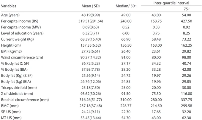
The mean age of the 62 women studied was 48.19 (8.99) years, mean monthly per capita income was R$ 319.51 or US$ 172.71 (291.64), which represents 0.69 (0.63) mini-mum wages and a mean of 6.32 (3.71) years of education (Table 1).
With regard to anthropometric variables, the mean BMI of women studied was in
the overweight category (BMI ≥ 25 Kg/m2),
higher than what is recommended, i.e. there were 42 (67.74%) interviewees with exces-sive weight, of which 28 (45.16%) patients
were overweight (BMI ≥ 25) and 14 (22.58%)
were obese (BMI ≥ 30) (Table 1).
Con-sequently, the mean waist circumference value was within the range of high risk for metabolic complications (> 88 cm) associ-ated with excessive weight.
In terms of percentage of body fat, a mean value of 37.93% (7.78) was found using the BIA, and of 36.72% (5.23), using the sum of skinfolds (ΣSF).
With regard to total body fat values (Kg), a mean value of 26.76 Kg (12.06) was found with the BIA, and of 25.56 Kg (9.14) with the sum of skinfolds ( SF). Thus, the mean percentage of body fat (%BF) was also increased in the classiication of risk for obesity-related disorders (> 32.0%) (Table 1). Of all 62 women interviewed, 36 (58.06%) had a much higher risk of metabolic com-plications, identiied by the waist
circumfer-ence values (≥ 88 cm), indicating increased
abdominal fat, which characterizes central obesity. The majority of women evaluated (n=50; 80.64%) had an increased percent-age of body fat, i.e. body adiposity assessed with two methods (sum of skinfolds and bioimpedance).
With regard to the correlation between the values of percentage (%) of body fat obtained with the sum of skinfolds and BIA, aiming to compare these two assessment methods, the concordance correlation icient (CCC=0.59) and determination coef-icient (r2=0.42; p<0.01) revealed a moderate
level of agreement between such methods (Figures 1A and 1B).
In contrast, the correlation between the values of total body fat (Kg) obtained
Tabela 1 - Medidas de tendência central e de dispersão das variáveis sociodemográicas e antropométricas das mulheres do estudo. Goiânia (GO), 2009
Table 1 – Central tendency and dispersion measurements of sociodemographic and anthropometric variables for women participating in the study. Goiânia (GO), 2009
Variables Mean ( SD) Median/ 50º Inter-quartile interval
75º
Age (years) 48.19(8.99) 49.00 43.00 54.00
Per capita income (R$) 319.51(291.64) 240.00 153.75 427.50
Per capita income (MW) 0.69(0.63) 0.52 0.33 0.92
Level of education (years) 6.32(3.71) 6.00 3.75 8.25
Current weight (Kg) 68.39(15.40) 66.90 58.48 73.22
Height (cm) 157.35(6.52) 156.50 153.00 162.25
BMI (Kg/m2) 27.73(6.61) 26.40 23.61 29.82
Waist circumference (cm) 90.27(14.32) 91.00 80.00 98.00
% Body fat (Σ SF) 36.72(5.23) 37.17 34.32 40.74
% Body fat (BIA) 37.93(7.78) 38.20 33.28 42.08
Body fat (Kg) (Σ SF) 25.56(9.14) 24.72 19.97 29.26
Body fat (kg) (BIA) 26.76(12.06) 24.85 19.96 29.85
Triceps skinfold (mm) 25.18(7.50) 25.00 20.00 30.00
Σ of skinfolds (mm) 93.62(30.26) 91.50 75.50 116.00
Brachial circumference (mm) 316.26(51.77) 310.00 280.00 337.75
BMC (mm) 237.18(37.48) 228.77 214.50 259.58
SF-US (mm) 24.24(9.11) 22.30 17.65 32.20
IAT-US (mm) 53.45(13.44) 54.70 43.00 62.30
DP: Standard-deviation; IMC: Body Mass Index; (%): percentage; DC: Skinfold; BIA: Bioimpedance; Σ: sum; CMB: Arm Muscle Circumference; ES-USG: Subcuta-neous thickness (Ultrasound); EIA-USG: intra-abdominal thickness (Ultrasound). Minimum Wage for the study period: R$ 465.00. Dollar for the study period: R$ 1.85
DP: Desvio-padrão; IMC: Índice de Massa Corporal; (%): percentual; DC: Dobras Cutâneas; BIA: Bioimpedância; Σ: somatório; CMB: Circunferência Muscular Braquial;
Figure 1A - Dispersion diagram of the concordance between body fat (%) as measured by the sum of skinfolds and bioimpedance (BIA) for women participating in the study. Goiania, GO, 2009
Figura 1A - Diagrama de dispersão da concordância entre a gordura corporal (%) avaliada pelo somatório das dobras cutâneas (ΣDC) e pela bioimpedância (BIA) das mulheres
participantes do estudo. Goiânia (GO), 2009
Figure 1B -Graph of the concordance between the mean and mean diference, and the calculation of the body fat percentage (%) concordance limit for women in the study (Bland and Altman). Goiânia,GO. 2009
Figura 1B - Gráico de concordância entre a média e a diferença da média e o cálculo do limite de concordância da porcentagem (%) de gordura corporal das mulheres do estudo (Bland e Altman). Goiânia (GO), 2009
Figure 2A -Dispersion diagram of the concordance between body fat (kg) as measured by the sum of skinfolds and bioimpedance (BIA) for women participating in the study. Goiânia, GO. 2009
Figura 2A - Diagrama de dispersão da concordância entre a gordura corporal (Kg) avaliada pelo somatório das dobras cutâneas (ΣDC) e pela bioimpedância (BIA) das mulheres
participantes do estudo. Goiânia (GO), 2009
Figure 2B - Graph of the concordance between mean and mean diference, and the calculation of the body fat (kg) concordance limit for women in the study (Bland and Altman). Goiânia, GO. 2009
Figura 2B - Gráico de concordância entre a média e a diferença da média e o cálculo do limite de concordância da gordura corporal (Kg) das mulheres do estudo (Bland e Altman). Goiânia (GO), 2009
with the above mentioned methods identi-ied a concordance correlation coeficient (CCC=0.91) and a determination coeficient
(r2=0.90; p<0.01) that showed an excellent
level of agreement between these methods (Figures 2A and 2B).
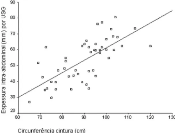
the US and waist circumference, aiming to compare one with the other, showed a
mod-erate coeficient of determination (r2=0.49;
p<0.01) between waist circumference and intra-abdominal thickness (Figure 3).
Discussion
The present study showed that the women evaluated had a mean weight and BMI higher than the recommendations for
ideal weight28 and an increased percentage
body fat29. This proile indicates the need
for a speciic health promotion interven-tion performed by a multidisciplinary team, aiming to reduce the risk factors of several
diseases associated with excessive weight3,35.
The comparison between the two meth-ods used to assess total body fat, which were proposed by this study (sum of skinfolds and bioelectrical impedance) indicated a
signiicant moderate level of agreement33,34.
This result was slightly lower than what was expected, when compared to recent studies conducted with a similar population (r2=0.90) 23, in addition to other populations,
such as female soccer players (r2=0.67)18
and non-institutionalized elderly women (r2=0.79)20.
The present study found a high level
of agreement between the two methods (bioimpedance and sum of skinfolds) used to assess body fat in kilograms (Kg). This has also been observed by other authors in studies conducted with women undergoing hemodialysis (r2=0.96)36 (r2=0.87)37 and with
overweight and obese women practicing walking (r2=0.83)19.
However, the results shown in this study differ from those found by
Rodrigues-Bar-bosa et al.22, who analyzed elderly women
and did not ind agreement (r2=0.25, p<0.05)
between the methods studied (BIA and sum of skinfolds). Such disagreement could sug-gest that the elderly population must require special attention when the body composi-tion assessment is performed. Nonetheless,
Justino et al.20 found a good level of
agree-ment (r2=0.79) between the methods when
assessing institutionalized elderly women as well, showing that these methods can be used even in such population.
In view of the indings of the present study, researchers believe that the use of bioimpedance and/or sum of skinfolds can beneit the body fat assessment and nutri-tional follow-up of the women evaluated.
Researchers have shown that bioimped-ance is an alternative method to estimate the percentage of body fat, when compared Figure 3 - Correlation between abdominal fat as measured by waist circumference and intra-abdominal fat thickness using intra-abdominal ultrasonography (USG) for women in the study. Goiânia, GO, 2009
to DXA, a gold standard method, as there is
a high level of agreement21. However, this
assessment must be performed in individu-als who are within the normal range of total body fat, because BIA tends to overestimate the percentage of body fat in about 4.40% in lean women and to underestimate it in
2.71% in obese women21.
As the methods were in disagreement with each other, body fat assessment per-formed with the sum of skinfolds, due to its
wide accessibility and inancial viability14,
was found to be a good resource, when a more accurate method could not be used.
This fact becomes very important in services with limited inancial resources, as both total body fat assessment methods (sum of skinfolds and bioimpedance) de-scribed in this study, due to their disagree-ment, can be useful in the follow-up of the nutritional state of women cared for in public health services, especially those who go to outpatient clinics aimed at women’s comprehensive health care. One limitation to the present study which should be con-sidered are the criticisms about the use of the sum of skinfolds in the assessment of obese patients13.
With regard to the comparison between ultrasonography and waist circumference measurement in visceral fat assessment, there were few studies that performed the same type of comparison proposed in this study. Some prioritized and performed more speciic comparisons between the methods considered to be standard in visceral fat as-sessment (computerized tomography and ultrasonography) and only few studies dealt
with anthropometric measures38,39.
Ultrasonography was found to be an excellent method to assess abdominal and/or visceral fat, when compared to computerized tomography and when the accuracy of anthropometric measures and that of ultrasonography were compared. Ultrasonography was a more accurate
tech-nique38,39 and it showed greater speciicity
and accuracy than waist circumference, even when compared to other methods used to estimate visceral fat, such as sagittal
abdominal diameter24.
Sagittal abdominal diameter shows a high correlation with the area of visceral fat assessed with CT, in addition to its good reli-ability, sensitivity and speciicity40. Of all the
methods with a slightly higher availability and lower cost, ultrasonography could be included in the body composition
assess-ment of the women evaluated25.
In addition, the present study found that the mean of intra-abdominal fat measure-ment was out of the ideal limits of estimated cardiovascular risk, as observed in a cross-sectional study conducted with 231 women, where authors identiied the value of 7.0 cm of intra-abdominal fat as cut-off point to estimate a moderate risk and 9.0 cm to estimate a high risk24,41.
It is known that waist circumference is a traditional method used to measure the metabolic risk, when values are higher
than 80 cm in the case of women28, and that,
regardless of the increased weight, abdomi-nal/visceral fat is an important risk factor for several chronic diseases, especially
cardiovascular diseases42.
In view of what has been described here, in cases when it is impossible to perform intra-abdominal fat thickness measurement with ultrasonography and when a more accurate method is not available, waist circumference can be used to assess body fat distribution.
Furthermore, as waist circumference measurement is a practical, non-invasive, simple, inexpensive and widely used method with assessment techniques that
have been standardized worldwide27, the
inclusion of this technique in the nutritional assessment of patients cared for in the ser-vices evaluated is also recommended as an essential part of the nutritional service protocol.
at the same theme used a similar sample size18-20,22,36. The fact that methods
consid-ered to be gold standard were not used, such as the DXA and CT, did not enable the direct comparison between these methods and anthropometry to be made. Nonethe-less, previous studies20-22,25,38,39 showed that
both bioimpedance and ultrasonography are accurate methods, allowing researchers to consider them as reference methods for comparison.
The inter- and intra-evaluator difference found when anthropometric measurements were collected could have been a bias of the present study. However, the fact that all anthropometrists were trained according to previously standardized techniques to reduce this possibility should be consid-ered. Few studies with the same design were found, especially those that used the same statistical analysis, thus hindering the comparison of the results obtained.
Consequently, based on the results achieved, the implementation of a mini-mum nutritional follow-up protocol, more adequate and complete for patients seeking care in outpatient clinics for women’s com-prehensive health care, is recommended.
Conclusions
There was a moderate level of agree-ment between the sum of skinfolds and bioimpedance in women with breast cancer and those with benign breast changes, who came from public health services. The level of agreement was also moderate between intra-abdominal thickness identiied with ultrasonography and waist circumference.
There was a high level of agreement between bioimpedance and body fat as-sessment (Kg).
Considering what has been described here and aiming to assess these women’s body composition, it is recommended that waist circumference assessment be includ-ed to evaluate body fat distribution and that the method of sum of skinfolds be used to evaluate percentage body fat (%) and body fat in kilograms (Kg). These should be per-formed until it becomes possible to assess such measurements with more precise and accurate methods (USG and BIA), as these are simple, low-cost, practical and reliable methods that can be used to implement the nutritional care protocol in the outpatient clinics analyzed.
References
1. Acuña K, Cruz T. Avaliação do estado nutricional de adultos e idosos e situação nutricional da população brasileira. Arq Bras Endocrinol Metab 2004; 48(3): 345-61.
2. Beghetto MG, Luft VC, Mello ED, Polanczyk CA. Avaliação nutricional: descrição da concordância entre avaliadores. Rev Bras Epidemiol 2007; 10(4): 506-16.
3. Bosy-Westphal A, Geisler C, Onur S, Korth O, Selberg O, Schrezenmeir J et al. Value of body fat mass vs anthropometric obesity indices in the assessment of metabolic risks factors. Int J Obes 2005; 1(2): 1-9.
4. Cassani RSL, Schmidt A, Rabito EI, Dutra-de-Oliveira JE, Marchini JS. Avaliação antropométrica e estado nutricional. In: Dutra-de-Oliveira JE, Marchini JS.
Ciências nutricionais.Aprendendo a aprender. São Paulo: Sarvier; 2008. p. 613-36.
5. Kim J, Meade T, Haines A. Skinfold thickness, body mass index, and fatal coronary heart disease: 30 year follow up of the Northwick Park heart study. J Epidemiol Community Health 2006; 60(2): 275-9.
6. Caan BJ, Kwan ML, Hartzell G, Castillo A, Slattery ML, Sternfeld B et al. Pre-diagnosis body mass index, post-diagnosis weight change, and prognosis among women with early stage breast cancer. Cancer Causes Control
2008; 19: 1319-28.
7. Irwin ML, McTiernan A, Baumgartner RN, Baumgartner KB, Bernstein L, Gilliland FD et al. Changes in body fat and weight after a breast cancer diagnosis: inluence of demographic, prognostic, and lifestyle factors. J Clin Oncol 2005; 23(4): 774-82.
8. Lerário AC, Bosco A, Rocha M, Santomauro AT, Luthold W, Giannella D, Wajchenberg BL. Risk factors in obese women, with particular reference to visceral fat component. Diabetes Metab 1997; 23: 68-74.
10. Carr DB, Utzschneider KM, Hull RL, Kodama K, Retzlaff BM, Brunzell JD et al. Intra-abdominal fat is a major determinant of the National Cholesterol Education Program Adult Treatment panel III. Criteria for the Metabolic Syndrome. Diabetes 2004; 53(8): 2087-94.
11. Faria AN, Ribeiro-Filho FF, Ferreira SRG, Zanella MT. Impact of visceral fat on blood pressure and insulin sensitivity in hypertensive obese women. Obesity Research 2002; 10(12): 1203-6.
12. Wajchenberg BL. Subcutaneous and visceral adipose tissue: their relation to the Metabolic Syndrome. Endocr Rev 2000; 21(6): 697-738.
13. Fontanive R, Paula TP, Peres WAF. Avaliação da composição corporal de adultos. In: Duarte, ACG.
Avaliação nutricional. Aspectos clínicos e laboratoriais. São Paulo: Atheneu; 2007. p. 41-63.
14. Rezende F, Rosado L, Franceschinni S, Rosado G, Ribeiro R, Marins JCB. Revisão crítica dos métodos disponíveis para avaliar a composição corporal em grandes estudos populacionais e clínicos. Arch Latinoam Nutr 2007; 57(4): 327-34.
15. Bottaro MF, Heyward VH, Bezerra RFA, Wagner DR. Skinfold method vs dual-energy x-ray absorptiometry to assess body composition in normal and obese women. J Exerc Physiol Online 2002; 5(2): 11-8.
16. Armellini F, Zamboni M, Robbi R, Todesco T, Rigo L, Bergamo-Andreis IAI, Bosello O. Total and intra-abdominal fat measurements by ultrasound and computerized tomography. Int J Obes Relat Metab Disord
1993; 17: 209-14.
17. Radominski RB, Vezozzo DP, Cerri GG, Halpern A. O uso da ultrassonograia na avaliação da distribuição de gordura abdominal. Arq Bras Endocrinol Metab 2000; 44(1): 5-12.
18. Buscariolo FF, Catalani MC, Dias LCGD, Navarro AM. Comparação entre os métodos de bioimpedância e antropometria para avaliação da gordura corporal em atletas do time de futebol feminino de Botucatu-SP. Rev Simbio-Logias 2008; 1(1): 122-9.
19. Fett CA, Fett WCR, Oyama SR, Marchini JS. Composição corporal e somatótipo de mulheres com sobrepeso e obesas pré e pós-treinamento em circuito ou caminhada. Rev Bras Med Esporte 2006; 12(1): 45-50.
20. Justino SR, Souza MH, Simeone G, Gomide PIC, Malafaia O. Correlação entre medidas antropométricas e massa corporal gorda avaliado por bioimpedância em mulheres idosas não institucionalizadas. Rev Med HEC/ FEMPAR 2005; 63(2): 18-21.
21. Sun G, French CR, Martin GR, Younghusband B, Green RC, Xie Y et al. Comparison of multifrequency bioelectrical impedance analysis with dual-energy X-ray absorptiometry for assessment of percentage body fat in a large, healthy population. Am J Clin Nutr 2005; 81(1): 74-8.
22. Rodrigues Barbosa A, Santarém JM, Jacob Filho W, Meirelles ES, Marucci MFN. Comparação da gordura corporal de mulheres idosas segundo antropometria, bioimpedância e DEXA. Arch Latinoam Nutr 2001; 51(1): 49-56.
23. Fett CA, Fett WCR, Marchini JS. Comparação entre bioimpedância e antropometria e a relação de índices corporais ao gasto energético de repouso e marcadores bioquímicos sanguíneos em mulheres da normalidade à obesidade. Rev Bras Cineantropom Desempenho Hum
2006; 8(1): 29-36.
24. Leite CC, Matsuda D, Wajchenberg BL, Cerri G, Halpern A. Correlação da medida de espessura intra-abdominal medida pela ultrassonograia com os fatores de risco cardiovascular. Arq Bras Endocrinol Metab 2000; 44(1): 49-56.
25. Stolk RP, Meijer R, Mali WPTM, Grobbee DE, Graaf Y. Ultrasound measurements of intra-abdominal fat estimate the metabolic syndrome better than do measurements of waist circumference. Am J Clin Nutr
2003; 77: 857-60.
26. Habicht JP. Estandardización de métodos
epidemiológicos cuantitativos sobre el terreno. Bol Oficina Sanit Panam 1974; 76: 375-84.
27. Lohman TG, Roche A, Martorell R (Ed.). Anthropometric standardization reference manual. Abridged Edition.
Champaign, IL: Human Kinetics; 1988.
28. WHO. Word Health Organization. Obesity: preventing and managing the global epidemic. Geneva: Report of a WHO Consultation on Obesity; 1998. 276p.
29. Kyle UG, Genton L, Slosman DO, Pichard C. Fat-free and fat mass percentiles in 5225 healthy subjects aged 15 to 98 years. Nutrition 2001; 17 (7/8): 534-41.
30. Durnin RVGA, Womersley J. Body fat assessed from total body density and its estimation from skinfold thicknesses: measurements on 481 men and women aged 16 to 72 years. Br J Nutr 1974; 32(1): 77-97.
31. Siri W.E. Body composition from luid spaces and density analysis of methods. In: Brozek J, Henschel A. Techniques for measuring body composition. Washington, DC: National Research Council; 1961. p. 223-44.
32. Chumlea WC, Guo SS. Bioelectrical impedance and body composition: Present status and future directions. Nutr Rev 1994; 52: 123-31.
33. Lin LI. A concordance correlation coeficient to evaluate reproducibility. Biometrics 1989; 45: 255-68.
34. Bland JM, Altman DG. Comparing methods of
35. Zhu S, Wang Z, Heshka S, Heo M, Faith MS, Heymsield SB. Waist circumference and obesity associated risk factors among whites in third National Health and Nutrition Examination Survey: clinical action thresholds. J Clin Nutr 2002; 76(2): 743-9.
36. Freitas ATVS, Filizola IM, Fornés NS. Gordura corporal de pacientes em hemodiálise. Brasília Med 2009; 46(2): 94-100.
37. Kamimura MA, Santos NSJ, Avesani CM, Canziani MEF, Draibe SA, Cuppari L. Comparison of three methods for the determination of body fat in patients on long term hemodialysis therapy. J Am Diet Assoc 2003; 103: 195-9.
38. Armellini F, Zamboni M, Casteli S, Micciolo R, Mino A, Turcato E et al. Measured and predicted total and visceral adipose tissue in women. Correlations with metabolic parameters. Int J Obes Relat Metab Disord
1994; 18: 641-7.
39. Tornaghi G, Raiteri R, Pozzato C, Rispoli A, Bramani M, Cipolat M et al. Anthropometric or ultrasonic measurements in assessment of visceral fat? A comparative study. Int J Obes Relat Metab Disord 1994; 18: 771-5.
40. Sampaio LR, Simões EJ, Assis AMOE, Ramos LR. Validity and reliability of the sagittal abdominal diameter as a predictor of visceral abdominal fat. Arq Bras Endocrinol Metab 2007; 51(6): 980-6.
41. Ribeiro Filho FF, Faria AN, Azjen S, Zanella MT, Ferreira SRG. Methods of estimation of visceral fat: advantages of ultrasonography. Obes Res 2003; 11: 1488-94.
42. Sharma AM. Adipose tissue: a mediator of
cardiovascular risk. Int J Obes Relat Metab Disord 2002; 26(S4): 5-7.