219
Rev Bras Med Esporte – Vol. 19, No 3 – May/Jun, 2013
ORIGINAL ARTICLE
EXERCISE AND SPORTS SCIENCES
EVALUATION OF HISTOMORPHOMETRIC PARAMETERS OF
RAT’S SOLEUS, SUBMITTED TO JUMP REMOBILIZATION IN
THE AQUATIC ENVIRONMENT
Lígia Inez Silva1 Anamaria Meireles1
Cassiane Merigo do Nascimento1 Bruno Pogorzelski Rocha1 Camila Thieime Rosa1
Lucinéia de Fátima Chasko Ribeiro2 Rose Meire Costa Brancalhão2 Gladson Ricardo Flor Bertolini1
1. Laboratory of Study of Injuries and Physiotherapeutic Resources from the State University of Western Paraná (Unioeste).
2. Laboratory of Cellular Biology from Unioeste.
Mailing address:
Gladson Ricardo Flor Bertolini Colegiado de Fisioterapia da Unioeste Rua Universitária, 2.069 – Jardim Universitário Caixa Postal: 711 85819-110 – Cascavel, PR, Brasil. [email protected]
ABSTRACT
Introduction: The muscular tissue is able to respond to stimuli such as immobilization that induces hypotrophy, altering muscle performance and it is important to find methods that aim to reverse these deleterious effects in the post-immobilization period. Objective: The aim of this study was to evaluate transverse and longitudinal histomorphometric parameters of the soleus muscle fibers of rats immobilized
in shortened position and submitted to remobilization by jumping in water. Methods: 24 rats divided into 3 groups were used: G1 – remobilized freely, G2 – remobilized with jumps daily, and G3 – jumps on alternate days. Immobilization and remobilization occurred in 2 weeks for the right limb. The variables analyzed were: muscle mass, muscle fiber diameter, length and sarcomeres in series estimate along the
muscle. Results: There was reduction in muscle mass for both groups. Concerning diameter, there was difference in G1 and G3. No significant differences were observed for muscle length; however, for the sarcomeres in series estimate significant changes were found in all groups. Conclusion: The protocol used presents partial activity against the deleterious effects of immobilization.
Keywords: muscles, immobilization, exercise.
INTRODUCTION
Muscle tissue is able to respond to different internal or external stimuli such as hormonal changes, exercises, nutrition, denerva-tion, electrostimuladenerva-tion, immobilizadenerva-tion, among others1, 2. Because
of this characteristic, muscle tissue is considered the most mutant among biological tissues in a way that it is able to respond to either normal or altered conditions by adapting itself morpho-logically and functionally3.
Focusing on immobilization effects on muscle tissue, we may observe that it results in reduction of sarcomeres in se-ries number as well as lesser muscle strength, complacency and loss of movement amplitude5. Short periods of time, from
seven to ten days, are enough to cause important changes in the morphometry of soleus and gastrocnemius muscles of rats and mice4,5. According to Kasper et al.6, fibers type I have lower
adaptation comparing to type II and that is the reason they are affected more. Then, we may say that soleus muscle for being predominantly constituted of tonic fibers, suffers more in terms of mobility restrictions7.
According to Koh and Tidball8, keeping the number of
sar-comeres in series in skeletal muscle is important to developing and muscle functioning. They state that the mechanical stimulus is responsible for such regulation and that it would be involved in releasing growth factors and enhancing the increasing of the number of sarcomeres in the muscle tissue. Besides that, immo-bilizing in stretched or shortened muscular position alters the number of sarcomeres in a way that changes the normal
length--tension relation producing maximum strength in an abnormal spot in the amplitude of the physiological movement which impairs the muscle performance9. Thus, the objective of the
present study is to evaluate transverse histomorphometric parameters as well as longitudinal aspects of soleus fibers in rats immobilized in stretched position and later remobilized submitted to jump remobilization in water medium.
MATERIALS AND METHODS
Characterization of samples and groups
In this study, 24 male Wistar rats aging 10 ± 2 weeks taken from the vivarium of State University of Western Paraná. This study followed international animal testing regulations and ethics and was approved by the Committee on Ethics and Animal Testing of State University of Western Paraná under no. 1.110. The 24 rats were grouped and kept in polypropylene cages placed in controlled environment with 12 hour cycle of darkness and light at 25 C degrees and provided with water and food ad libitum. The rats had their right hind leg
immobilized in maximum plantar flexion for 15 consecutive days in order to keep the soleus muscle in maximum shortened position. Later, they were divided in three groups of 7 rats each to undergo therapeutic intervention:
• Group 1 (G1; n = 10): after the immobilization, rats were remobilized
without any intervention.
• Group 2 (G2; n = 10): after the immobilization, rats were submitted
220 Rev Bras Med Esporte – Vol. 19, No 3 – May/Jun, 2013 Table 1. Results of muscular mass and muscle fiber diameter relating to the group studied and comparing values from right soleus muscle (RSM) and left soleus mus-cle (LSM).
RSM LSM
Muscular Mass
G1 0.1249 ± 0.0118 g 0.1561 ± 0.0235 g*
G2 0.1340 ± 0.0224 g 0.1678 ± 0.0224 g*
G3 0.1217 ± 0.0241 g 0.1525 ± 0.0283 g*
Muscle Fiber Diameter
G1 14.05 ± 1.98 μm 23.01 ± 3.94 μm*
G2 18.53 ± 2.34 μm 18.56 ± 4.87 μm
G3 13.37 ± 3.06 μm 19.49 ± 4.82 μm*
* Significant statistical difference.
• Group 3 (G3; n = 10): remobilization similar to group 2, but the
jumps were done every other day for two weeks.
Immobilization Protocol
In this study, immobilization apparatus designed by Coutinho
et al.10 was used to shorten the soleus muscle. The joint ankle joint
was immobilized in maximum plantar flexion. Animals were obser-ved on a daily basis during 15 days of immobilization to avoid any damage to the apparatus.
Remobilization Protocol
After taking the immobilization apparatus out, the G1 animals only touched water to guarantee the same treatment among the experimental groups and minimize stress caused by the contact with the water. G2 animals were submitted to jump remobiliza-tion in water medium with overload of 50% of their weight as pre-sented in studies by Rogatto et al11. To make the overload, two
lead weighs were joined by a Velcro® band which was positioned on the anima thorax because this device would not impair any movements. The jumps were performed in 4 series of 5 jumps each, with an interval of 3 minutes among each series daily during 15 days in the afternoon. Concerning G3 animals, interventions were similar to G2 but jumps were performed every other day during 15 days. The jumps were performed in a 200-liter round water tank, 50 cm depth and water temperature kept between 31°C and 33°C and checked by Incoterm®
thermometer. Before the training, all animals were weighd to verify how much weigh would be used as overload.
Preparation of microscope slides and histological analysis
At the end of the experimentation, animals were weighed and euthanized by decapitation in guillotine. Then, right and left soleus muscles were dissected, cleaned and weighed on an analytical scale Shimadzu®. After that, the soleus muscles were fixed on a flat board on which the length of the relaxed muscles was evaluated through a vernier caliper(Mitutoyo®
).
Later, muscles were submerged to 10% formol and cut longitu-dinally which medial part was analyzed transversally and the lateral part was analyzed longitudinally.
Before preparing the microscope slides to analyze the transver-sal variables, soleus muscles were dehydrated and the samples were paraffin-embedded. Then, transversal cuts were made using rotary microtome at 7µm thickness and were stained with hematoxylin and eosin (HE stain). After that, the samples were viewed through an optical microscope Opton®
, 10x magnification, and images were captured and digitalized to have the fibers diameters measured by program Image-Pro-Plus 3.0®
.
Lateral part of soleus muscle was taken out from the 10% formol solution after 3 hours to perform the longitudinal analysis. They were submerged in 30% nitric acid for 72 hours and after that they were stored in 50% glycerol solution. The histological slides con-sisted of five isolated fibers from tendon to tendon e mounted on slides containing varnish. These slides were viewed through an optical microscope Opton®
, 10x magnification, and images were captured and digitalized to check an estimated number of
sarco-meres in series along the muscle. So, the counting was done along 50μm in 6 distinct grids reaching up to 300μ. The calculus to reach the total estimate of sarcomeres in series along the muscle was made through cross-multiplication.
Analytical Statistics
The variables in the study were evaluated by comparing results from left and right soleus muscles among animals belonging to the same experimental group and among other groups using ANOVA testing with repeated and unidirectional measures res-pectively. p < 0.05 was considered significant.
RESULTS
Based on the analysis of transversal parameters of immobili-zed soleus muscles and their muscle mass, there was a significant difference for both groups when comparing left muscle (control) to right muscle (experimental) (p< 0.05). Comparing the diameter of muscle fiber, G1 and G3 presented the right muscles of soleus smaller than the left ones (p< 0.05).That was not found in G2 ani-mals (p>0.05) (table 1).
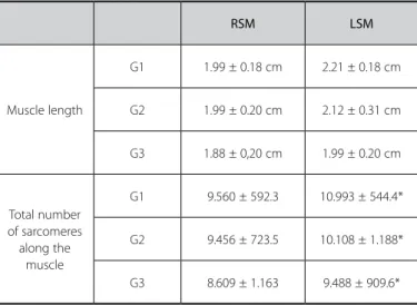
Comparing the longitudinal parameters found relating to mus-cle length, there was no significant difference (p> 0.05). However, the total estimate of sarcomeres along the muscle has shown sig-nificant difference in all groups when comparing to right and left muscles (p< 0.05) (table 2).
DISCUSSION
Skeletal muscle atrophy has been a great problem for patients that undergo movement restrictions caused by surgeries, joint di-seases or cast immobilization12 resulting muscle hypotrophy,
re-duction of muscle cross-section area and muscle strength even during short periods of time like fifteen days13-15. Tonic muscles like
soleus present a greater hypotrophy. That is the reason why devising resources to help people have their optimal movements back is extremely important besides the existing electrical stimulation and physical exercises17-22. Physical exercise is beneficial to muscle
221
Rev Bras Med Esporte – Vol. 19, No 3 – May/Jun, 2013
Table 2. Results on muscle length and total number of sarcomeres in soleus muscle relating to the group studied and comparing values from right soleus muscle (RSM) and left soleus muscle (LSM).
RSM LSM
Muscle length
G1 1.99 ± 0.18 cm 2.21 ± 0.18 cm
G2 1.99 ± 0.20 cm 2.12 ± 0.31 cm
G3 1.88 ± 0,20 cm 1.99 ± 0.20 cm
Total number of sarcomeres along the
muscle
G1 9.560 ± 592.3 10.993 ± 544.4*
G2 9.456 ± 723.5 10.108 ± 1.188*
G3 8.609 ± 1.163 9.488 ± 909.6*
* Significant statistical difference.
diabetic Wistar rats that present lesion on the sciatic nerve and were submitted to exercises on a treadmill. However, there is not enough data on scientific literature about using resistant anaerobic exercises for recovery after immobilization. Ju et al.24 analyzed the jump effects
in sandbox on Wistar rats remobilization after two-week hindlimb suspended plus intermittent weight support. They observed that five weeks of exercises were enough for the total recovery of the trabecular microarchitecture of the femoral metaphysis and that did not happen with free remobilization.
Results in this study show that functional overload induced by physical exercise in a daily basis may reverse partially the deleterious effects caused by immobilization of medium transverse diameter of muscle fibers but the exercises performed every other day did not have that same effect. So, we may state that this study reinforces research findings on muscular tissue recovery after immobilization as Kannus et al.25 who observed that high intensity exercises as
walking on treadmill caused beneficial effects on recovery of fiber cross-sectional area of soleus muscle in rats. Sakakima et al.26 also
observed that three-week immobilization period and a six-week remobilization period on a treadmill the effects on recovery of fiber cross-sectional area of soleus muscle were enhanced compared to free remobilization. According to Brito et al.27 100 samples of muscle
fibers are enough to evaluate properly the medium trophism. Ho-wever, another variable related to muscular trophism, that is, muscle mass has no change in values similar to the non-immobilized sides. According to Butterfield et al.28 the addition of sarcomeres in
series have a great impact on muscular performance because they enhance the contracting speed and potency. As immobilization promotes reduction of sarcomeres29, in this study we tested the
hypothesis of jump as a way to recover longitudinal morphological characteristics of soleus muscle in rats. Even though there are no
REFERENCES
1. Tidball JG. Mechanical signal transduction in skeletal muscle growth and adaptation. J Appl Physiol 2005;98:1900-8.
2. Carmeli E, Haimovitch TG. The expression of MMP-2 following immobilization and high-intensity run-ning in plantaris muscle fiber in rats. Scientific World Journal 2006;5:542-50.
3. Pattison JS, Folk LC, Madsen RW, Booth FW. Identification of differentially expressed genes between young and old rat sóleo muscle during recovery from immobilization induced atrophy. J Appl Physiol 2003;95:2171-9.
4. Williams PE. Effect of intermittent stretch on immobilised muscle. Ann Rheum Dis 1988;47:1014-6. 5. Lima SC, Caierão QM, Durigan JLQ, Schwarzenbeck A, Silva CA, Minamoto VB, Guirro RRJ. Curto
período de imobilização provoca alterações morfométricas e mecânicas do músculo de rato. Rev Bras Fisioter 2007;11:297-302.
6. Kasper CE, Talbot LA, Gaines JM. Skeletal muscle damage and recovery. AACN Clin Issues 2002;13:237-47. 7. Tanaka T, Kariya Y, Hoshino Y. Histochemical study the effects of aging on recovery from muscular
atrophy caused by disuse in rats. J Orthop Sci 2004;9:76-85.
differences either on immobilized or non-immobilized sides rela-ting to muscle length, and that result found for the croup control, and that finding may suggest that the remobilization period was effective to reverse some deleterious effects of immobilization. Koh and Tidbal9 reported positive effects of three-week remobilization
after four-week immobilization period. Estimated analysis of sarco-meres in series along the muscle has not found advantages on the protocol of physical exercises by jump comparing to free remobi-lization, though. Losses of sarcomeres in series along the muscular microarchitecture are caused by adjustments in fiber extremities for ideal actin and myosin overposition in myofibrils to perform maximum tension29, but the optimal length was not reached based
on the differences found. Thus, all the groups presented reduction on estimated sarcomeres along the soleus muscle of rats.
Those findings comply with previous studies that used the same model and presented reduction of sarcomeres along the muscle when animals were submitted to remobilization by swimming, low intensity walk on treadmill or series of static stretch19-21. Butterfield
and Herzog30 highlighted that tension in the muscle-tendon unit
is not the primary source of sarcomerogenesis but it may occur in some cases of eccentric exercise28 like contractions and disregarded
in the present study.
The absence of one group immobilized and having no period of remobilization may be seen as one of the limitations of this stu-dy. However, that group was disregarded due to the reduction of studies using animals because the designers of the immobilization method have pointed it as a producer of mass reduction and mus-cular length10 and a short period of immobilization around 7 days
is enough to decreasing muscular mass, fiber area and number of sarcomeres in series.
CONCLUSION
Physical exercise performed daily acts against deleterious effects caused by immobilization and that was not observed in exercises made every other day relating to muscular mass and muscular fiber diameter. Such effect was also observed in the muscular length of soleus muscles of both groups. However, findings in this study show that the protocol used was not effective on muscle recovery of longitudinal parameters relating to estimated quantity of sarco-meres along the soleus muscle.
ACKNOWLEDGEMENTS
National Council for Scientific and Technological (CNPq) and State University of Western Paraná for their support and research scholarship.
222 Rev Bras Med Esporte – Vol. 19, No 3 – May/Jun, 2013
8. Koh TJ, Tidball JG. Nitric oxide synthase inhibitors reduce sarcomere addition in rat skeletal muscle. J Physiol 1999;519:189-96.
9. De Deyne PG. Application of passive stretch and its implications for muscle fibers. Phys Ther 2001;81:819-27.
10. Coutinho EL, Gomes AR, Franca CN, Salvini TF. A new model for the immobilization of the rat hind limb. Braz J Med Biol Res 2002;35:1329-32.
11. Rogatto GP, Oliveira CAM, Faria MC, Luciano E. Respostas metabólicas agudas de ratos Wistar ao exercício intermitente de saltos. Motriz 2004;10:61-6.
12. Caron Az, Drouin G, Desrosiers J, Trensz F, Grenier G. A novel hindlimb immobilization procedure for studying skeletal muscle atrophy and recovery in mouse. J Appl Physiol 2009;106:2049-59. 13. Christensen B, Dyrberg E, Aagaard P, Kjaer M, Langberg H. Short-term immobilization and recovery
affect skeletal muscle but not collagen tissue turnover in humans. J Appl Physiol 2008;105:1845-51. 14. Min K, Smuder AJ, Kwon OS, Kavazis AN, Szeto HH, Powers SK. Mitochondrial-targeted antioxidants pro-tect skeletal muscle against immobilization-induced muscle atrophy. J Appl Physiol 2011;111:1459-66. 15. HVID, L. G. Ørtenblad N, Aagaard P, Kjaer M, Suetta C. Effects of ageing on single muscle fibre contractile
function following short-term immobilization. J Physiol 2011;589:4745-57.
16. Sato S, Suzuki H, Tsujimoto H, Shirato K, Tachiyashiki K, Imaizymi K. Casted-immobilization down-regulates glucocorticoid receptor expression in rat slow-twitch soleus muscle. Life Sci 2011;89:962-7. 17. Ishihara A, Kawano F, Ishioka N, Oishi H, Higashibata A, Shimazy T, et al. Effects of running exercise during recovery from hindlimb unloading on soleus muscle fibers and their spinal motoneurons in rats. Neurosci Res 2004;48:119-27.
18. Cornachione A, Cação-Benedini LO, Shimano MM, Volpon JB, Martinez Ez, Mattiello-Sverzut AC. Mor-phological comparison of different protocols of skeletal muscle remobilization in rats after hindlimb suspension. Scand J Med Sci Sports 2008;18:453-61.
19. Konno EAB, Alves EPB, Bertolini GRF, Barbieri CH, Mazzer N. Remobilização por alongamento estático cíclico em músculo sóleo de ratos imobilizados em encurtamento. Rev Bras Med Esporte 2008;14:122-5.
20. Natali LH, Silva TS, Ciena AP, Padoin MJ, Alves EPB, Aragão FA, et al. Efeitos da corrida em esteira em músculos sóleos de ratos encurtados por imobilização. Rev Bras Med Esporte 2008;14:490-3. 21. Volpi FS, Casarolli LM, Pudell C, Menon T, Ciena AP, Alves EPB, et al. Efeitos da remobilização em duas
semanas com natação sobre o músculo sóleo de ratos submetidos à imobilização. Rev Bras Med Esporte 2008;14:168-70.
22. Fujita N, Murakami S, Arakawa T, Miki A, Fujino H. The combined effect of electrical stimulation and resistance isometric contraction on muscle atrophy in rat tibialis anterior muscle. Bosn J Basic Med Sci 2011;11:74-9.
23. Malysz T, Ilha J, Nascimento PS, Faccioni-Heuser MC, De Angelis K, Schaan BD, et al. Exercise training improves the soleus muscle morphology in experimental diabetic nerve regeneration. Muscle Nerve 2011;44:571-82.
24. Ju Y, Sone T, Okamoto T, Fukunaga M. Jump exercise during remobilization restores integrity of the trabecular architecture after tail suspension in young rats. J Appl Physiol 2008;104:1594-600. 25. Kannus P, Jozsa L, Järvinen TL, Kvist M, Vieno T, Järvinen TA, et al. Free mobilization and low- to
high-intensity exercise in immobilization-induced muscle atrophy. J Appl Physiol 1998;84:1418-24. 26. Sakakima H, Yoshida Y, Sakae K, Morimoto N. Different frequency treadmill running in
immobilization-induced muscle atrophy and ankle joint contracture of rats. Scand J Med Sci Sports 2004;14:186-92. 27. Brito MKM, Camargo Filho JCS, Vanderlei LCM, Tarumoto MH, Pai VD, Giacometti JA. Dimensões
geométricas das fibras do músculo sóleo de ratos exercitados em esteira rolante: a importância da análise por meio de imagens digitalizadas. Rev Bras Med Esporte 2006;12:103-7.
28. Butterfield TA, Leonard TR, Herzog W. Differential serial sarcomere number adaptations in knee extensor muscles of rats is contraction type dependent. J Appl Physiol 2005;99:1352-8.
29. Tabary JC, Tabary C, Tardieu C, Tardieu G, Goldspink G. Physiological and structural changes in the cat’s soleus muscle due to immobilization at different lengths by plaster casts. J Physiol 1972;224:231-44. 30. Butterfield TA, Herzog W. The magnitude of muscle strain does not inluence serial sarcomere number