S
CIENTIFICA
RTICLES Revista Brasileira de FisioterapiaEffects of stretching and resistive exercise
in rat skeletal muscle
Efeito do alongamento e do exercício contra-resistido no
músculo esquelético de rato
Secchi KV1, Morais CP1, Cimatti PF1, Tokars E1, Gomes ARS2
Abstract
Objective: To analyze the effect of stretching and resistive exercise on the soleus muscle in rats. Methods: Twenty-four Wistar rats (380±50g) were evaluated, divided into four groups (n=6): C, intact controls; S, left soleus muscle stretched for 40 minutes twice a week; RE, resistive exercise consisting of four series of ten jumps three times a week; and RE+S, resistive exercise plus stretching. After eight weeks, the animals were sacrificed and their soleus muscles were evaluated regarding muscle weight, muscle fiber cross-sectional area (MFCSA), muscle length, number of sarcomeres in series, sarcomere length and percentage of connective tissue. The statistical analysis consisted of comparisons between the groups using the analysis of variance (ANOVA) post-hoc Tukey tests, with a significance level set at ≤0.05. Results: The MFCSA in RE and S were greater than in C. Muscle length and the number of sarcomeres in series in RE+S were less than in S and RE. The number of sarcomeres in series in S was greater than in C. No changes were found in sarcomere length or percentage of connective tissue. Conclusions: Resistive exercise associated with stretching caused a decrease in muscle length and in the number of sarcomeres in series, probably due to the daily frequency of exercises. Stretching alone, performed twice a week, or resistive exercise performed three times a week, was sufficient to induce muscle hypertrophy.
Key words: muscle stretching exercises; exercise; musculoskeletal system; rats.
Resumo
Objetivo: Analisar o efeito do alongamento e do exercício resistido no músculo sóleo de rato. Materiais e métodos: Foram avaliados 24 ratos Wistar (380±50g) divididos em quatro grupos (n=6): C, controle-intacto; Along, alongamento do músculo sóleo esquerdo durante 40 minutos; ER, exercício resistido, quatro séries de dez saltos; ER+Along, exercício resistido e alongamento. Após oito semanas, foi realizada a eutanásia dos animais e os músculos sóleos foram avaliados quanto ao peso muscular, área de secção transversa das fibras musculares (ASTFM), comprimento muscular, número de sarcômeros em série, comprimento dos sarcômeros e porcentagem de tecido conjuntivo. A análise estatística foi realizada pela comparação entre grupos, por meio do teste de análise de variância (ANOVA) post hoc Tukey, com significância ≤0,05. Resultados: As ASTFM dos grupos ER e Along aumentaram quando comparadas ao grupo C. O comprimento muscular e o número de sarcômeros em série do ER+Along foram inferiores aos dos grupos Along e ER. O número de sarcômeros em série do Along foi superior ao C. Não foram encontradas alterações no comprimento dos sarcômeros e na porcentagem de tecido conjuntivo. Conclusões: O exercício resistido associado ao alongamento causou diminuição no comprimento muscular e no número de sarcômeros em série, provavelmente devido à freqüência diária de exercícios. A realização isolada do alongamento, duas vezes por semana, ou do exercício resistido, três vezes por semana, foi suficiente para induzir hipertrofia muscular.
Palavras-chave: exercícios de alongamento muscular; exercício; sistema musculoesquelético; ratos.
Received: 16/08/2007 – Revised: 14/12/2007 – Accepted: 08/04/2008
1 Physical Therapy Undergraduate Program, Universidade Tuiuti do Paraná (UTP) – Curitiba (PR), Brazil
2 Physical Therapy Undergraduate Program, Universidade Federal do Paraná (UFPR), Setor Litoral – Matinhos (PR), Brazil
Financial support: UTP, Faculdade Evangélica do Paraná (Fepar) and Graduate Program in Cellular Biology, UFPR (Curitiba)
Correspondence to: Anna Raquel Silveira Gomes, Curso de Fisioterapia, Universidade Federal do Paraná, Setor Litoral, Rua Jaguariaíva, 512, CEP 83260-000, Matinhos (PR), Brazil, e-mail: [email protected]
Introduction
Sports training and rehabilitation programs are usually made up of a set of exercises which include stretching before and/or after counter resistance exercises1,2. Many of the
stre-tching protocols are recommended to enhance movement amplitude, as well as to prevent and treat musculoskeletal problems. Several studies with humans evaluated muscle adaptations induced by diferent stretching protocols applied to normal and/or shortened muscles1,3-8.
Bandy and Iron5 tested the efects of stretching sessions
sustained for 15, 30, and 60 seconds, carried out once a day with humans. he results indicated that 30 seconds of daily stretching, was enough to increase the range of motion (ROM) of the hamstrings of young adults5. No signiicant diferences
were observed upon carrying out two series of exercises a day6.
Grady and Saxena7 also showed that humans who performed
30 seconds of stretching every day increased and kept their ROM. Elderly humans submitted to ive weekly sessions for ha-mstrings muscle stretching also had increases in ROM. hese sessions, consisted of four repetitions, lasted 60 seconds each and were carried out over the course of six weeks4.
Nonethe-less, upon testing the stretching times shorter than 60 seconds, the ROM gains and maintenance were not as efective4.
Some researchers claim that as far as clinical use is concer-ned, the stretching sessions must be carried out ive times a week, so that the ROM can be enhanced in a more signiicant way8. However, two-three weekly sessions are enough to
ge-nerate ROM gains8. After the gains, one stretching session per
week is enough for maintenance8.
he animal model, in which it is possible to analyze the mus-cular ibers, showed that 30 minutes of daily passive stretching was suicient to prevent a loss in the number of sarcomeres in series and muscular atrophy, in muscles shortened by im-mobilization9. Coutinho et al.10 showed that stretching sessions
sustained for 40 minutes and carried out three times a week on a rat’s soleus muscle was enough to increase the muscular length, the number of sarcomeres in series, and the muscular iber transverse sectional area of non-shortened muscles.
Another method often used for muscle mass gains, with consequent hypertrophy, are counter-resistance exercises, both in humans and animals11. he muscular hypertrophy resulting
from the practice of counter-resistance exercises may be trans-lated as an increase in the transverse sectional area of the mus-cle ibers (radial growth), as well as an increase in the number of sarcomeres in series (longitudinal growth), derived from an increments in protein synthesis11,12.Still, it has been shown that
gains in muscular strength may not involve an increase in the transverse section of the muscle ibers, but only a gain in the sarcomeres in series, or still, just neural adaptations12,13.
he hypertrophy mechanisms can be activated both in hu-mans and in animals, depending on the intensity, duration, and frequency of the stretching and resistance exercises. In spite of this, the response of the skeletal muscle to this combination is still unknown, both for enhancing training in sport as well as speeding up the rehabilitation process. hus, the purpose of the present study was to investigate the adaptations of the muscular ibers after the resistance exercises with stretching.
Materials and methods
Animal and experimental groups
Twenty-four albino Wistar lineage, six-month old (380±50g) rats were used, obtained from the Central Biotherium and kept in a private biotherium located in the laboratory of Pharmacol-ogy of the Universidade Tuiuti do Paraná (UTP). he animals were grouped together and kept in standard plastic cages, un-der controlled environmental conditions (luminosity: 12-hour light/dark cycle), with free access to water and pellet rations. he project was carried out following the international ethical norms for animal experimentation14 and received the approval
of the Committee for Ethics in Research of the UTP, under pro-tocol number 019/2005. In order to perform the passive stretch-ing, the dissection of the soleus muscle and the euthanasia, the animals were previously weighed and anesthetized with an intraperitoneal injection of ketamine (Dopalen, Sespo, Jacareí, São Paulo), 95mg/kg of body weight, and xylazine (Anasedan, Sespo, Jacareí, São Paulo), 12mg/kg of body weight.
he animals were randomly divided into four groups: 1) Con-trol (C, n=6): the rats were not submitted to any procedure; 2) Stretching (Stretch, n=6): the rats underwent the stretching of the left soleus muscle; 3) Resistance Exercise (RE, n=6): the rats were submitted to a protocol of counter-resistance trai-ning; 4) Resistance Training and Stretching (RE+Stretch, n=6): the rats were submitted to muscle stretching, twice a week (tuesday and thursday), and to counter-resistance exercises three times a week (monday,wednesday and friday), for eight consecutive weeks. he stretching and resistance exercise pro-tocols were carried out from 13h30 onwards every day, in an ascending order from rat 1 to 6 for the RE+Stretching group. In the Stretching groups, all rats were anesthetized and concomi-tantly positioned in the stretching position.
Stretching protocol
he animals had their left tibio-tarsic joint manually posi-tioned in maximal dorsal lexion, keeping the soleus muscle stretched with the aid of scotch tape for 40 minutes10. he
protocol was carried out with the anesthetized animal twice a week (tuesday and thurday), for eight consecutive weeks.
Counter-resistance exercise training protocol
15he counter-resistance exercise was carried out as descri-bed by Renno et al.15. he training protocol consisted of four
se-ries of ten leaps each, with a 60 seconds interval between them (Stopwatch Cal. H545, Tokyo, Japan). he training was carried out three times a week (monday, wednesday and friday), for eight consecutive weeks.
After that, both soleus muscles (left and right) of the RE+Stretching group, and only the left soleus muscle of the other groups were removed and weighed, in isolation (Mark Bell Engineering, Italy). Next, they were longitudinally divided, into two equal parts; one part was submitted to the routine procedures in order to evaluate the number of sarcomeres in series10 and the other part was ixed in Zenker’s luid for later
morphological and morphometrical evaluations.
Identification of the number of sarcomeres in series
he muscle ibers were isolated and ixed as described by Coutinho et al.10. For each muscular iber, the number of
sar-comeres in series was identiied along 300µm, under a light microscope (Nikon, Tokyo, Japan), with a 100x objective. he measurements were carried out with a 14’’video monitor with a video-image system (Adler CCTV) itted to the microscope (Nikon,Tokyo, Japan), with the aid of a counter (Veeder-Root, Washington, USA), in the laboratory of pathology of the Facul-dade Evangélica do Paraná (Fepar).
he total number of sarcomeres and the length of each muscular iber in isolation were estimated by the correla-tions between the number of sarcomeres identiied along the 300µm of the iber and the total muscle length, as described by Williams et al.16. Despite the controversies in the literature,
this study considered the length of the sarcomeres along the muscular ibers as being homogeneous (Coutinho et al.10).
Procedures for morphological analyses
he fragment taken out from the medial half of each so-leus muscle belly was dyed with Harris hematoxiline and eosin (H&E) for the histomorphometric analysis of the transverse section of the muscular ibers. In other histological slides, other cuts were dyed with Mallory’s trichrome to evaluate the percen-tages of conjunctive tissue (perimysium and endomysium).
he photomicrographs of the histological cuts were taken by a light microscope (Axyophot, Carl Zeiss, Oberkochen, Ger-many) and captured in a video-image system (Applied Spectral
Imaging, Migdal Ha’emek, Israel) by means of the Case Data Ma-nager Expo Software (Applied Spectral Imaging, Migdal Ha’emek, Israel, version 4.0) in the Postgraduate Department in Celular Biology of the Universidade Federal do Paraná (UFPR) at Curi-tiba. In each muscle, the transverse section area of the muscular ibers, as well as the percentages of conjunctive tissue were me-asured, with the aid of the UTHSCSA Image Tool 3.0 (developed by the University of Texas Health Science Center at San Antonio, and available at http://ddsdx.uthscsa.edu/dig/itdesc.html).
A transverse sectional area of one hundred muscular i-bers of each muscle was measured, chosen randomly from the muscular belly area of the histological section as described by Coutinho et al.10. he conjunctive tissue area percentage
analysis was irst carried out by selecting the whole area of the cut dyed with Mallory’s trichrome and photographing it with a 10x objective, equivalent to 100%. Next, the transverse section areas of all of the muscular ibers were excluded, and only the conjunctive tissue (that is, perimysium and endomy-sium) was isolated. After analyzing these results, they were expressed as percentages17.
Statistical analysis
In order to check out whether each variable was normally distributed, the Shapiro Wilks test was conducted. he conclu-sion of the test took into account the associated p value , with a reliability level of 99%. After the conirmation of normality, the initial body weight values were compared to the weight after eight weeks of testing, in each experimental group (intragroup), by using the paired t-test.
In the RE+Stretch group the paired t-test was used to compare the right paw (resistance exercise) and the left one (submitted to stretching and to resistance exercise). he results of all of the analyzed variables (body weight, muscular weight, muscular length, number and length of sarcomeres, trans-verse section area of the muscular ibers, and percentages of conjunctive tissue) were compared between the experimental groups (intragroups) with ANOVA, the post hoc Tukey test. he values were considered signiicant when p≤0.05.
Results
Body and muscular weight
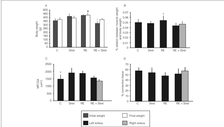
he RE + Stretch group had an increase in its inal body wei-ght when compared to the initial weiwei-ght (p=0.007). he inal body weight was superior in the RE group when compared with the C group (p=0.002) and with the RE+Stretch group (p=0,002; Figure 1A). he weight of the left soleus muscle of the group
Figure 1. Effect of stretching and resistive exercise in the body weight and in the radial analyses of the rat soleus muscle.
Initial weight 2500
C D
B A
MFCSA (um
2)
Body weight
(g)
% r
elation between muscle weight
and body weight
% connective tissue
0 60 100 160 200 260 300 360 400 460 600
70 0 0.01 0.02 0.03 0.04 0.05 0.06 0.07
C Stret RE
C Stret RE
RE + Stret
C Stret RE RE + Stret
RE + Stret
C Stret RE RE + Stret
60 50 40
30 20 10
0 2000
1500
1000
500
0
Left soleus
Final weight
Right soleus +
+ #
*
C= intact control group; Stret=stretching group; RE= Resistance Exercise group; RE + Stret= Stretching + Resistance Exercise group.
1A: *p<0.05 when compared to the final body weight; final body weight, #p<0.05 when compared to C and ER+Along groups; 1B: muscle weight, +p<0.05 compared to C, Along and ER+Along groups; 1C: muscle fibers cross sectional area (MFCSA), +p<0,05 compared to Along and ER groups; 1D: connective tissue percentage. C=intact control group; Along=stretching group; ER=resistive exercise group; ER+Along=resistive exercise and stretching group.
that underwent the resistance exercise (RE) had an increase (Figure 1B) when compared with the remainder of the groups: C (p=0.003); Stretch (p=0.01), RE+Stretch (p=0.0003).
Transverse sectional areas of the
muscular fibers (TSAMF)
An increase in the TSAMF was observed in the Stretch group (p=0.037), as well as in the RE group (p=0.02), when compared to the C group (Figure 1C).
Conjunctive tissue percentages
No signiicant diferences were found in the percentages of the conjunctive tissue in any of the groups evaluated (Figure 1D).
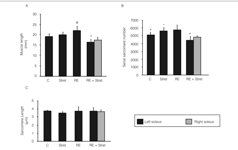
Muscular length
A reduction was observed of the left soleus muscle length in the RE+Stretch group when compared to the other groups: C (p=0.04); Stretch (p=0.005), and RE (p=0.0003). In addition to this, an increase in the resistance exercise group (RE) was
also noticed in relation to the control group (C) (p=0.05), as shown in Figure 2A.
Total number of sarcomeres in series and
length of sarcomeres
he total number of sarcomeres in series of the Stretch group was greater than that demonstrated by the C group. Yet, the RE+Stretch was inferior to the Stretch (p=0.002) and RE (p=0.001) groups, as shown in Figure 2B. No diferences in the sarcomere length values of the soleus muscle were observed in any of the analyzed groups (Figure 2C).
Discussion
he present study showed that both the stretching pro-tocol, carried out twice a week, and the counter-resistance exercises, performed three times a week, applied in isolation, for eight consecutive weeks, were enough to induce muscle hypertrophy, and an increase in the transverse section area of the muscular ibers was observed. Coutinho et al.10 tested
Figure 2. Effect of stretching and resistive exercise in the longitudinal analyses of the rat soleus muscle. +
+
+ #
*
C Stret RE RE + Stret C Stret RE RE + Stret
C Stret RE RE + Stret
Left soleus Right soleus
Muscle length
(mm)
Serial sar
comer
e number
Sar
comer
e Length
(
m)
30
7000
6000
5000
4000
3000
2000
1000
0 25
20
15
10
5
0
5
4
3
2
1
0 A
C
B
C= intact control group; Stret=stretching group; RE= Resistance Exercise group; RE + Stret= Stretching + Resistance Exercise group.
2A. Muscle Length, #p<0.05 when compared to C; +p<0.05 compared to C, Along and ER groups. 2B. Serial sarcomere number, *p<0.05 when compared to C, +p<0.05 when compared to Along and ER groups, (ANOVA), 2C Sarcomere Length. For a description of the groups see the legend to Figure 1.
the efects of the stretching of the soleus muscle in rats, with a normal length, that is, non-shortened, carried out three times a week, for 40 minutes, and perceived an increase in the transverse sectional area, that is, radial growth, and also a rise in the number of sarcomeres in series (sarcomeroge-nesis), longitudinal growth, without altering the muscular weight. hus, it can be inferred that even when the frequency of the stretching was reduced to only twice a week, as in the present study, it is also possible to induce radial and longitu-dinal muscular hypertrophy.
Russell, Motlagh and Ashley18 have suggested that the
direction of the muscular growth, both longitudinally (in series) and radially (in parallel), is not controlled by the transcription, but by a post-transcription mechanism, just the same as in translation. Studies have shown that the longitudinal growth occurs by the increase in protein syn-thesis, particularly in the distal extremities of the muscle fiber19,20. The parallel muscle growth, in turn, which occurs
in the densest regions of the muscle, takes place through the diffusion of neo-synthesized proteins, by means of microtu-bules18. In spite of these indications, the mechanisms that
regulate the muscular growth, both in series and in parallel, remain unknown.
In the present study, the stretching of normal muscles carried out twice a week did not alter the percentages of con-junctive tissue. hus, as it had already been noted by other au-thors, the skeletal muscle responds diferently to the exercise and according to the dysfunction ( for example, lesions21, use
reduction22, immobilization11, and sarcopenia15), as
demons-trated by Williams16, who evaluated the efects of stretching
in shortened muscles for 15 minutes every other day, and did not notice any increases in the number of sarcomeres in series16. Nevertheless, the percentages of conjunctive tissue
were inferior when compared to muscles that were shortened only by immobilization16. his suggests that the plasticity of a
normal muscle is diferent from that of a shortened one. Hornberger and Farrar23 carrried out resistance training
in rats for a period of eight weeks, and observed an increase of the muscle mass and of the amount of protein of the long hallux lexor muscle (LHF), but with no changes in the soleus. he authors attribute the LHF’s speciic hypertrophy to the kind of training (climbing), which put that particular muscle
under stress23. Moreover, it was detected that ovariectomized
rats submitted to the same resistance training protocol used in this study had an increase in their muscular weight, both in the anterior tibial and in the soleus, but showed no dife-rences in the transverse sectional area of the muscular ibers of either muscle15. So, it can be inferred that the increases of
both the muscular weight and of the transverse sectional area of the muscular ibers in the RE group may be related to sexual hormones, since the animals of the present study were males, as well to the type and frequency of the training. Furthermore, it could be stated that the increases in muscular mass was not directly proportional to the enlargement of the TSAMF, in that in the Stretch group, no muscular weight increases were ob-served. Only in the TSAMF, for that kind of muscle, whether there was a predominance of fast or slow twitch ibers, also interfered with the adaptations to exercise.
Both the training protocol applied in this study and the levels of hypertrophy observed in the muscles submitted to the resistance exercises (27% greater than control) were comparable to those found in humans24. It came as a surprise
that when the resistance exercises and the stretching were associated, there was a reduction of the muscular length and in the number of sarcomeres in series. his result suggests that the frequency with which the protocols were carried out, that is, on a daily basis, reduced sarcomerogenesis. However, when the stretching was performed in isolation twice a week, there was an increase in the number of sarcomeres. Other au-thors also reported the inluences of the stretching frequency in muscular plasticity10,25. It could, therefore, be inferred that
the association of the stretching with the resistance exercises regulated the number of sarcomeres in series and the trans-verse TSAMF through diferent mechanisms, for no change was observed in the TSAMF. he nitric oxide derived from the neuronal isoform of the sintase nitrous oxide is a positive modulator of the addition of sarcomeres in series26,27, whereas
the calcineurine, a Ca+2/calmoduline dependent phosphatase
protein and the growing factor similar to that of insuline I (IGF-I), activating the phosphatidil-inositol 3-kinase (PI3K)-AKT (a serine threonine kinase) seems to regulate the mus-cular hypertrophy28,29.
hus, the reduction in the number of sarcomeres in series and the absence of alterations in the TSAFM in the RE+Stretch group corroborated the indings of some authors who suggest an inter-val of at least 36 to 48 hours between the training sessions, since
this is the peak period of protein synthesis after exercise2.
Ano-ther relevant aspect which must be taken into account was the daily manipulation of the animals belonging to the RE+Stretch group. his may have contributed to the animal’s stress, resulting in the loss of sarcomeres. hus, the exercise frequency must be emphasized, especially for the rest period between the training sessions, so that catabolism is not increased.
he present study is not exempt from limitations, such as the applied stretching protocol, which could have been main-tained for a time-span more typically used in clinical practice, which was maintained for only a few seconds, in spite of some physical therapy techniques, such as the global postural ree-ducation (GPR), which advocates gradual increases in move-ment amplitude for 20 to 30 minutes30. More research will be
necessary to shed some light on the cellular, molecular, and functional mechanisms of the efects derived from the asso-ciation of stretching and resistance exercises, so widely used in clinic physical therapy practice. It would also be advisable that randomized clinic studies should be carried out followed by magnetic resonance imaging evaluations and/or biopsies to measure the transverse sectional areas of the muscle and ibers, as well as analyses of functional muscular performance by means of an isokinetic dynamometer, electromyography, and force platforms.
Conclusions
he association between stretching and resistance exerci-ses caused a reduction in the muscular length and in the num-ber of sarcomeres in series, probably due to the daily frequency of training. he stretching and resistance exercise protocols, when applied in isolation, were suicient to induce muscular hypertrophy.
Acknowledgements
We would like to thank the inancial support provided by Elisangela Poppin for the manufacturing of the histological sli-des, and by Carlos Barbosa, for carrying out the experimental procedures. We were also aided by Rosinei Valle, Carolina Por-tella, and Priscilla Franco Cimatti, from the Fepar, who helped us calculate the number of sarcomeres in series.
References
Gajdosik RL. Passive extensibility of skeletal muscle: review of the literature 1.
with clinical implications. Clin Biomech (Bristol, Avon). 2001;16(2):87-101.
Kraemer WJ, Adams K, Cafarelli E, Dudley GA, et al. Progression models in 2.
resistance training for health adults. American College of sports medicine. Med Sci Sports Exercise. 2002;34(2):364-80.
Wallin D, Ekblom B, Grahn R, Nordenborg T. Improvement of muscle flexibility: 3.
a comparison between two techniques. Am J Sports Med. 1985;13(4):263-8.
Feland JB, Myrer JW, Schulthies SS, Fellingham GW, Measom GW. The effect 4.
of duration of stretching of the hamstring muscle group for increasing range of motion in people age 65 years or older. Phys Ther. 2001;81(5):1110-7.
Bandy WD, Iron JM. The effect of time on static stretch on the flexibility of 5.
hamstring muscles. Phys Ther. 1994;74(9):845-52.
Bandy WD, Iron JM, Briggler M. The effect of time and frequency of 6.
static stretching on flexibility of the hamstring muscles. Phys Ther. 1997;77(10):1090-6.
Grady JF, Saxena A. Effects of stretching the gastrocnemius muscle. J Foot 7.
Ankle Surg. 1991;30(5):465-9.
Frontera WR, Dawson DM, Slovik DM, editors. Exercise in rehabilitation 8.
medicine. USA: Human Kinetics; 1999.
Williams PE. Use of intermittent stretch in the prevention of serial sarcomere 9.
loss in immobilised muscle. Ann Rheum Dis. 1990;49(5):316-7.
Coutinho EL, Gomes ARS, França CN, Oishi J, Salvini TF. Effect of passive 10.
stretching on the immobilized soleus muscle fiber morphology. Braz J Med Biol Res. 2004;37(12):1853-61.
Timson BF. Evaluation of animal models for the study of exercise-induced 11.
muscle enlargement. J Appl Physiol. 1990;69(6):1935-45.
Koh TJ. Do adaptations in serial sarcomere number occur with strength 12.
training? Hum Mov Sci. 1995;14:61-77.
Wilmore JH, Costill DL, editores. Fisiologia do esporte e do exercício. 2
13. a
ed. Barueri: Manole; 2001.
National Research Council. Guide for the care and use of laboratory 14.
animals. Washington (DC): National Academy Press; 1996.
Renno AC, Gomes ARS, Nascimento RB, Salvini T, Parizoto N. Effects of 15.
a progressive loading exercise program on the bone and skeletal muscle properties of female osteopenic rats. Exp Gerontol. 2007;42(6):517-22.
William PE, Catanese T, Lucey EG, Goldspink G. The importance of stretch 16.
and contractile activity in the prevention of connective tissue accumulation in muscle. J Anat. 1988;158:109-14.
Mattiello-Sverzut AC, Carvalho LC, Cornachione A, Nagashima M, 17.
Neder L, Shimano AC. Morphological effects of electrical stimulation and intermittent muscle stretch after immobilization in soleus muscle. Histol Histopathol. 2006;21(9):957-64.
Russel B, Motlagh D, Ashley WW. Form follows function: how muscle 18.
shape is regulated by work. J Appl Physiol. 2000;88(3):1127-32.
Williams PE, Goldspink G. The effect of immobilization on the longitudinal 19.
growth of striated muscle fibres. J Anat. 1973;116(Pt 1):45-55.
Dix DJ, Eisenberg BR. Myosin mRNA accumulation and myofibrillogenesis 20.
at the myotendinous junction of stretched muscle fibers. J Cell Biol. 1990; 111(5 Pt 1):1885-94.
Hwang JH, Ra YJ, Lee KM, Lee JY, Ghil SH. Therapeutic effect of passive 21.
mobilization exercise on improvement of muscle regeneration and prevention of fibrosis after laceration injury of rat. Arch Phys Med Rehabil. 2006;87(1):20-6.
Trappe S, Trappe T, Gallagher P, Harber M, Alkner B, Tesch P. Human 22.
single muscle fiber function with 84 day bed-rest and resistance exercise. J Physiol. 2004;557(Pt 2):501-13.
Hornberger TA, Farrar RP. Physiological hypertrophy of the FHL muscle 23.
following 8 weeks of progressive resistance exercise in the rat. Can J Appl Physiol. 2004;29(1):16-31.
Widrick JJ, Stelzer JE, Shoepe TC, Garner DP. Functional properties of 24.
human muscle fibers after short-term resistance exercise training. Am J Physiol Regul Integr Comp Physiol. 2002;283(2):R408-16.
Gomes ARS, Coutinho EL, França CN, Polonio J, Salvini TF. The 25.
effect of one stretch a week applied to the immobilized soleus muscle on rat muscle fiber morphology. Braz J Med Biol Res. 2004;37(10): 1473-80.
Tidball JG, Lavergne E, Lau KS, Spencer MJ, Stull JT, Wehling M. 26.
Mechanical loading regulates NOS expression and activity in developing and adult skeletal muscle. Am J Physiol. 1998;275(1 Pt 1):C260-6.
Koh TJ, Tidball JG. Nitric oxide synthase inhibitors reduce sarcomere 27.
addition in rat skeletal muscle. J Physiol. 1999;519 Pt (1):189-96.
Dunn SE, Burns JL, Michel RN. Calcineurin is required for skeletal muscle 28.
hypertrophy. J. Biol. Chem. 1999;274(31):21908-12.
Glass DJ. Molecular mechanisms modulating muscle mass. Trends Mol 29.
Med. 2003;9(8):344-50.
Souchard PE. Reeducação postural global: método do campo fechado. 30.
São Paulo: Ícone; 1998.