ABSTRACT
http://dx.doi.org/10.1590/1678-775720130031
evaluation of cytotoxicity, antimicrobial activity
and physicochemical properties of a calcium
aluminate-based endodontic material
Emmanuel João Nogueira Leal SILVA1, Daniel Rodrigo HERRERA2, Tiago Pereira ROSA2, Thais Mageste DUQUE2, Rogério Castilho JACINTO2,3, Brenda Paula Figueiredo de Almeida GOMES2, Alexandre Augusto ZAIA2
1- Health and Science Center, Grande Rio University (UNIGRANRIO), Rio de Janeiro, RJ, Brazil.
2- Department of Restorative Dentistry, Endodontics Division, Piracicaba Dental School, State University of Campinas, Piracicaba, SP, Brazil. 3- Endodontics Division, Federal University of Pelotas, Pelotas, RS, Brazil.
Corresponding address: Emmanuel J. N. L. Silva - Escola de Ciências da Saúde, Universidade Grande Rio (UNIGRANRIO) - Rua Herotides de Oliveira, 61/902 - Icaraí - Niterói - RJ - Brasil - e-mail: [email protected]
Submitted: January 11, 2013 - Modiication: August 18, 2013 - Accepted: November 1, 2013
A
calcium aluminate-based endodontic material, endoBinder, has been developed inorder to reduce MTA negative characteristics, preserving its biological properties and clinical applications. Objectives: The aim of this study was to evaluate the cytotoxicity, antimicrobial activity, pH, solubility and water sorption of endoBinder and to compare them with those of white MTA (WMTA). Material and Methods: Cytotoxicity was assessed through a multiparametric analysis employing 3T3 cells. Antimicrobial activity against
Enterococcus faecalis (ATCC 29212), Staphylococcus aureus. (ATCC 25923) and Candida
albicans (ATCC 10556) was determined by the agar diffusion method. pH was measured at periods of 3, 24, 72 and 168 hours. Solubility and water sorption evaluation were performed following ISO requirements. Data were statistically analyzed by ANOVA and Tukey`s test
with a signiicance level of 5%. Results: EndoBinder and WMTA were non-cytotoxic in all
tested periods and with the different cell viability parameters. There was no statistical differences between both materials (P>.05). All tested materials were inhibitory by direct contact against all microbial strains tested. endoBinder and WMTA presented alkaline pH in all tested times with higher values of pH for WMTA (P<.05). Both materials showed values complying with the solubility minimum requirements. However, endoBinder showed lower solubility than WMTA (P<.05). No statistical differences were observed regarding water sorption (P>.05). Conclusion: Under these experimental conditions, we concluded that the calcium aluminate-based endodontic material endoBinder demonstrated suitable biological and physicochemical properties, so it can be suggested as a material of choice
in root resorption, perforations and root-end illing.
Keywords: endodontics. Biocompatible materials. Dental materials.
INTRODUCTION
Mineral trioxide aggregate (MTA) is a material that has been developed at Loma Linda University14
initially as a root-end illing material and later has been used for pulp capping, pulpotomy, apexogenesis, apical barrier formation in teeth with open apexes, repair of root perforations, and as a root canal illing material25. MTA is a powder that
consists of ine hydrophilic particles that set in the presence of moisture34. MTA has been recognized as
a bioactive material13 that is hard tissue conductive,
hard tissue inductive, and biocompatible17,23.
Mineral trioxide aggregate (MTA) has been shown to induce mineralization and to have favorable sealing properties17,22,25,26. Nevertheless,
MTA remains subject to some concerns, such as its long setting time11,25, poor handling characteristics,
low resistance to compression, low low capacity11,
high cost, and presence and release of arsenic12,28.
A calcium aluminate-based endodontic material, endoBinder (Binderware, São Carlos, SP, Brazil), has been developed with the intention of preserving the properties and clinical applications of MTA trying to reduce its negative characteristics1,24.
endoBinder is mostly composed of Al2O3 (≥68.5%), CaO (≤31.0%), SiO2 (0.3-0.8%), MgO (0.4-0.5%),
and Fe2O3 (<0.3%). The cement is produced by the process of calcining Al2O3 and CaCO3 at temperatures between 1315°C and 1425°C to achieve a uniform composition. The product resulted of this process is cooled and then triturated until an adequate particle size is obtained. The inal product is a result of the following chemical reaction: CaCO3+Al2O3=Ca(AlO2)2+CO221,24. endoBinder has
good cell response, allowing greater development of cells at an advanced state of osteoblastic differentiation than the one obtained with MTA7, it
has less tissue reaction than MTA, it is biocompatible when tested in rat subcutaneous tissue and showed no gelatinolytic activity of MMP-2 and MMP-91,30.
However, up to now, there are limited publications about the physicochemical and biological properties of this calcium aluminate-based material and its possible use in clinical practice.
Thus, the aim of the present study was to evaluate the cytotoxicity, antimicrobial capability, pH, solubility and water sorption of endoBinder and to compare them with those presented by WMTA (Angelus Indústria de Produtos Odontológicos, Londrina, PR, Brazil).
MATERIAL AND METhODS
Cytotoxicity evaluation was performed according to ISO 10993-5 specifications (2009)20. The
agar diffusion method was used to measure the antimicrobial activity. pH was measured at periods of 3, 24, 72 and 168 hours. Solubility and water sorption evaluations were performed according to ISO 6876/2001 speciications (2001)19. Both materials were mixed according to
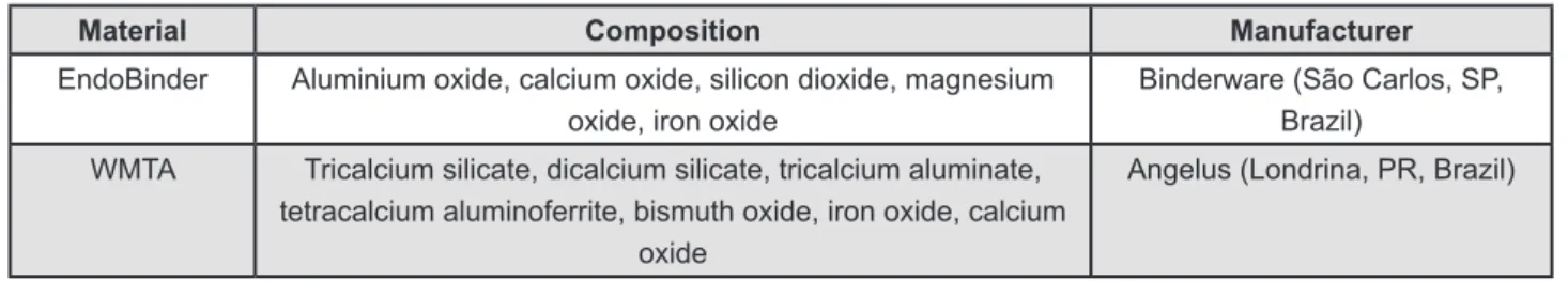
the manufacturer’s instruction, with a powder-to-liquid ratio of 3:1. The composition of the evaluated materials is shown in Figure 1.
Cytotoxicity
Under aseptic conditions, the materials were mixed on a glass slab for 1 min, and placed in
Telon rings (5 mm in diameter, 2 mm high). The specimens were allowed to set completely for 24 h at 37°C and 100% humidity under sterile conditions. After setting, the materials were placed in Dulbecco`s Modiied Eagle`s Medium (DMEM) (Gibco, Life Technologies Corporation, Grand Island, NY, USA) with 10% Foetal Bovine Serum (Gibco, Life Technologies Corporation, Grand Island, NY, USA) using a 1.25 cm2/mL ratio between the
sample surfaces and the medium volume. Undiluted extracts were used for the test.
Cytotoxicity was evaluated with a commercial kit (Cytotox, Xenometrix AG, Allschwill, Switzerland) that evaluates three different cell viability parameters sequentially on the same cell culture: XTT, neutral red (NR), and crystal violet dye elution (CVDe). The XTT test is based on the ability of mitochondrial enzymes from metabolically active cells to reduce 2,3-bis(2-methoxy-4-nitro-5-sulphophenyl)-2H-tetrazolium-5-carboxanilide (XTT) molecules to a soluble salt of formazan, detectable by its absorbance at 480 nm, as measured by a spectrophotometer (Urit 660; URIT Medical electronic CO, Guangxi, China). The same cells submitted to the XTT test were washed and assayed with the neutral red uptake test (NR), which determines the levels of viable cells through their membrane integrity. The vital dye NR is incorporated through endocytosis and accumulates preferentially on the lysosomes of membrane intact viable cells. After 3 h of exposure to the dye, cells were ixed and the NR was extracted and measured by the optical density (OD) of the supernatant at 540 nm, which directly relates to the proportion of viable cells. After the NR test, ixed cells were washed and evaluated for the total density of cells adhered, as estimated by the crystal violet dye exclusion test (CVDe). CVDe is a simple assay that evaluates cell density by staining DNA; after elimination of excess dye, the absorbance at 540 nm is proportional to the amount of cells in the well.
Fibroblast cells (lineage 3T3) were obtained from the American Type Culture Collection (ATCC) and cultivated in DMeM supplemented with 10% Foetal Bovine Serum (FBS) (Gibco, Life Technologies Corporation, Grand Island, NY, USA), 100 µg/ml streptomycin, and 100 mg/mL penicillin at 37°C in a humidiied incubator under an ambient pressure air atmosphere containing 5% CO2. Conluent cells
Material Composition Manufacturer
EndoBinder Aluminium oxide, calcium oxide, silicon dioxide, magnesium oxide, iron oxide
Binderware (São Carlos, SP, Brazil)
WMTA Tricalcium silicate, dicalcium silicate, tricalcium aluminate, tetracalcium aluminoferrite, bismuth oxide, iron oxide, calcium
oxide
Angelus (Londrina, PR, Brazil)
were detached with 0.25% trypsin and 0.05% ethylenediaminetetraacetic acid (Gibco, Life Technologies Corporation, Grand Island, NY, USA) for 5 min, and aliquots were subcultured. For the experimental set, 5x10³ cells were cultured in
96-well culture plates and allowed to achieve 80% conluence. After 24 h, the medium was removed from each well and replaced by 200 µl of one of the materials eluted in triplicate, as described above, for further 24 h.
Antimicrobial activity
The agar diffusion method was used to measure the antimicrobial activity of endoBinder and WMTA against E. faecalis (ATCC 29212), S. aureus. (ATCC 25923) and C. albicans (ATCC 10556). Isolated for 24 h, colonies of pure culture of each microorganism were grown on Brain Heart Infusion (BHI; Oxoid, Basingstoke, U.K.) agar plates. Then, they were inoculated into tubes containing 5 mL of BHI broth (Oxoid Microbiology Products, Thermo Fisher Scientific, Basingstoke, U.K.). The suspension was adjusted spectrophotometrically at 800 nm to match the turbidity of 1.5×10⁸ CFU mL-1 (equivalent
to 0.5 McFarland turbidity standard). Five hundred μL of each test microorganism suspension was inoculated into glass bottles containing 50 mL of BHI agar at 46°C, vortexed, and poured onto 130-mm plates containing a previously set layer of Mueller Hinton Agar (MHA, Oxoid Microbiology Products, Thermo Fisher Scientiic, Basingstoke, U.K.).
Sterilized stainless steel tubes of 8.0×1.0×10 mm (inner diameter 6 mm) were added to the surfaces of the media and illed with each tested substance. The plates were maintained for 2 h at room temperature in the appropriate gaseous conditions to allow the diffusion of the agents through the agar and then incubated at 37°C again under the appropriate gaseous conditions for an appropriate period of time: aerobe, 24 h; facultatives, 24–48 h in a CO2 incubator (Jouan, Thermo Fisher Scientiic, Saint Herblain, France), in an atmosphere of 10% CO2. Zones of inhibition of microbial growth around the cylinder containing the tested substances were measured using a digital caliper and recorded after the incubation period. The inhibitory zone was considered to be the shortest distance (mm) from the outer margin of the cylinder to the initial point of the microbial growth. Three replicates were made for each microorganism.
ph analysis
Shortly after manipulation, the materials were carefully placed in plastic tubes (polyethylene) measuring 1.0 mm in internal diameter and 10.0 mm in length with only one open end with the aid of a lentulo spiral. eight samples were used
for each material. After being illed and weighed, each specimen was immediately immersed in test glass tubes containing 10 mL of deionized water, which were then sealed with parailm (American National Can Company, Menasha, WI, USA) and placed in oven at 37°C, being kept throughout the study period. The pH was measured with pH meter (QM-400; Quimis Aparelhos Cientíicos, Diadema, SP, Brazil) previously calibrated with solutions of known pH (4, 7, 10). Previously to the immersion of specimens, pH of the deionized water was veriied, showing pH 6.5. After removal of the specimens, the test tubes were shaken for 5 seconds before pH measurement. pH evaluations were performed always in fresh tubes containing deionized water at each evaluation period.
Solubility and water sorption
To determine the solubility (SL) and water sorption (WS), ISO 687619 (2001)speciication
was used. Five samples were prepared for each tested material, using telon ring molds of 20 mm in diameter and 1.5 mm high. A nylon thread was inserted into the material before setting, allowing the sample to be hung and immersed in distilled water throughout the experimental period. The samples were kept on a cellophane-lined glass plate, and another cellophane-wrapped glass plate was placed on the top of the illed rings. The assembly was placed in a chamber with 95% relative humidity at 37°C for 24 hours. After setting, the specimens were removed from the rings and the residues and lose particles were removed. Samples were weighed in an analytical balance with 0.001 g precision (dry mass, m
1) and then placed in closed
lasks with 50 mL of distilled water. Care was taken to avoid any contact between the samples and the inner surface of the container and the liquid. After 24 hours, the samples were removed from the lasks and weighted again to obtain the mass after saturation with water (m
2). The specimens were
then placed in a desiccator at 37°C for 48 h and reweighed again (m
3). SL was calculated as:
Solubility = m
3− m1 x 100
m 1
WS was calculated as:
Water Sorption = m
2– m3 x 100
m 3
Statistical analysis
RESULTS
Cytotoxicity
Figure 2 shows the cell viability, evaluated by three different assays, in the periods of 24 and 48 hours. No significant difference was found among endoBinder and WMTA in any experimental time (P>.05). No statistical difference was found
between the different assays (P>.05).
Antimicrobial activity
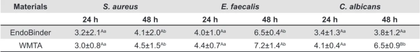
The mean area of the zones of antimicrobial activity (mm) provided by endoBinder and WMTA are presented in Table 1. All tested materials showed antimicrobial activity against all microbial strains tested. No statistical difference was observed between endoBinder and WMTA against E.faecalis
and S.aureus (P>.05). However, C. albicans was
more susceptible to WMTA than to endoBinder in 48 hours (P<.05).
ph
pH values at the different evaluation periods are shown in Table 2. endoBinder and WMTA presented alkaline pH in all experimental times, with a maximum pH value at the 3 h evaluation. Both materials showed a decrease in pH values along the experimental times. WMTA showed higher pH values with statistical differences in all tested periods (P<.05).
Solubility and water sorption
endoBinder showed an average weight loss of 1.47%, while WMTA showed a loss of 2.5%. Although both materials were in agreement with ISO 6876 statement23, endoBinder presented
solubility lower than WMTA (P<.05). Regarding WS,
endoBinder and WMTA had an increase in mass of 9.47% and 9.17%, respectively, without statistical difference between them (P>.05).
Materials S. aureus E. faecalis C. albicans
24 h 48 h 24 h 48 h 24 h 48 h
EndoBinder 3.2±2.1Aa 4.1±2.0Ab 4.0±1.0Aa 6.5±0.4Ab 3.4±1.3Aa 3.8±1.2Aa
WMTA 3.0±0.8Aa 4.5±1.5Ab 4.4±0.7Aa 7.2±1.4Ab 4.1±0.4Aa 6.5±0.9Bb
Table 1- Mean and standard deviation of the zones of microbial growth inhibition (mm) provided by the materials as well as statistical signiicance*
Values are means of microbial growth inhibition (mm) from triplicate experiments. *Different capital letters represent signiicant differences between the materials in the same experimental time (P<.05). Different lowercase letters represent signiicant differences between the same material in different time points (P<0.05)
3 hours 24 hours 48 hours 72 hours 168 hours
EndoBinder 8.96±0.42a 8.94±0.43a 8.80±0.50a 8.78±0.33a 8.46±0.45a
WMTA 10.22±0.46b 10.13±0.49b 10.12±0.79b 9.99±1.08b 9.76±1.52b
Control 6.50 6.50 6.50 6.50 6.50
*Values followed by different superscript letters indicate statistically signiicant differences (P<.05) in comparison between materials in the same experimental time
Table 2- Means and standard deviations of pH values at the different experimental times as well as statistical signiicance* Figure 2- Cytotoxic effects of materials elutes on 3T3 cells by XTT, NR, and crystal violet tests, expressed as percentage of control (cells exposed to culture medium). Bars indicate mean±SD
DISCUSSION
According to the manufacturer, endoBinder has been developed to preserve the properties and clinical applications of MTA, without its negative characteristics24. The present study assessed the
cytotoxicity, antimicrobial activity, pH, solubility and water sorption of endoBinder and compared them with those presented by WMTA. Thereby, this study evaluated some of the main properties that should be considered for a suitable endodontic material. These tests must attend international standards. The International Organization for Standardization, also known as ISO, is the world’s largest international standards developer. Cytotoxicity evaluation was performed according to ISO 10993-45 speciications and solubility and water sorption evaluation was carried out according to ISO 6876/200120.
Cytotoxicity was tested by employing a multiparametric assay, which evaluates in the same sample three different cell viability parameters, namely mitochondrial activity, membrane integrity and cell density. This method increases the chance of detection of cytotoxic effects, allowing correlation of different parameters, and provides a better understanding about toxicity mechanisms of biomaterials10,27. According to the present results,
both materials were highly biocompatible in every parameter studied. These indings are in agreement with previous studies that demonstrated excellent biological properties of MTA, such as the ability to enhance proliferation of periodontal ligament ibroblasts, to induce differentiation of osteoblasts, to stimulate mineralization of dental pulp cells, to have a good biocompatibility and to be nontoxic to several cells linages9,10,30. In relation to endoBinder,
recent works have also showed good in vitro and
in vivo biological properties, biocompatibility in tissues and absence of gelatinolytic activity for MMP-22,31. One methodological aspect that needs to
be discussed is the fact that sealers were exposed to cell culture media after 24 hours of manipulation. endodontic cements are used in a freshly mixed condition in an incompletely polymerized stage. Thus, the results of the cytotoxicity test of the present study should not directly extrapolate to the clinical situation. However, previous studies demonstrated similar results using short time periods for comparative purposes of cytotoxicity10.
In the present study, a modiied agar diffusion test was used, which has been widely employed to assess the antimicrobial activity of several endodontic materials in vitro and allows direct comparisons between endodontic substances3,4,15,32,33. The
microorganisms chosen were selected due to their known resistance to the endodontic procedures.
E. faecalis and C. albicans are considered two of most resistant species in the oral cavity and are
frequently associated with failure of root canal treatment16. Furthermore, S. aureus has also been
isolated from primary and secondary or persistent endodontic infections31. Our results showed similar
antimicrobial activity for endoBinder and WMTA against E. faecalis, S. aureus and C. albicans
at 24 h evaluation. A recent study6, showed a
higher susceptibility of S. aureus and C. albicans
for MTA than endoBinder, at 24 h evaluation, and no differences between materials for E. faecalis. The discrepant results could be explained by methodological differences. The antibacterial and antifungal properties of MTA have been extensively evaluated, with conflicting reports4,24,32,33. The
differences in the results could be attributed not only to the bacterial source, difference between strains, amount of the bacteria inoculated, incubation time, metabolic activity of the microorganisms tested, but also to the molecular size, solubility, and diffusion of the materials through the aqueous agar medium, among others3,4,15,32,33. The antimicrobial activity
of endoBinder and MTA might be due to their high and constant pH. The inluence of the composition of endoBinder and MTA on antimicrobial activity requires further study.
Alkalinization of the medium occurs through the dissociation of calcium ions and hydroxyl ions when the material comes into contact with water. In this experiment, tubes of 1.0 mm in internal diameter were used to limit the contact surface of the materials to the surrounding water, simulating a clinical condition. Our results showed an alkaline pH for both materials, WMTA pH was signiicantly higher in all tested periods (P<.05). One possible
explanation to the differences observed in the present study is related to endoBinder synthesis. Phases with low Ca+ ions content are privileged in
endoBinder synthesis and the material releases a smaller quantity of Ca+ ions7,24. Although a higher
pH promotes a better antimicrobial activity and a lower cytocompatibility, the present results showed that both materials are nonirritant to cells and have good antimicrobial capability.
High solubility of endodontic materials is undesirable because dissolution may cause release of the materials, allowing formation of gaps between them and the dental structure. Regarding the solubility test, both materials were within the recommended values of ISO 6786/200119,
according to which the tested material should not have solubility greater than 3%. However, EndoBinder showed a signiicantly lower SL than WMTA (P<.05). Our results are in agreement with
previous studies that showed MTA as a material with low solubility5,18,25. On the other hand, the
higher solubility of MTA favored a higher pH level8,
has been evaluated for endoBinder. It is important to point out that the solubility testing standards recommend immersion of the materials in water only after complete setting. However, this situation is impossible to be achieved clinically, since the materials are immediately placed in contact with luids and blood. Therefore, solubility values in a clinical scenario are probably higher than the ones found in vitro5. Recently, a novel method
was described to evaluate the solubility by the volumetric measurements of the cements using Micro-CT images8. This method could overcome the
limitations of the ISO methodology and could be closed to simulate a clinical condition.
Water diffusion into cements may result in deterioration of their physical/mechanical properties, decreasing the life expectancy of the interfaces by hydrolysis and microcrack formation29.
However, water sorption could be beneic as it promotes an expansion of the material, which may promote a proper sealing. Being both materials hydrophilic, a high water sorption was anticipated and confirmed by the results. No statistical differences were observed between the two tested materials in this aspect (P<.05). As the values of
water sorption were similar between both materials, and as WMTA is a gold standard material, we might afirm that EndoBinder has good water sorption properties.
CONCLUSION
The present indings demonstrated the suitable cytotoxicity, physicochemical properties and the antimicrobial capability of this new calcium aluminate-based cement, which is known as endoBinder.
ACKNOwLEDgMENTS
The authors deny any conlicts of interest related to this study.
REFERENCES
1- Aguilar FG, Garcia LF, Rossetto HL, Pardini LC, Pires-de-Souza FC. Radiopacity evaluation of calcium aluminate cement containing different radiopacifying agents. J endod. 2011;37(1):67-71. 2- Aguilar FG, Roberti Garcia LF, Panzeri Pires-de-Souza FC. Biocompatibility of new calcium aluminate cement (endoBinder). J endod. 2012;38(3):367-71.
3- Al-Hezaimi K, Al-Shalan TA, Naghshbandi J, Simon JH, Rotstein I. MTA preparations from different origins may vary in their antimicrobial activity. Oral Surg Oral Med Oral Pathol Oral Radiol endod. 2009;107(5):e85-8.
4- Al-Nazhan S, Al-Judai A. evaluation of antifungal activity of mineral trioxide aggregate. J endod. 2003;29(12):826-7.
5- Bortoluzzi eA, Broon NJ, Bramante CM, Felippe WT, Tanomaru Filho M, Esberard RM. The inluence of calcium chloride on the setting time, solubility, disintegration, and pH of mineral trioxide aggregate and white Portland cement with a radiopaciier. J Endod. 2009;35(4):550-4.
6- Carvalho Panzeri Pires-de-Souza F, Moraes PC, Fonseca Roberti Garcia L, Aguilar FG, Watanabe e. evaluation of pH, calcium ion release and antimicrobial activity of a new calcium aluminate cement. Braz Oral Res. 2013;27(4):324-30.
7- Castro-Raucci LM, Oliveira IR, Teixeira LN, Rosa AL, Oliveira PT, Jacobovitz M. effects of a novel calcium aluminate cement on the early events of the progression of osteogenic cell cultures. Braz Dent J. 2011;22(2):99-104.
8- Cavenago BC, Pereira TC, Duarte MA, Ordinola-Zapata R, Marciano MA, Bramante CM, et al. Inluence of powder-to-water ratio on radiopacity, setting time, pH, calcium ion release and a micro-CT volumetric solubility of white mineral trioxide aggregate. Int endod J. 2013:10.1111/iej.12120. epub ahead of print. 9- Damas BA, Wheater MA, Bringas JS, Hoen MM. Cytotoxicity comparison of mineral trioxide aggregates and endoSequence bioceramic root repair materials. J endod. 2011;37(3):372-5. 10- De-Deus G, Canabarro A, Alves G, Linhares A, Senne MI, Granjeiro JM. Optimal cytocompatibility of a bioceramic nanoparticulate cement in primary human mesenchymal cells. J endod. 2009;35(10):1387-90.
11- Duarte MA, Alves de Aguiar K, Zeferino MA, Vivan RR, Ordinola-Zapata R, Tanomaru-Filho M, et al. evaluation of the propylene glycol association on some physical and chemical properties of mineral trioxide aggregate. Int endod J. 2012;45(6):565-70. 12- Duarte MA, Oliveira Demarchi AC, Yamashita JC, Kuga MC, Campos Fraga S. Arsenic release provided by MTA and Portland cement. Oral Surg Oral Med Oral Pathol Oral Radiol endod. 2005;99(5):648-50.
13- enkel B, Dupas C, Armengol V, Akpe Adou J, Bosco J, Daculsi G, et al. Bioactive materials in endodontics. expert Rev Med Devices. 2008;5(4):475-94.
14- Ford TR, Torabinejad M, McKendry DJ, Hong CU, Kariyawasam SP. Use of mineral trioxide aggregate for repair of furcal perforations. Oral Surg Oral Med Oral Pathol Oral Radiol endod. 1995;79(6):756-63.
15- Gomes BP, Ferraz CC, Garrido FD, Rosalen PL, Zaia AA, Teixeira FB, et al. Microbial susceptibility to calcium hydroxide pastes and their vehicles. J endod. 2002;28(11):758-61.
16- Gomes BP, Lilley JD, Drucker DB. Associations of endodontic symptoms and signs with particular combinations of speciic bacteria. Int endod J. 1996;29(2):69-75.
17- Gomes-Filho Je, Faria MD, Bernabé PF, Nery MJ, Otoboni-Filho JA, Dezan-Júnior E, et al. Mineral trioxide aggregate but not light-cure mineral trioxide aggregate stimulated mineralization. J endod. 2008;34(1):62-5.
18- Hungaro Duarte MA, Minotti PG, Rodrigues CT, Zapata RO, Bramante CM, Tanomaru Filho M, et al. effect of different radiopacifying agents on the physicochemical properties of white Portland cement and white mineral trioxide aggregate. J endod. 2012;38(3):394-7.
19- International Organization for Standardization. ISO 10993: Biological evaluation of medical devices – Part 5: Tests for in vitro cytotoxicity. Geneva: ISO; 2009.
23- Moretton TR, Brown Ce Jr, Legan JJ, Kafrawy AH. Tissue reactions after subcutaneous and intraosseous implantation of mineral trioxide aggregate and ethoxybenzoic acid cement. J Biomed Mater Res. 2000;52(3):528-33.
24- Pandolfelli VC, Oliveira IR, Rossetto HL, Jacobovitz M, inventors; Fundação Universidade Federal de São Carlos, assignee. Aluminous cement-based composition for application in endodontics and cementitious product obtained thereof. BR patent INPI 0704502-6. 2007 November 29.
25- Parirokh M, Torabinejad M. Mineral trioxide aggregate: a comprehensive literature review - Part I: chemical, physical, and antibacterial properties. J endod. 2010;36(1):16-27.
26- Parirokh M, Torabinejad M. Mineral trioxide aggregate: a comprehensive literature review - Part III: Clinical applications, drawbacks, and mechanism of action. J endod. 2010;36(3):400-13.
27- Scelza MZ, Linhares AB, Silva Le, Granjeiro JM, Alves GG. A multiparametric assay to compare the cytotoxicity of endodontic sealers with primary human osteoblasts. Int endod J. 2012;45(1):12-8.
28- Schembri M, Peplow G, Camilleri J. Analyses of heavy metals in mineral trioxide aggregate and Portland cement. J endod. 2010;36(7):1210-5.
29- Sideridou I, Tserki V, Papanastasiou G. Study of water sorption, solubility and modulus of elasticity of light-cured dimethacrylate-based dental resins. Biomaterials. 2003;24(4):655-65.
30- Silva eJ, Accorsi-Mendonça T, Almeida JF, Ferraz CC, Gomes BP, Zaia AA. evaluation of cytotoxicity and up-regulation of gelatinases in human ibroblast cells by four root canal sealers. Int Endod J. 2012;45(1):49-56.
31- Silva eJ, Herrera DR, Almeida JF, Ferraz CC, Gomes BP, Zaia AA. evaluation of cytotoxicity and up-regulation of gelatinases in ibroblast cells by three root repair materials. Int Endod J. 2012;45(9):815-20.
32- Siqueira JF Jr, Rôças IN. exploiting molecular methods to explore endodontic infections: Part 2 - Redeining the endodontic microbiota. J endod. 2005;31(7):488-98.
33- Stowe TJ, Sedgley CM, Stowe B, Fenno JC. The effects of chlorhexidine gluconate (0.12%) on the antimicrobial properties of tooth-colored ProRoot mineral trioxide aggregate. J endod. 2004;30(6):429-31.