ABSTRACT
The oroantral istula is one of the complications most common after dental extractions in posterior maxillary, mainly in the region of second and third molar. The diagnosis is based on clinical signs and symptoms, which may be present in pain, fever, hyposmia and drainage of purulent discharge; additional examinations such as computed tomography and sinus nasal endoscopy improve diagnostic precision. It is therefore of paramount importance to correct early diagnosis and treatment avoiding in this way, the symptons of sinusitis, infections and dysphonia. Several surgical techniques have been proposed for the closure of the bucosinusal istula; the use of Bichat´s fat pad has become a successful alternative among them. Many surgical treatments have been to propose for closure of bucosinusal istula; Bichat fat pad has been a good alternative. The propose for this article is report two cases with closure of bucosinusal istulas through Bichat’s fat pad, as well as to discuss the surgical techniques, characteristics, indications and peculiarities.
Indexing terms: Oroantral istula. Oral surgical procedures. Surgical laps.
RESUMO
A fístula bucosinusal ou oro-antral é uma das complicações mais comuns, após extrações dentárias na região posterior de maxila, principalmente em região de segundo e terceiro molares permanentes. O diagnóstico se baseia em sinais e sintomas clínicos, nos quais pode-se destacar: dor, febre, hiposmia e drenagem de secreção purulenta. Os exames complementares como a endoscopia sinonasal e a tomograia computadorizada melhoram a precisão diagnóstica. Assim, é de suma importância, o correto diagnóstico e tratamento precoce, evitando dessa maneira a instalação de sinusites, infecções e disfonia. Diversas técnicas cirúrgicas têm sido propostas na literatura, para o fechamento da fístula bucossinusal, dentre elas, a utilização do corpo adiposo de Bichat tem se tornado uma alternativa bem sucedida. O objetivo deste trabalho é relatar dois casos clínicos de fechamento de fístulas bucossinusais através do retalho da bola de Bichat, realizados pelo Departamento de Cirurgia e Traumatologia Bucomaxilofacial do Hospital da Face e Hospital da Restauração em Recife, Pernambuco. Ainda, descrever acerca de sua técnica cirúrgica, características, indicações e peculiaridades.
Termos de indexação: Fistula bucoantral. Procedimentos cirúrgicos bucais. Retalhos cirúrgicos.
Oroantral istulas closure using Bichat’s fat pad
Fechamento de fístulas bucossinusais através do uso da bola de Bichat
Marcelo Fernando do AMARAL1
Luiz Antonio Portela GUERRA2
Marleny Elizabeth Márquez de Martínez GERBI3
Audemir Rocha MELO1
David Gomes de Alencar GONDIM1
Rui MEDEIROS JUNIOR1
Suzana Lubambo de MELO3
Alexandrino Pereira dos SANTOS NETO3
1 Hospital da Restauração, Residência em Cirurgia e Traumatologia Buco-Maxilo-Facial. Recife, PE, Brasil.
2 Universidade de Pernambuco, Faculdade de Odontologia, Departamento de Cirurgia e Traumatologia Buco-Maxilo-Facial. Camaragibe, PE, Brasil. 3 Universidade de Pernambuco, Faculdade de Odontologia. Av. General Newton Cavalcanti, 1650, Tabatinga, 54753-901, Camaragibe, PE, Brasil.
Correspondência para / Correspondence to: MEMM GERBI. E-mail: <[email protected]>.
decreasing the alveoli and carrying the tooth apex to a very straight conexion with the sinus cavity3. The proximity of the
root apices to the sinus cavity makes more susceptible the dental element extraction to occur oroantral communication and it is being obeyed by the following decreasing order related to the risk: second molar, irst molar, third molar, second premolar and irst premolar4. Cystic and tumor lesion as well as local traumas
are also found as etiological factors for such occurrence. Several methods have been proposed in the literature for oral sinus communication closures such as local laps, remote laps, grafting and the adipose tissue of the cheek. Among the mucoperiosteal laps from the labial area which is described by Rehrmann as the most used
INTRODUCCION
Maxillary sinuses are spaces illed with air, located bilaterally in the maxilla. These are lined by a respiratory epithelium, secreting mucus and these are the largest among the paranasal sinuses1. An oroantral communication generally
occurs for some reason after dental extractions; and, it creates a way among the maxillary sinus and the oral cavity that due to tissue proliferation. Both from sinus and oral mucosa develop an epithelialised way (istula) among the cavities2.
Local anesthesia initially was given through superior alveolar nerve blocks: previous, intermediate, posterior and major palatine nerve; incision around the istula subsequently was performed with n°15 scalpel blade and excision of this. The cleaning of maxillary sinus was performed with 0,9% saline solution (SS) and then a quadrangular oral mucoperiosteal lap was made through 0,9% saline solution irrigation (100 ml) with relaxing incisions, previous and posterior to the istula, at the level of posterior region, which would be the second molar. Incisions in the periosteum of the lap was made with Bichat fat pad herniation through divulsion and the traction of this was performed to the local of communication. The suture with 4-0 silk thread and the lap reposition to the original repositioning were made (Figure 2). The removal of the suture happened after 15 days of the surgical procedure (Figure 3).The patient authorized the publication of his case after signing written consent form. She was clinical followed-up during 18 months and showed no signs of recurrence of istula. because it has a broad base. The blood supply of the lap
is guaranteed and the mobility of itself can be improved through the performance of incisions in the periosteum at lap base. The palatal laps are designed based on the largest palatal vessels and these are classiied as rotational laps. This type of lap has good mobility and thickness of tissue5-6.
The closure of oroantral istula can yet be done through the use of monocortical bone grafts or tooth transplant7.
The adipose tissue of the cheek was used by Abuabara et al.6 for closure of oroantral istula in 28
patients, obtaining success index of 100% in the cases. The objective of this paper is to report two clinical cases of oroantral istula closure through Bichat fat pad as well as to describe about it surgical technique, characteristics, indication and peculiarities.
CASE REPORT
Clinical case 1
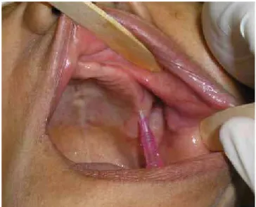
Patient, 47 years, female, sought the Oral and Maxillofacial Traumatology Surgical Service in the ‘Hospital da Face’ (Recife-PE), complaining of liquid outlow through nasal cavity by feeding himself. About the clinical intra-oral examination, it was observed opening on the superior second molar (Figure 1) and the maxilla showed up edentulous.
Figure 1. Clinical aspect: introduction of a catheter to identify a communication
Patient reported dental extraction in that region for the past 12 months and not complaining about pain. The curse of sinusitis disappeared after following a clinical and image examination. The treatment proposed was the oroantral istula closure with Bichat’s fat pad.
Figure 2. Immediate postoperative period. Observe the arrow pointing to the adi-pose tissue in position.
The patient authorized the publication of the case upon signature of an informed consent form and is currently in outpatient follow-up for 14 months without signs of recurrence.
DISCUSSION
Several methods for closure of oroantral communication have been reported in the literature as the palatine and buccal lap8-9. However, these methods not
always bring signiicant results.
The Valsalva maneuver is a type of physical evaluation and it is important in the diagnosis of oroantral istula, however, it might not be used for communication diagnosis soon after dental element extraction because it can cause or increase communication.
The adipose tissue of the cheek was described for the irst time in 1802 by Pichat6 but only in 1977 Egyedi10
used it in oroantral and oronasal communication as a pediculated graft associated to a skin graft. Its use as a free graft was described by Neder11 and as a pediculated graft
by Tideman et al.12 . These same scholars observed that the
epithelialisation of the graft occurs during 2 or 3 weeks without necessity to cover this with skin grafts.
The Bichat’s fat pad anatomically has elongated formation like as an ice cream cone which the Bichat’s fat pad is a supericial extremity that separate buccinators and masseter muscle and the cone separates its deep extension that extends to infratemporal fossa, separating pterygoid muscles and serving as cushion to facilitate the muscle movements related to the other13.
As advantages of the Bichat’s fat pad are cited: it causes less disturbances and scars in the vestibule than a slippage of laps14, allows adjustments after one week15
and is considered a simple and speedy procedure16-17 Clinical case 2
Patient 22 years old, male, attended the Surgery and Oral maxillofacial Traumatology Department at the ‘Hospital da Restauração’ in Recife (PE). The main complaint was a “hole in the mouth” and water low by the nostrils during oral hygiene or performance of mouthwashes. During the anamneses the patient related to have done an exodontia of the element 17 for 4 months. There was a istula in the previous extraction region conirmed by x-ray image (Figure 4) which can be observed solution continuity from the loor of the right maxillary sinus, veiling absence of the maxillary sinus which would suggest chronic sinusitis and element included in the region.
Figure 4. Panoramic radiograph. Observe the Arrow identifying communication and associated element included.
Fistula closure through Bichat’s fat pad rotation was proposed and the maintenance of the element was included (Figure 5 and 6).The surgical procedure differentiated itself from the clinical case 1 because it is a triangular mucoperiosteal lap only with an anterior relaxing incision to the istula. The synthesis was performed with 4-0 nylon thread.
Figure 5. Immediate post-surgical aspect.
without removal of tooth or bones; At least, postoperative discomfort to the patient15-17,low morbidity, possibility to
be associated to other laps16 and it does not lose furrow
depth2,15. There was not any complication observed in the
two cases presented herein. Patients did not report any signiicant painful symptomatology; however, we did not agree in relation to the maintenance of the furrow depth related to the use of Bichat´s fat pad. We observed loss of furrow depth, however to a lesser extent as compared to that observed with the techniques of using vestibular laps.
The fact that only can be used just once as a main disadvantages18, possibility of trismus in the postoperative
period, limitation of its use for small and medium defects, possibility of causing esthetic alterations perceived in the speech16. There was not any patient presenting trismus,
disturbance of speech or signiicant esthetic alteration in postoperative period in the cases presented.
We believe that in cases of chronic sinusitis might perform previous antibiotic therapy and postponement of surgical treatment until infectious process stabilization. Postsurgical antibiotic therapy(amoxicillin, 500 mg, 8-8 hours for 7 days) was performed in the clinical cases described herein, since in any case was presented chronic sinusitis nasal descongestant was also prescribed(Sorine®Drops) 3 times a
day for 7 days, analgesic (dipyrone 500mg) for 2 days and mouthrinses of chlorhexidine digluconate 0,12%, 2 time a day for 7 days as collutory. Postoperative recommendations were passed on as: not to sneeze with closed mouth, not
blow the nose, avoid passing the tongue on the surgical site, maintain good oral hygiene and mild diet.
We consider that copious irrigation with saline solution(0,9%) of the involved maxillary sinus is an important step for a reduced possibility of infection with lap loss and consequently a istula recurrence.
CONCLUSION
It concludes that the use of Bichat´s fat pad for closure of oroantral istulas should be included more frequently in the surgical possibilities because it is a procedure relatively simple with low complication index, comfortable postoperative for the patient and it can be used for closure of small and medium defects in the molar region up to the canine.
Collaborators
MF AMARAL and LAP GUERRA performed surgery for closure of oroantral istula related to the irst clinical case cited and conducted in the ‘Hospital da Face’ and participated of the paper writing. AR MELO, DGA GONDIM and R MEDEIROS JUNIOR performed surgery for closure of oroantral istula related to the second clinical case cited and conducted in the ‘Hospital da Restauração’ and participated of the paper writing MEMM GERBI, SL MELO and AP SANTOS NETO took part of the writing paper.
REFERENCES
1. Peterson LJ. Cirurgia oral e maxilofacial contemporânea. 3ª ed. Rio de Janeiro: Guanabara Koogan; 2000.
2. Garcia RR, Rabêlo LRS, Moraes M, Moreira RWF, Albergaria-Barbosa JR. Utilização de enxerto pediculado do corpo adiposo da bochecha no tratamento de comunicações oro-antrais. Rev Port Estomatol Cir Maxilofac. 2000;41:17-24. doi.10.1590/ S1980-65232010000100021
3. Marzola C. Acidentes e complicações da endodontia: proilaxia e tratamento. In: Marzola C. Técnica exodôntica. 2ª ed. São Paulo: Pancast; 1994. p. 284-9.
4. Faig-Leite H. Topograia dento-alveloar. In: Madeira MC. Anatomia da face: bases anátomo-funcionais para a prática odontológica. 5ª ed. São Paulo: Sarvier; 2004. p. 29-34.
5. Eppley B, Scaroff A. Oro-nasal istula secondary to maxillary augmentation. Int J Oral Surg. 1984;13(6):535-8. doi:10.1016/ S0300-9785(84)80026-5
6. Abuabara A, Cortez AL, Passeri LA, De Moraes M, Moreira RW. Evaluation of different treatments for oroantral/oronasal communications: experience of 112 cases. Int J Oral Maxillofac Surg. 2006;35(2):155-8. doi:10.1016/j.ijom.2005.04.024
7. Haas R, Watzak G, Baron M, Tepper G, Mailath G, Watzek G. A preliminary study of monocortical bone grafts for oroantral istula closure. Oral Surg Oral Med Oral Pathol Oral Radiol Endod. 2003;96(3):263-6. doi: 10.1016/ S1079210403003755
8. Killey HC, Key LW. An analysis of 250 cases of oro-antral istula treated by the buccal lap operation. Oral Surg Oral Med Oral Path. 1967; 24(6):726-39. doi:10.1016/0030-4220(67)90506-3
9. Yih WY, Merrill RG, Howerton DW. Secondary closure of oroantral and oronasal istulas: a modiication of existing techniques. J Oral Maxillofac Surg. 1988;46(5):357-64. doi:10.1016/0278-2391(88)90218-2
11. Neder A. Use of buccal fat pad for grafts. Oral Surg Oral Med Oral Pathol. 1983;55(4):349-50. doi:10.1016/0030-4220(83)90187-1
12. Tideman H, Bosanquet A, Scott J. Use of the buccal fat pad as a pedicled graft. J Oral Maxillofac Surg. 1986;44(6):435-40. doi: 10.1016/S0278-2391(86)80007-6
13. Madeira MC. Anatomia da face. São Paulo: Editora Sarvier; 1995.
14. Baumann A, Ewers R. Application of the buccal fat pad in oral reconstruction. J Oral Maxillofac Surg. 2000;58(4):389-92. doi:10.1016/j.ajoms.2011.05.001
15. Hanazawa Y, Itoh K, Mabashi T, Sato K. Closure of oroantral communications using a pedicled buccal fat pad graft. J Oral Maxillofac Surg. 1995;53(7):771-5. doi:10.1016/0278-2391(95)90329-1
16. Dean A, Alamillos F, García-López A, Sánchez J, Penãlba M. The bucal fat pad lap in oral reconstruction. Head Neck. 2001;23(5):383-8. doi: 10.1002/hed.1048
17. Martín-Granizo R, Naval L, Costa A, Goizueta C, Rodriguez F, Monje F, et al. Use of buccal fat pad to repair intraoral defects: review of 30 cases. Br J Oral Maxillofac Surg. 1997;35(2):81-4. doi:10.1016/S0266-4356(97)90680-X
18. Rapidis AD, Alexandridis CA, Eleftheriadis E, Angelopoulos AP. The use of the buccal fat pad for reconstruction of oral defects: review of the literature and report of 15 cases. J Oral Maxillofac Surg. 2000;58(2):158-63. doi:10.1016/S0278-2391(00)90330-6