ABSTRACT
This report aims to present a clinical case of endodontic re-treatment performed on a left inferior premolar with two canals of dificult localization. The importance of anatomical knowledge and the use of an operating microscope, especially in complex reintervention are discussed. An endodontic re-treatment was performed on element 34, which had a prosthetic restoration and vestibular istula. In clinical examination, it was observed a negative result to the sensitization test and a positive result to vertical percussion test. After all the obturating material was removed, an operating microscope was requested to assess a possible fracture or the presence of another canal. It was noticed the presence of another canal that was treated and obturated as the existing canal. After 12 months, a new radiographic exam was performed, whose result was found asymptomatic and radiographically normal. In complex peculiar anatomy cases, the operating microscope is vital for the localization of extra canal to guarantee treatment success.
Indexing terms: Endodontics. Microscopy. Premolar tooth.
RESUMO
O artigo tem como objetivo apresentar um caso clínico de retratamento endodôntico realizado em um pré-molar inferior esquerdo com dois canais de difícil localização, onde é discutida a importância dos conhecimentos anatômicos e o uso do microscópio operatório, sobretudo em reintervenções complexas. Realizou-se um retratamento endodôntico no elemento 34, possuidor de restauração protética e fístula por vestibular. No exame clínico, constatou-se resultado negativo ao teste de sensibilidade e resultado positivo ao teste de percussão vertical. Após remoção de todo o material obturador existente, o microscópio operatório foi solicitado para avaliação de uma possível fratura ou presença de outro canal. Foi constatada a presença de outro canal, o qual foi tratado e obturado como o já existente. Após 12 meses, novo exame radiográico foi feito e constatou-se normalidade radiográica e assintomatologia. Em casos mais complexos e com anatomia peculiar torna-se imprescindível a utilização do microscópio operatório no auxílio à localização de um canal extra para o sucesso do tratamento.
Termos de indexação: Endodontia. Microscopia. Dente pré-molar.
Using an operating microscope to re-treat an inferior premolar with
two canals
Uso do microscópio operatório no retratamento de um pré-molar inferior com dois canais
Janaína Fernandes SANTOS1
Gustavo Moreira ALMEIDA1
Eduardo Fernandes MARQUES1
Carlos Eduardo da Silveira BUENO2
1 Faculdade Ciodonto, Departamento de Endodontia. Praça Ruy Barbosa , 185, Centro, 45653-400, Ilhéus, BA, Brasil. Correspondência para /
Corres-pondence to: GM ALMEIDA. E-mail: <[email protected]>.
2 Faculdade São Leopoldo Mandic, Curso de Odontologia, Programa de Pós-Graduação em Endodontia. Campinas, SP, Brasil.
lesion. The degree of success of these cases, described in the literature, is lower in re-treatment with chronic apical periodontitis cases. The internal anatomy of the irst inferior premolars is particularly complex due to the variation in the number of canals and their coniguration5-6. Introduction of
operating microscope in Endodontics redeined visualization concepts, providing image magniication7 and a higher
rate of success in endodontic treatments. Variation in the number of canal treatments has changed throughout time, given that in 1955 only 8.9% of inferior premolars with more than one canal were found8. Moreover, in 1984 the
authors3 found 27.8% of two-canal teeth; and in a research9
that was carried out in 1999 with 100 in vitro diaphanous inferior premolars, they concluded that 49% of the elements bore two radicular canals.
INTRODUCTION
A discussion topic for a number of scholars, the anatomy of root canals is one of the major challenges of endodontic treatments. The lower premolar teeth get most attention given the signiicant variation in the number of canals1-2.
The endodontist must be diligent with regard to the presence of an extra canal; given that even if it is not visible in the radiograph, it doesn’t mean that it doesn’t exist3.
Treatment becomes more complicated when the radicular canal is infected with microorganisms4. The latter
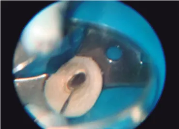
In the second session, temporary seal was removed with total isolation, and canal was irrigated with 5.25% hypochlorite. In addition to the latter, K 10 iles (Dentsply/ Maillefer, Ballaigues, Switzerland) were used to explore the canal under an operating microscope, 8X magniication (Figure 2); and posteriorly, 20X magniication at which a vestibular canal was localized. In order to conirm the exam result, a radiograph was taken with the positioned iles (Figure 3) that allowed veriication of the point where the canals were divided in the meso-apical direction. Dealing with this canal was dificult, given that it was obstructed with debris from previous treatments.
Ação, São Paulo, Brazil) associated with aqueous vehicle – isotonic sodium chloride solution - (Fórmula & Ação, São Paulo, Brazil) was applied as intracanal remedy sealed with a temporary obturator (Villevie, Joinville, Brazil). Another appointment was scheduled within 7 days.
Castelucci10 reported the use of a microscope as
one of the most important factors in the signiicant upturn in chances to save teeth, both surgically and non-surgically, especially in complex cases and endodontic re-interventions.
The purpose of the reported case aimed to emphasize the importance of dental anatomy knowledge and the use of an operating microscope in the identiication and localization of extra canals among others. The procedure inal result brings clinical success.
CASE REPORT
Patient D.R.B.O, female, 44 years old, leucoderma, came to the dentist’s ofice with a complaint about pain in the 34th element (1st left inferior premolar). Based on
the patient’s report, it was found that the tooth had been submitted twice to unsuccessful endodontic treatment. Two years after the last intervention, vestibular istula and pain to mastication reappeared.
Anamnesis was performed and no signiicant alteration was observed. In clinical examination, it was observed a negative result to the sensitization test and a positive result to vertical percussion test. Clinically, the patient had a prosthetic restoration and a vestibular istula was found in the apical palpation exam. A complementary periapical radiograph exam (Kodak Rochester, New York, USA) (Figure 1) showed a periradicular lesion with periapical bone rarefaction and a possible unobturated canal. Given the clinical-radiographic exam, endodontic re-treatment of the element was indicated, leaving periapical surgery as a last treatment option. The procedure started with the administration of anesthesia (regional block of the left inferior alveolar nerve) with articaine 4% (DFL Indústria e Comércio S.A, Rio de Janeiro, Brazil). Surgical access was performed with the aid of ultrasound with adapted tips, TRA 24 D and TRA 15C D (Dental Trinks, São Paulo, Brazil), followed by complete isolation (rubber dam Madeitex, São José dos Campos, Brazil). Removal of gutta-percha from the canal was done with 1ml of eucalyptol (Fórmula & Ação, São Paulo, Brazil) and instruments D1, D2 and D3 (ProTaper - Dentsply/Maillefer, Ballaigues, Switzerland). Instrument D1 is employed to unclog the canal cervical third; D2 is used to unclog the mid third and D3 to unclog the apical third using 2ml of sodium hypochlorite solution at 5.25% (Fórmula & Ação, São Paulo, Brazil) in every ile substitution. The operating microscope with a 20 times magniication (Alliance, São Paulo, Brazil) was employed to search for another possible canal, fracture or issure, but nothing was detected. Therefore, calcium hydroxide PA (Fórmula &
Figure 1. First X-ray.
After 12 months, another radiographic exam was made, showing the restoration process in the periradicular region (Figure 6).
Hence, pre-enlargement of both canals using rotary-device limes 35/10 and 20/07 ProDesign (Easy Endo, Belo Horizonte, Brazil), Gates Glidden 4, 3 and 2 (Dentsply/Maillefer, Ballaigues, Switzerland), followed by odontometry using a foraminal localizer Root ZX (J. Morita, Kyoto, Japan). After the real working extent was measured, crown-apical technique instrumentation was initiated, following the manufacturer (Easy Endo, Belo Horizonte, Brazil) recommended sequence: white ile (20/03); yellow ile (15/05); red ile (22/04), blue ile (25/04); green (20/06), and the last ile, black (20/07). At each lime exchange, 2 ml of sodium hypochlorite 5.25% was used as irrigator. After the two radicular canals were cleaned and molded, a cone test was carried out (Figure 4) using FM cones (Sparttan/ Precise-Jalisco, Mexico). It was possible to conirm by means of radiographic exam the working length measurement. Then irrigation was performed to remove the smear layer with EDTA 17% (Formula & Ação, São Paulo, Brazil) for 3 minutes with ultrasonic cavitations and inal toilet with 5 ml of sodium hypochlorite at 5.25%. Canals were dried with sterilized absorbent paper cones (Dentsply/Maillefer, Ballaigues, Switzerland) after the inal rinsing. Subsequently, the radicular canal system was obturated with endodontic cement AHPlus (Dentsply/Maillefer, Ballaigues, Switzerland) by continuous wave of condensation technique11 (Termo
Pack-Easy Endo, Belo Horizonte, Brazil). Following obturation, a provisional double cervical sealing (Villevie, Santa Catarina, Brazil) and glass ionomer Vidrion F (White, Rio de Janeiro, Brazil) were applied. Final radiography was made and directed to prosthetic restoration. Treatment was conirmed and assessed after the inal radiograph was taken (Figure 5). The patient freely signed an informed consent regarding the publication and divulgence of this study case.
Figure 4. Cone test.
Figure 5. Last radiograph.
REFERENCES
1. Robinson S, Czerny C, Gahleitner A, Bernhart, Kainberger FM, Dental CT evaluation of mandibular irst premolar root conigurations and canal variations. Oral Surg Oral Med Oral
DISCUSSION
Anatomical variations in the irst inferior premolar are described in the literature11. In 1973, the analysis of
1393 irst inferior premolars was performed through distal and vestibular-lingual radiographic exams. The results showed the presence of one canal in 63% of the examined teeth; two canals in 22.7% of the cases, and 0.4% of the teeth presented three radicular canals12. In 1978, it was
found that 74% of the 400 irst inferior premolars had a sole canal, 25.5% of the samples had two canals, and only 0.5% presented three radicular canals13. These data served
as basis for this report. Therefore, the presence of a second canal should be always considered, given the signiicant variation of internal anatomy of inferior premolars14.
Missing the presence of an extra canal may negatively affect the case outcome15. The latter was
observed in this study, given that it was reported in the anamnesis that the tooth was submitted to endodontic treatment twice, with symptoms such as positive vertical percussion test, presence of vestibular istula and periradicular lesion with periapical osseous rarefaction. This case is similar to a clinical case published in 199116.
The existence of longitudinal issures on the tooth root mesial face and/or vestibular face is related to radicular bifurcation3,9,17. In 1998, it was reported that
the main bifurcations usually occur at the mid third or apical level18 which is in accordance with the present
study. Notwithstanding, it disagrees with other authors who showed that bifurcations can occur at any root level19. Racial factor seems to exert some inluence, and
prevalence of two canals in the irst inferior premolar is signiicantly greater in black people2,8.
Multiple canals can be present in a tooth whose radiographic image may be similar to a sole root; whereas multiple roots can be present and poorly deined in the radiographic exam20. However, critical radiograph
analysis must be performed21 and changes to the x-ray
beams angulations must be done in order to detect any radiographic density alterations, which suggest the presence of additional canals. In this study, it was noticed that there was no sharpness in the presence of additional canal. However, the obturated canal was medially projected
and the other projection was distally projected, suggesting the presence of an extra canal.
In the tactile exam of the main canal walls, a previously curved small ile (30 to 40 degree) must be employed with progressive stride and carefully rotated to the right and to the left with a quarter of a turn until the second canal is penetrated16. After submitting the tooth
to this technique, radiograph should be made. Particular attention should be given to vestibular and lingual walls, since they cannot be viewed in the radiograph and most of the extra canals are originated on these walls with terminations at acute angles22-23. In this study, the extra
canal was located distally on the lingual wall and was radiographically conirmed.
Certainly, dentistry has undergone a technological revolution in the past decades, but the real revolution is yet to come. True revolution will come when we realize that we need to keep our eyes open, given that our standard vision without the aid of magnifying glass, microscope or any other available method is unitting to provide excellence in dentistry in every stage of clinical diagnosis up to the most complex clinical or surgical procedure 24. These study indings
prove, whenever it was possible to observe improvement in endodontic treatment prognosis by clinically employing an operating microscope, in agreement with other authors25
who mentioned the importance of such technology in the localization of canals that are dificult to access.
CONCLUSION
It was concluded based on the reported case and relevant literature review that inferior premolars have a wide anatomical variation, which favors the presence of extra canals. Experience and anatomy knowledge are vital prerequisites to the dental surgeon, combined with the utilization of the operating microscope that allows better lighting and visual magniication.
Collaborators
JF SANTOS, GM ALMEIDA, EF MARQUES and CES BUENO participated in all stages of this article elaboration.
Pathol Oral Radiol Endod. 2002;93(3):328-32. doi:10.1067/ moe.2002.120055
3. Leite HF, Madeira MC, Simoes S, Filho Niccoli WD, Oliveira JA, Martins AA. Estudo radiográico da ocorrência de dois canais radiculares em primeiros pré-molares inferiores. Rev Odontol UNESP. 1984;13(1/2):113-8.
4. Kakehashi S, Stanley HR, Fitzgerald RJ. The effects of surgical exposure of dental pulps in germ-free and conventional laboratory rats. Oral Surg Oral Med Oral Pathol. 1965;20(3):340-9. doi:10.1016/0030-4220(65)90171-4
5. Gorni FGM, Gagliani MM. The outcome of endodontic retreatment: a 2-yr follow - up. J Endod. 2004;30(1):1-4. doi:10.1097/00004770-200401000-00001
6. Pécora JD, Saquy PC, Neto Souza MD, Woelfel JB. Root form and canal anatomy of maxillary irst premolars. Braz Dental J. 1991;2(2):87-94.
7. Carr GB. Microscopes in endodontics. J Calif Dent Assoc. 1992;20(11):55-61.
8. Amos ER. Incidence of bifurcated canals in mandibular bicuspids. J Am Dent Assoc. 1955;50(1):70-1. doi.org/10.14219/jada. archive.1955.0011
9. Silva FSP, Koga K, Silveira JCF, Nascimento CM. Variações na coniguração radicular interna de pré-molares inferiores. Rev APCD, 1999;53(1):64-9.
10. Castelucci A. Magniication in endodontics: the use of operating microscope. Endoc Pract. 2003;15(5):377-84.
11. Buchanan LS. The continuous wave of condensation technique: a convergence of conceptual and procedural advances in obturation. Dent Today. 1994;13(10):80-2.
12. Zillich R, Dowson J. Root canal morphology of mandibular irst and second premolars. Oral Surgery Oral Med Oral Pathol. 1973;36(5):738-44. doi:10.1016/0030-4220(73)90147-3 13. Vertucci FJ. Root canal morphology of mandibular premolars.
J Am Dent Assoc. 1978;97(1):47-50. doi.org/10.14219/jada. archive.1978.0443
14. Baisden MK, Kulild JC, Weller RN. Root canal coniguration of the mandibular irst premolar. J Endod. 1992;18(10):505-8. doi:10.1016/S0099-2399(06)81352-X
15. Cleghorn B, Christie W, Dong C. Root and root canal morphology of the human permanent maxillary irst molar: a literature review. J Endod. 2006;32(9):813-21. doi:10.1016/j. joen.2006.04.014
16. England Jr NC, Hartwell GR, Lance JR. Detection and treatment of multiple canals in mandibular premolars. J Endod. 1991;17(4):174-8. doi:10.1016/S0099-2399(06)82012-1 17. Madeira MC, Leita HF, Oliveira JA, Simões S, Niccoli Filho WD.
Anatomia exterior da raiz do primeiro pré-molar inferior. Arq Cent Est Cur Odont UFMG. 1984;21(1):33-46.
18. Rios LV. Coniguração interna dos pré-molares. RGO - Rev Gaúch Odontol. 1998;36(2):101-5.
19. Cohen S, Burns R. Caminhos da polpa. 6ª ed. Rio de Janeiro: Guanabara Koogan; 1997.
20. Cleghorn BM, Christie WH, Dong CC. The root and root canal morphology of the human mandibular irst premolar: a literature review. J Endod. 2007;33(5):509-16. doi:10.1016/j. joen.2006.12.004
21. Pereira MGC, Baca GA, Botella LMC. Estudio de la evolución morfológica del foramen mentale y la anatomia interna de los premolares inferiores: resultados de un estúdio sobre población prehistórica, medieval y moderna. Barcelona: Editora E. T. D. S. A. Multipublicaciones; 1997.
22. Yoshioka T, Villegas JC, Kobayashi C, Suda H. Radiographic evaluation of root canal multiplicity in mandibular irst premolars. J Endod. 2004;30(2):73-4. doi:10.1097/00004770-200402000-00002 23. Sert S, Bayirli GS. Evaluation of the root canal conigurations
of the mandibular and maxillary permanent teeth by gender in the Turkish population. J Endod. 2004;30(6):391-8. doi:10.1097/00004770-200406000-00004
24. Worschech CC, Murgel CAF. Micro-odontologia: visão e precisão em tempo real. Maringá: Dental Press; 2008.
25. Alaçam T, Tinaz AC, Genç Ö, Kayaoglu G. Second mesiobuccal canal detection in maxillary irst molars using microscopy and ultrasonics. Aust Endod J. 2008;34(3):106-9. doi: 10.1111/j.1747-4477.2007.00090.x