CASE REPORT
J of Evidence Based Med & Hlthcare, pISSN- 2349-2562, eISSN- 2349-2570/ Vol. 2/Issue 23/June 08, 2015 Page 3491
UNUSUAL CASE OF TORSION GALL BLADDER WITH GANGRENE
PRESENTING AS PERITONITIS
A. Sai Datta1, K. Srinivas Rao2
HOW TO CITE THIS ARTICLE:
A. Sai Datta, K. Srinivas Rao. ”Unusual Case of Torsion Gall Bladder with Gangrene Presenting as Peritonitis”. Journal of Evidence based Medicine and Healthcare; Volume 2, Issue 23, June 08, 2015; Page: 3491-3494.
ABSTRACT: Torsion of Gall bladder is extremely rare condition and poses diagnostic challenge. The incidence of this condition is uncommon in children. We report a case of torsion of Gall bladder in a child presenting as peritonitis which was only detected at laparotomy. The gallbladder was found to be twisted around its pedicle and to be gangrenous. Cholecystectomy was performed, and the patient had an uneventful postoperative course. We also reviewed 245 cases reported in the Japanese literature.
KEYWORDS: Torsion, Gangrene, Gall bladder, Peritonitis, Mesentry, Perforation, rebound tenderness.
INTRODUCTION:
An unusual case of torsion of the gall bladder presenting with peritonitis in a 14yr old boy. Torsion of gall bladder has been reported in the literature suggesting the age of
presentation in the seventh decade of life and uncommon in paediatric age group.
The condition is very difficult to diagnose preoperatively and most often established at laparotomy.
Torsion of G.B. with gangrene is extremely uncommon, unusual condition presenting as acute abdomen.
The aetiology of this condition is mainly attributed to the long mesentry of the gall bladder and non-attachment of the gall bladder to the undersurface of the liver-floating gall bladder.
CASE REPORT:
A 14 yr old boy reported to this institution with diffuse severe acute abdominal pain for the last 1 day.
Associated with vomiting’s, fever and anorexia.
Past history of mild attacks of colicky abdominal pain which subsided spontaneously.
Examination: The patient was in a state of shock with severe dehydration, tachycardia, tachypnoea, hypotension.
Abdominal examination revealed diffuse tenderness, rebound tenderness with rigidity. Bowel sounds were absent. Normal hernial orifices. All these features were suggestive of peritonitis due to hollow viscusperforation? Appendicular Perforation.
Investigations:
CASE REPORT
J of Evidence Based Med & Hlthcare, pISSN- 2349-2562, eISSN- 2349-2570/ Vol. 2/Issue 23/June 08, 2015 Page 3492
Ultrasound abdomen - free fluid in the pelvis, paracolic gutters with internal echoes suggestive of peritonitis.
MANAGEMENT:
The case posted for exploratory laparotomy & procedure under GA through a midline incision.
On opening the peritoneal cavity, the following findings were appreciated.
1. Gangrenous gall bladder with twisted mesentry –meso-gallbladder around 360 degree.[1] 2. About 700 ml of bile stained fluid in Morrisonsperisplenic paracolic gutters and pelvis
present.
3. Perforation of gall bladder of size 0.5cm X 0.5cm at fundus.
4. Meckel’s diverticulum with narrow base was a coincidental finding.
5. Appendix, rest of the abdominal viscera are normal.
Cholecystectomy & resection and anastomosis of Meckel’s diverticulum was performed.
Abdomen was closed in layers and skin wound secured with Clips.
Post-operative period was uneventful and patient was discharged on 9th POD.
Histopathology:
Inflammed congested hemorrhagic mucosa of the G.B. with chronic inflammation- hemorrhagic cholecystitis
Follow up-the patient was asymptomatic after 6 months follow up period.
DISCUSSION:
Torsion of the G.B. with gangrene and perforation is unusual condition causing peritonitis The common age incidence is in 7th decade of life.[1][2]
There are specific aetiologies for this condition, the commonest being lengthy gall bladder mesentry with floating G.B.- congenital.[3]
Other factors – old age leading to resorption of fat in G.B. fossa causing apparent lengthening of the duct and vascular pedicle making it vulnerable to torsion and gangrene.[1][2]
The condition is extremely difficult to diagnose pre-operatively and can only be suspected in CT scan abdomen with contrast.[2]
Gallbladder torsion observed by ultrasonography or computed tomography is a markedly enlarged "floating" gallbladder with a continuous hypoechoic line indicating edematous change in the wall.[4]
MRI & MRCP may be useful in definitive diagnosis.[5]
Several case series have been reported in Japanese literature. Cholecystectomy is the treatment option.[3]
CASE REPORT
J of Evidence Based Med & Hlthcare, pISSN- 2349-2562, eISSN- 2349-2570/ Vol. 2/Issue 23/June 08, 2015 Page 3493
REFERENCES:
1. Wendel AV. A case of floating gallbladder and kidney complicated by cholelithiasis and perforation of the gallbladder. Ann Surg. 1898; 27: 199. [PMC free article] [PubMed]
2. Yeh H, Weiss M, Gerson C. Torsion of the gallbladder: the ultrasonographic features. J Clin Ultrasound. 1989; 17: 123–125. doi: 10.1002/jcu.1870170211. [PubMed] [Cross Ref]
3. Shaikh AA, Charles A, Domingo S, Schaub G. Gallbladder volvulus: report of two cases and review of the literature. Am Surg. 2005; 71: 87. [PubMed]
4. Nakao A, Matsuda T, Funabiki S, Mori T, Koguchi K, Iwado T, Matsuda K, Takakura N, Isozaki H, Tanaka N. Gallbladder torsion: case report and review of 245 cases reported in the Japanese literature. J Hepatobiliary Pancreat Surg. 1999; 6: 418–421.
doi: 10.1007/s005340050143.[PubMed] [Cross Ref]
5. Matsuhashi N, Satake S, Yawata K, Asakawa E, Mizoguchi T, Kanematsu M, Kondo H, Yasuda I, Nonaka K, Tanaka C, Misao A, Ogura S. Volvulus of the gallbladder diagnosed by ultrasonography, computed tomography, coronal magnetic resonance imaging and magnetic resonance cholangio-pancreatography. World J Gastroenterol. 2006; 12: 4599–4601. [PubMed]
6. Aibe H, Honda H, Kuroiwa T, Yoshimitsu K, Irie H, Shinozaki K, Mizumoto K, Nishiyama K, Yamagata N, Masuda K. Gallbladder torsion: a case report. Abdom Imaging. 2002; 27: 51– 53. doi: 10.1007/s00261-001-0050 [PubMed] [Cross Ref]
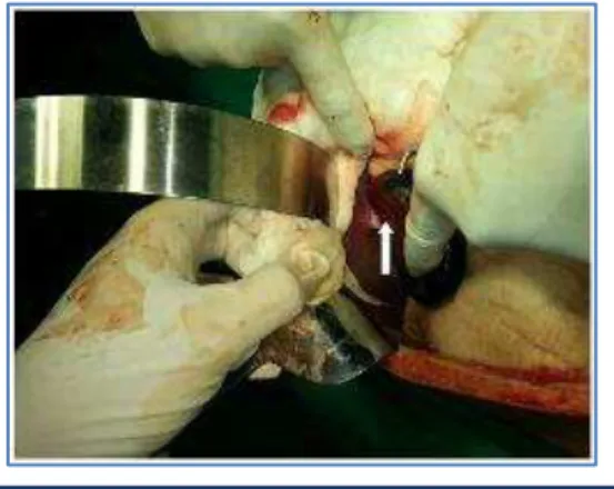
Fig. 1: Showing gangrenous gall bladder
CASE REPORT
J of Evidence Based Med & Hlthcare, pISSN- 2349-2562, eISSN- 2349-2570/ Vol. 2/Issue 23/June 08, 2015 Page 3494
NAME ADDRESS EMAIL ID OF THE CORRESPONDING AUTHOR:
Dr. A. Sai Datta,
Flat No. 9, Teja Apartments, 4/13, Brodipet, Guntur-522002, Andhra Pradesh.
E-mail: [email protected]
Date of Submission: 19/05/2015. Date of Peer Review: 20/05/2015. Date of Acceptance: 25/05/2015. Date of Publishing: 08/06/2015.
AUTHORS:
1. A. Sai Datta 2. K. Srinivas Rao
PARTICULARS OF CONTRIBUTORS:
1. Associate Professor, Department of General Surgery, Katuri Medical College.
2. Consultant Pulmonologist, Department of Pulmonary Medicine, Katuri Medical College.