J. Evolution Med. Dent. Sci./ eISSN- 2278-4802, pISSN- 2278-4748/ Vol. 5/ Issue 57/ July 18, 2016 Page 3929
EVALUATION OF SCROTAL LESIONS BY GRAY SCALE ULTRASONOGRAPHY AND COLOUR DOPPLER
Punya Pratap Singh1, Kavita Gahlot2, Vivek Agrawal3, Manoj Sharma4, Himanshu Sharma5
1Assistant Professor, Department of Radiodiagnosis, Bundelkhand Medical College, Sagar. 2Medical Officer, Department of Emergency Medicine, Bundelkhand Medical College, Sagar. 3Senior Consultant & Intervention Radiologist, Max Super Speciality Hospital, Saket, Delhi. 4Senior Consultant, Max Super Speciality Hospital, Saket, Delhi.
5Assistant Professor, Department of Pharmacology, Bundelkhand Medical College, Sagar.
ABSTRACT
BACKGROUND
Until the mid 1970 s, scrotal examination was limited to palpation and transillumination. Miskin and Bain (1974) first reported the use of Ultrasonography to examine testes and scrotum. With advancement like colour Doppler, ability to assess testicular vascularity increased. Although other radiological investigations have roles in specific situations, present study was undertaken to evaluate the usefulness and accuracy of sonography and colour Doppler in scrotal abnormalities.
METHODS
The study was conducted in the Department of Radiodiagnosis, Max Super Speciality Hospital, Saket, New Delhi, between May 2012 and May 2013. Prospective study carried out in 100 patients after proper informed and written consent with inclusion criteria of All age patients with clinical suspicion of scrotal pathology and exclusion criteria of History of operative or therapeutic procedures on the scrotum with exception of vasectomy. These patients scrotums were scanned in various plan on Voluson 730 PRO (GE Healthcare) system and LOGIC E-9 (GE Healthcare) using linear probe (As applicable) with comparison from asymptomatic side. Relevant other radiological and pathological investigations were done wherever indicated.
RESULTS
Majority of patients were between 21-40 years of age (62%) with commonest presenting complaint was scrotal swelling in 73%. Fluid collections were the commonest abnormality detected on sonography. Hydrocele was the most frequent fluid collection seen in 38 cases (26.3%). In acute inflammation, hypoechogenicity of the testes was most common sonographic feature. In our study, ultrasound showed 99% accuracy to distinguish between Intratesticular and Extratesticular pathology.
CONCLUSIONS
High-resolution USG with colour Doppler can reliably define the morphological features and vascularity of scrotal lesions. USG is highly accurate in evaluating the consistency of scrotal mass and in localizing scrotal abnormality. Ultrasound evaluation of the scrotum must include colour Doppler imaging of testis and epididymis with comparison from the asymptomatic side.
KEYWORDS
High resolution ultrasonography, Colour Doppler, Hydrocele, Varicocele, Scrotal Wall Oedema.
HOW TO CITE THIS ARTICLE: Singh PP, Gahlot K, Agrawal V, et al. Evaluation of scrotal lesions by gray scale ultrasonography and
colour Doppler. J. Evolution Med. Dent. Sci. 2016;5(57):3929-3940, DOI: 10.14260/jemds/2016/899
)NTRODUCT)ON
Until s examination of the scrotal contents was limited to palpation and transillumination. Miskin and Bain.[ ] first
reported the use of B Brightness -mode Ultrasonography USG to examine testes and scrotum. USG have following advantages-(igh frequency transducers, haemodynamic information, low cost and rapidity in examination and freedom from radiation hazards.
A)MS AND OBJECT)VES
. To evaluate spectrum of ultrasonographic findings in various scrotal pathologies.
Financial or Other, Competing Interest: None. Submission 26-01-2016, Peer Review 02-03-2016, Acceptance 07-03-2016, Published 16-07-2016. Corresponding Author:
Dr. Punya Pratap Singh, Flat -9, Type–IV Quarters, Block–B, 2nd Floor,
B.M.C. Campus, Sagar-470002, Madhya Pradesh.
E-mail: drpunya@gmail.com DOI: 10.14260/jemds/2016/899
. To evaluate value of colour Doppler in distinguishing and characterizing the blood flow patterns in scrotal pathologies.
. To assess the role of high frequency real time ultrasonography to accurately distinguish between Testicular and Extratesticular scrotal masses.
MET(ODS
The study will be conducted in a tertiary referral teaching hospital; patients were enrolled in the study after proper informed and written consent during the period of study with the following inclusion and exclusion criteria.
)nclusion Criteria
Patients with all age groups.
Patients with strong clinical suspicion of scrotal pathology referred for scrotal duplex sonography.
Exclusion Criteria
J. Evolution Med. Dent. Sci./ eISSN- 2278-4802, pISSN- 2278-4748/ Vol. 5/ Issue 57/ July 18, 2016 Page 3930 contralateral side and findings are analysed. )n patients with
suspicion of testicular tumours, other regions were scanned to look for the presence of secondaries. )n case of varicocele, especially the left side, the kidneys were scanned to rule out renal mass.
Chest Radiograph Postero-Anterior View , Computed Tomography and Laboratory )nvestigations
)t was taken in those cases which were suspected to have testicular tumour to detect retroperitoneal lymph nodes and metastases and in suspected tubercular epididymis with relevant histopathological investigations.
RESULTS
Commonest age group is - years, which is % of patients.
Fig. : Age Distribution of Cases
Commonest presentation was scrotal swelling in % cases.
Fig. : Clinical Symptomatology
Malposition testes
Testicular tumours Testicular trauma
Testicular & epididymal cysts Simple cyst
Epididymal cyst Testicular cyst Testicular atrophy Varicoceles
Testicular microlithiasis (ernias
Omentocele Enterocele
Scrotal wall oedema
. .
. . . . . .
. .
.
Total 5
Table : Nature of Lesion
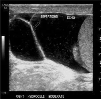
Fluid collections were the commonest abnormality with hydrocele was most common type [Table - ].
Fig. : Right Moderate (ydrocele with )nternal Echoes and )nternal Septations
J. Evolution Med. Dent. Sci./ eISSN- 2278-4802, pISSN- 2278-4748/ Vol. 5/ Issue 57/ July 18, 2016 Page 3931
Sonographic Features Non seminomatous Germ Cell Tumour
n= Seminoma n= Teratocarcinomas Ca n= Leukaemia n= Azzopardi Tumour n= TESTES Size Normal Enlarged )nvolve pattern
Focal Diffused
Echo texture
Tunica inversion
Calcification
Present Absent
Contralateral Testes -- (eterogeneous; Cystic & (yperechoic areas -normal -- Relatively homogenous, cystic areas -Normal -(ypoechoic - microlithiasis Testicular microlithiasis -(ypoechoic - -normal - -(yperechoic -normal ADNEXAL STRUCTURE Epididymis
Spermatic cord Thickened n=Thickened n= NormalNormal NormalNormal NormalNormal NormalNormal
(YDROCELE Minimal n=
Septated n= Septated Minimal anechoic Minimal Absent Absent
Associated Findings
Lymph nodes S)TE
Para-aortic Peripancreatic Periportal )liac
S)ZE Bulky confluent
> cm Bulky confluent> cm
Liver metastases - - -
-Lung metastases - - -
(ydroureteronephrosis - - -
-Pneumonia - - - -
Table : Sonographic Features & Associated Findings of Tumours
Six cases of testicular tumours were encountered in the study. Tumour lesions are comparatively hypoechoic in % of cases [Table - ].
Fig. : Lt. Testicular Tumour A - Left Testis enlarged in size with markedly increased diffuse vascularity. B Showing increased vascularity of Lt. epididymis with increased peak systolic velocity > cm/sec and reduced
J. Evolution Med. Dent. Sci./ eISSN- 2278-4802, pISSN- 2278-4748/ Vol. 5/ Issue 57/ July 18, 2016 Page 3932
Fig. : Left Testicular Tumour C&D T W) Transverse )mage showing )ncreased Size of Left Testis with )sointense Signals Post contrast T W Fat Suppressed )mage was showing Marked (omogeneous Contrast Enhancement
Fig. : E&F : Left Testicular Tumour-Multiple Rounded (ypoechoic Lesions seen involving Liver Parenchyma Liver Metastasis . Enlarged well-defined (ypoechoic RT Para-aortic Lymph Node also noted
J. Evolution Med. Dent. Sci./ eISSN- 2278-4802, pISSN- 2278-4748/ Vol. 5/ Issue 57/ July 18, 2016 Page 3933
Fig. ( : Lt. Testicular Tumour turned out to be Yolk Sac Tumour on (istopathological Examination Micrograph was showing Yolk Sac Tumour with
Schiller-Duval Bodies, (&E Stain, x
Associated findings were lymph nodes involvement, lung metastasis, liver metastasis, hydronephrosis, pneumonia, etc. [Table - ]. Unevenly distributed pattern of flow were noted in % cases of tumour except a case of leukemic infiltration showing even distribution [Table - ].
Features NonseminomatousGerm Cell
Tumour Seminoma Teratocarcinoma Leukaemia
Azzopardi Tumours
S)ZE OF LES)ON Less than . cm
More than . cm - - - - -
Colour Doppler )ncreased-Normal- )ncreased Normal )ncreased Decreased
PATTERN OF FLOW distributed vesselsUnevenly distributed vesselsUnevenly Evenly distributed vessels
SPECTRAL ANALYS)S Peak Systolic Velocity > . cm/sec
Resistivity )ndex < . -- - -
-Table : Colour Doppler Findings of Tumours
)n cases of acute inflammation, % were young and sexually active age group; . % was showing hypoechogenicity of the testes. Epididymis was the commonest anatomical structure involved. CD could demonstrate increased flow in % testes and % epididymis.
Fig. : Focal Orchitis seen as focal altered (ypoechoic )ntratesticular Lesion showing increase Vascularity. )n Colour Doppler Study Right Testis showing )ncreased Diastolic Flow and Reduced Resistivity )ndex < . .
J. Evolution Med. Dent. Sci./ eISSN- 2278-4802, pISSN- 2278-4748/ Vol. 5/ Issue 57/ July 18, 2016 Page 3934
Fig. A : Right Testis has )ncreased in Size and More (ypoechoic in Echotexture as compared to Left Testis. Right Testis showing more Vascularity as compared to Left Testis
Fig. B&C : Right Testis showing )ncreased Vascularity, More Peak Systolic Velocity and )ncreased Diastolic Flow
J. Evolution Med. Dent. Sci./ eISSN- 2278-4802, pISSN- 2278-4748/ Vol. 5/ Issue 57/ July 18, 2016 Page 3935
Fig. E&F : Right Submandibular Gland was showing (eterogeneous Echotexture with )ncreased Vascularity suggestive of Acute )nflammatory Changes
% cases of orchitis demonstrate a PSV of more than cm/sec and % cases demonstrate R) Resistivity )ndex less than . . Chronic inflammation of scrotal structures, in majority of cases involvement of epididymis noted and showing hypoechoic echotexture. One testes . % normal on gray scale showed increased vascularity on CD study and was considered abnormal.
Fig. : Left Chronic Orchitis – A Left Testis was increased in size with altered (eterogeneous Echotexture. B On
colour Doppler study, Left Testis showing increased Vascularity Peak Systolic Velocity > cm/sec and
increased Diastolic Flow Resistivity )ndex < .
One case . % of epididymitis was also noted on CD, which was not apparent on gray scale sonography. Cases of chronic testicular torsion, commonest pattern was enlarged testes . % with heterogeneous echopattern . % . With
CD blood flow in symptomatic testes was absent in all three cases % . )n four cases of testicular trauma, hyperechoic echopattern is noted in % cases. Discrete fracture identified in % cases with haematocele in % cases.
Fig. : A Right Testicular Torsion – (ypoechoic Echotexture of Right Testis without )ntratesticular
J. Evolution Med. Dent. Sci./ eISSN- 2278-4802, pISSN- 2278-4748/ Vol. 5/ Issue 57/ July 18, 2016 Page 3936
Fig. B&C : Right Testicular Torsion – (ypoechoic Echotexture of Right Testis without )ntratesticular Vascularity. Extratesticular Vessels were showing Vascular Flow
Testicular atrophy cases, most common heterogeneous pattern noted in . % cases and reduced flow signals in
% cases.
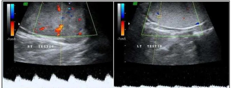
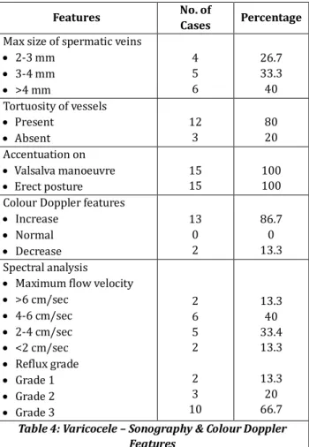
Varicocele was seen on left side in . % cases and . % on right side. All cases demonstrated accentuation on Valsalva manoeuvre with . % cases show reflux.
Fig. : Right Varicocele A Multiple Dilated Anechoic Serpiginous Structure seen in Left Scrotal Sac. B On colour Doppler study, Serpiginous Structure was showing Reflux as change in Colour signal Red to Blue and Prominence as )ncreased
J. Evolution Med. Dent. Sci./ eISSN- 2278-4802, pISSN- 2278-4748/ Vol. 5/ Issue 57/ July 18, 2016 Page 3937
Features No. of Cases Percentage
Max size of spermatic veins - mm
- mm > mm
. .
Tortuosity of vessels Present
Absent Accentuation on Valsalva manoeuvre Erect posture Colour Doppler features )ncrease
Normal Decrease
.
. Spectral analysis
Maximum flow velocity > cm/sec
- cm/sec - cm/sec < cm/sec Reflux grade Grade Grade Grade . . . . .
Table : Varicocele – Sonography & Colour Doppler Features
Seven percent of cases showed spermatocele as compared to % of epididymal cysts were identified and confirmed on aspiration. Omentocele was seen in % cases, while enterocele in four cases. Thus hernial incidence of % was noted.
)n our study, testicular microlithiasis was encountered in % cases. Four cases had bilateral testicular microlithiasis with associated teratocarcinoma on one side was found in one case.
The incidence of cryptorchidism was % in our series. Most common position was in inguinal canal and smaller than their contralateral counterpart in % cases. On sonography, % were homogeneous and hypoechoic in echotexture with torsion noted in one case. On sonography two testes % were homogeneous and hypoechoic in echotexture.
Features No. of Cases Percentage
POS)T)ON OF TESTES )nguinal
Deep inguinal ring Other
S)ZE )ncreased Normal Decreased EC(OPATTERN Normal (ypoechoic (yperechoic (eteroechoic
ASSOC)ATED FEATURES Torsion
)nguinal hernia (ydrocele
Table : Undescended Testes Features
Two cases depicted a thickened scrotal wall with normal testes. On CD increased vascularity were identified in the scrotal wall, which had high resistance blood flow with R) values more then , . and . respectively.
Omentocele was % cases, while enterocele in % cases. Thus, an incidence of % was noted. )n our study, sonography revealed a highly echogenic mass separated from the testes in omental hernia and anechoic mass in inguinoscrotal region of in cases of enterocele. On CD, vascular signals were demonstrated within the bowel wall and within the omentum.
J. Evolution Med. Dent. Sci./ eISSN- 2278-4802, pISSN- 2278-4748/ Vol. 5/ Issue 57/ July 18, 2016 Page 3938 and % cases respectively. )n one case, we cannot identify
testis A case of traumatic haematocele . So, in our study high frequency ultrasound showed % accuracy to distinguish between )ntratesticular and Extratesticular pathology.
D)SCUSS)ON
patients with scrotal lesions were included in this study. Predominant group in the study was to years comprising of % cases. Commonest presenting complaint was that of scrotal swelling in % followed by . % scrotal pain.
Fluid Collections
Fluid collections were the commonest abnormality with hydrocele as most frequent fluid collection, same reported by Langer et al.[ ] Thus, accuracy of % was achieved in
diagnosing (ydrocele same as reported by Gutman et al.[ ]
All cases of haematocele showed fluid with internal echoes and septations considered diagnostic of haematocele also by Stewart et al.[ ] A case of lymphocele with lymphatic
collection in inguinoscrotal region and upper thigh. Chung et al.[ ] have described these features in case of lymphocele.
Testicular Tumours
)n the present study, all cases of testicular tumours were encountered and diagnosed in all cases with nearly same accuracy reported by Fowler et al.[ ]
The sonographic characteristic of testicular tumours was the heterogeneous appearance of the testes. Tumour lesions appeared less echogenic than normal testes in % of cases, also reported by Arger et al.[ ]
The seminoma was hypoechoic, homogeneous and had sharply circumscribed margins, while Nonseminomatous Germ Cell Tumours NSGCT were characterized by heterogeneous echotexture, irregular margins and cystic spaces. Similar observations were made by Nachtscheim et al.[ ]
A case of Azzopardi tumour was noted. Grantham et al.[ ]
also reported hyperechoic foci in six out of seven regressed germ cell tumours of the testes.
Testicular microlithiasis was noted in one case . % of testicular tumour. Berger et al.[ ] reported microlithiasis to
be present in % of their patients with testicular tumours. The distribution of blood vessels within the tumour was random and disorganized in hypervascular tumours. These findings were similar to those observed by (orstman et al.[ ]
Enlarged para-aortic lymph nodes were the most common site of metastases detected in . % cases of tumours. This feature was also observed by Mostofi FK et al.[ ] Acute )nflammation
% patients were in young sexually active males. Testes were involved in % cases. Most common sonographic feature was
(orstman WC et al.[ ]
Chronic )nflammation
Eighteen cases of chronic inflammation of scrotal structures were included in the study. Majority of age group of to years noted.
Testes were involved in . % cases as compared to . % of epididymis. Epididymis enlargement was diffuse
. % with hypoechoic echotexture . % .
)nvolvement of spermatic cord was noted in . % cases. Evidence of tuberculosis in lung was associated in five cases . % . Epididymal calcification was noted in . % cases. Strikingly similar observations were noted by Kim et al. [ ]
On CD, . % of patients with chronic inflammation showed increased vascular signals; . % testes and . % of epididymitis Normal on gray scale showed increased vascularity on CD and were considered abnormal.
Thus, undiagnosed cases of orchitis and epididymis One each were detected, which were normal on gray scale sonography which was detected on CD. )ncreased sensitivity and specificity of CD to assess scrotal inflammation has been asserted by Barton JW.[ ]
Testicular Torsion
Two cases of acute and one case of chronic testicular torsion were included in our study with all was under years. Tumeh et al.[ ] described torsion to occur commonly between the
ages of to years. Features indistinguishable to those of torsion were noted in five cases of acute inflammation on gray scale sonography alone. Bird et al.[ ] also remarked same.
On sonography, the commonest pattern was enlarged testes . % with heterogeneous echopattern as most common pattern . % . Bird et al.[ ] found similar findings
in testicular torsion.
Changes in peritesticular tissue were also noted. The epididymis was enlarged in two cases . % with hypoechoic echopattern in one case . % . )n a single case of chronic torsion, the epididymis was enlarged and heterogeneous. Tumeh et al.[ ] noted similar features in their
series. )n one case of testicular torsion, spiral twist of spermatic cord was noted. Baud et al.[ ] described it to be a
reliable sign of testicular torsion. With CD, blood flow in symptomatic testes was absent in all three cases % . (owever, CD could demonstrate flow signals on asymptomatic side only in two post-pubertal testes . % . No colour signal was identified on the asymptomatic side in a child aged four years with torsion.
)n two patients, spectral analysis revealed decrease in R) with dampened flow, while in one patient the waveform was nearly venous. Baud et al.[ ] described similar waveforms in
J. Evolution Med. Dent. Sci./ eISSN- 2278-4802, pISSN- 2278-4748/ Vol. 5/ Issue 57/ July 18, 2016 Page 3939
Testicular Trauma
)n the present study, four cases of testicular trauma were diagnosed. Sonography demonstrated hyperechoic echopattern in % cases. )ncreased size of testes was observed in two patients % . Discrete fracture could be identified in one patient % . Jeffrey et al.[ ] noted similar
findings.
Echogenic fluid suggestive of haematocele was noted in three patients % . Jeffrey et al.[ ] noted presence of
haematocele in % cases in their series.
On CD, no vascular signal was identified in one case % of blunt testicular trauma. )n remaining three cases, normal intratesticular flow was seen.
Testicular Atrophy
% patients were showing testicular atrophy. The testes were noted to be hypoechoic in . % and heterogeneous in . % cases. The epididymis in all cases was small in size. Similar findings were found by Cross et al.[ ]
Varicoceles
They comprised % of total number of cases comparable to % to % cases by Berger et al.[ ]
They were seen more commonly on left side, as in our study also found by McClure et al.[ ] All cases demonstrated
accentuation on Valsalva manoeuvre and on erect posture. % cases were diagnosed on colour Doppler, which were undiagnosed by clinical examination suggesting colour Doppler more sensitive.
Greenberg et al.[ ] found reflux in all cases, clinical
varicocele as in our study. No significant difference in PSV in relation to presence or absence of varicocele and the degree of reflux was noted.
Malpositioned Testes
The incidence of cryptorchidism was % in our series. )ts most common position was in inguinal canal, % cases also noted by Kleinteich et al.[ ]
On sonography, % were homogeneous and hypoechoic in echotexture. Sizes of % testes were smaller than their contralateral counterpart. Torsion of undescended testes were noted in one case also noted by Nguyen and (ricak.[ ]
Testicular Cysts
(amm et al.[ ] had an incidence of % of testicular cysts, while
in our study it is %. )n all cases they were seen as well circumscribed, anechoic lesions with thin smooth walls and posterior acoustic enhancement. Malignant cysts were usually multilocular with shaggy, thick, poorly marginated walls and surrounded by neoplastic parenchyma with tumour vascularity. (orstman WG.[ ] stressed on similar features to
differentiate these two conditions. A case of tunica albuginea cyst was seen.
Epididymal Cysts and Spermatoceles
% cases of spermatocele and % cases of epididymal cysts were identified and confirmed on aspiration.
While the cyst contents were echogenic in . % cases of spermatocele, it was anechoic in % cases of epididymal cysts. Septation were noted in . % cases of spermatocele and in % cases of epididymal cyst. Doherty et al.[ ] noted
similar findings. On CD, blood flow in septae was seen in . % cases of spermatocele and % cases of epididymal cyst.
Scrotal (ernias
Omentocele was % cases, while enterocele in % cases. Thus, an incidence of % was noted. An incidence of . % was noted by Subramanyam BR et al.[ ] in their study. )n our study,
sonography revealed a highly echogenic mass separated from the testes in omental hernia and anechoic mass in inguinoscrotal region of in cases of enterocele. Subramanyam BR et al.[ ] noted similar findings.
On CD, vascular signals were demonstrated within the bowel wall and within the omentum.
One postoperative case of herniorrhaphy was studied, which had multiseptated collections in the spermatic cord. Another patient had thickened spermatic cord with no traceable vascular signals in the cord or ipsilateral testes. The testes had atrophied in this case.
Testicular Microlithiasis
)n our study testicular microlithiasis have incidence of % with % cases had bilateral testicular microlithiasis with associated teratocarcinoma on % case. Doherty et al.[ ] described
similar findings with a reported incidence of . %.
Scrotal Wall Oedema
Scrotal wall oedema was found in two patients, one due to heart failure and second due to filariasis. Scrotal wall thickened with multiple layers like onion peel. Thickening of penile skin was also noted in both cases. Grainger et al.[ ]
described similar findings.
ACKNOWLEDGEMENT
We acknowledge Dr. R. K. Mathur for guiding us in every step of research work. We also acknowledge Dr. Vijay Bahadur Singh, Dr. Anuja Patil, Dr. Mukesh Patidar, Dr. Parul Gupta, Dr. Bhagyashree Patil, Dr. Viral Shah and Dr. Manohar Singh Rathore for her assistance with the radiologic findings and Mr. )ndal Singh for his assistance in statistics work. Mr. Shri Jalim Singh, Smt. Pushpa Singh, Smt. Pooja Singh, Mr. Rahul Shrivastava and Mr. Ravikant Gupta for mental support and technical help during study.
REFERENCES
. Miskin M, Bains J. B-mode ultrasonic examination of the testes. J Clin Ultrasoun ; : – .
. Langer JE. Ultrasound of scrotum. Seminars in Roentgenology ; : - .
. Gutman (, Golimbu M, Subramanyam BR. Diagnostic ultrasound of scrotum. Urology ; : - .
. Stewart R, Caro)) BA. The scrotum: in diagnostic ultrasound. St. Louis. Rumack CM, Wilson SR, Charboneau JW. Elsevier Mosby; ; nd edition.
. Chung SE, Frush DR, Fordham LA. Sonographic appearances of extratesticular fluid and fluid containing scrotal masses in infants and children due to diagnosis.
AJR ; : - .
. Fowler RC, Chennells PM, Ewing R. Scrotal ultrasonography: a clinical evaluation. Br J Radiol
J. Evolution Med. Dent. Sci./ eISSN- 2278-4802, pISSN- 2278-4748/ Vol. 5/ Issue 57/ July 18, 2016 Page 3940 . (orstman WG, Melson GL, Middleton WD, et al. Testicular
tumours: findings with colour doppler US. Radiology
; : - .
. Mostofi FK. Testicular tumours: epidemiologic, etiologic and pathologic features. Cancer ; : - . . (orstman WC, Middleton WD, Melson CL. Scrotal
inflammatory disease: colour doppler ultrasound findings. Radiology ; : - .
. Kim (S, Yang DM, Yoon M(, et al. Comparison of tuberculous and pyogenic epididymal abscesses: clinical, gray-scale sonographic, and colour doppler
sonographic features. AJR Am J
Roentgenol ; : - .
. Barton JW, Brown JM, (ammers LW, et al. Quantitative doppler assessment of acute scrotal inflammation.
Radiology ; : - .
. Tumeh SS, Benson CB, Richie JP. Acute diseases of the scrotum. Semin US CT MR ; : - .
. Bird K, Rosenfield AT, Taylor KJW. Ultrasonography in testicular torsion. Radiology ; : - .
. Baud RO, Kennelly MJ, Adler RS, et al. Nonpulsatile arterial waveforms experimental study during graded testicular torsion in an animal model. Radiology
; : - .
on subclinicalvaricocele : diagnosis by doppler ultrasonic stethoscope. J Reprod Med ; : - . . Kleinteich B, Popp W, Grahl KO. Congenital testicular
dystopias and concomitant abnormalities. Kinderarztl
Prax ; : - .
. Nguyen (T, (ricak (. Cryptorchidism: strategies in detection. European Radiology ; : - . . (amm B, Foboe F, Loy V. Testicular cysts: differentiation
with ultrasound and clinical findings. Radiology ; : - .
. Doherty FJ. Ultrasound of the nonacute scrotum. Semin Ultrasound CT MR ; : - .
. Subramanyam BR, Balthazar EJ, Raghavendra BN, et al. Sonographic diagnosis of scrotal hernia. AJR
; : - .
. Doherty FJ, Mullins )L, Sant GR, et at. Testicular microlithiasis: a unique sonographic appearance. J U Med
; : - .
. Grainger AS, (ide )G, Elliot ST. The ultrasound appearance of scrotal odema. Eur J U)trasound