Development of an Extracorporeal Perfusion
Device for Small Animal Free Flaps
Andreas M. Fichter1*, Lucas M. Ritschl1, Anna Borgmann1, Martin Humbs1, Peter B. Luppa2, Klaus-Dietrich Wolff1, Thomas Mücke1
1Department of Oral and Maxillo-Facial Surgery, Technische Universität München, Klinikum Rechts der Isar, München, Germany,2Institute for Clinical Chemistry and Pathobiochemistry, Technische Universität München, Klinikum Rechts der Isar, München, Germany
Abstract
Background
Extracorporeal perfusion (ECP) might prolong the vital storage capabilities of composite free flaps, potentially opening a wide range of clinical applications. Aim of the study was the development a validated low-cost extracorporeal perfusion model for further research in small animal free flaps.
Methods
After establishing optimal perfusion settings, a specially designed extracorporeal perfusion system was evaluated during 8-hour perfusion of rat epigastric flaps followed by microvas-cular free flap transfer. Controls comprised sham-operation, ischemia andin vivoperfusion. Flaps and perfusate (diluted blood) were closely monitored by blood gas analysis, com-bined laser Doppler flowmetry and remission spectroscopy and Indocyanine-Green angiog-raphy. Evaluations were complemented by assessment of necrotic area and light
microscopy at day 7.
Results
ECP was established and maintained for 8 hours with constant potassium and pH levels. Subsequent flap transfer was successful. Notably, the rate of necrosis of extracorporeally perfused flaps (27%) was even lower than afterin vivoperfusion (49%), although not statisti-cally significant (P= 0,083). After sham-operation, only 6% of the total flap area became necrotic, while 8-hour ischemia led to total flap loss (98%). Angiographic and histological findings confirmed these observations.
Conclusions
Vital storage capabilities of microvascular flaps can be prolonged by temporary ECP. Our study provides important insights on the pathophysiological processes during extracorporeal tissue perfusion and provides a validated small animal perfusion model for further studies.
OPEN ACCESS
Citation:Fichter AM, Ritschl LM, Borgmann A, Humbs M, Luppa PB, Wolff K-D, et al. (2016) Development of an Extracorporeal Perfusion Device for Small Animal Free Flaps. PLoS ONE 11(1): e0147755. doi:10.1371/journal.pone.0147755
Editor:Helge Bruns, University Hospital Oldenburg, GERMANY
Received:October 19, 2015
Accepted:January 7, 2016
Published:January 25, 2016
Copyright:© 2016 Fichter et al. This is an open access article distributed under the terms of the
Creative Commons Attribution License, which permits unrestricted use, distribution, and reproduction in any medium, provided the original author and source are credited.
Data Availability Statement:A minimal dataset containing all relevant data underlying this study, including laser Doppler flowmetry and remission spectroscopy results, areas of necrosis, blood gas analysis, as well as the results from preliminary studies, can be found athttp://datadryad.org(doi:10. 5061/dryad.0p7vm).
Funding:This study was funded by the Deutsche Forschungsgemeinschaft (WO 507/4-1),http://www. dfg.de.
Introduction
Prolonged vital storage of organs by temporary extracorporeal perfusion (ECP) was first dem-onstrated by Alexis Carrel [1] in the 1930s and is today routinely used for the vital storage of isolated organs [2–4]. As opposed to isolated organs, however, composite flaps (or extremities) are composed of different tissues with a varying tolerance towards ischemic damage. While bone, tendons skin and fat usually tolerate intervals of prolonged ischemia quite well, muscle, blood vessels and neuronal tissue have a low tolerance towards ischemia [5]. The warm ische-mia time, defined as the time a certain tissue can tolerate the lack of oxygen without special measures like cooling, should not exceed six hours in composite tissues [6]. A second reason why flap perfusion differs from organ perfusion is the fact that microvascular free flaps are no isolated, confined organs. Instead, flaps are harvested from their environment resulting in a large wound surface, promoting microbial contamination and excessive bleeding during pro-longed extracorporeal perfusion. Nevertheless, existing studies indicate that ECP may hold the potential for prolonged vital storage of composite tissues [7–10], but fundamental questions, like optimal perfusion pressure and flow settings, oxygen/ carbondioxide ratio, perfusate tem-perature and the composition of an ideal perfusate still remain unanswered. Moreover, clinical use of such a system has not yet been established for free flap perfusion.
Studies aimed at the perfusion of living tissues depend on expensive and resource intensive large animal models since perfusion is usually achieved using commercially available systems borrowed from human cardiac bypass circuits. Due to the small size and low blood volumes, these circuits are not suited for small animal free flaps [11]. Our study group could already demonstrate the suitability of an extracorporeal perfusion device in the training of microsur-geons to simulate realistic conditions in cadavers [12]. The objective of the present study was to develop and evaluate a low-cost extracorporeal perfusion device for the prolonged vital stor-age of small animal free flaps that should serve as a model for future free flap perfusion studies.
Materials and Methods
All animals were treated and housed in accordance with the EU-guidelines. The study was approved by the regional government (Regierung von Oberbayern, AZ55.2-1-54-253-86-08) and was conducted in accordance with the German Animal Welfare Act. A total of 64 male Fischer-344 rats (250–300g, Fa. Charles River, Kißlegg, Germany) were used. Food and water were providedad libitum. All surgical procedures were performed under intravenous general anesthesia [ketamine 100mg/kg (Narketan1, Fa. Vétoquinol GmbH, Ravensburg, Germany) und xylazine 5 mg/kg (Rompun1, Fa. Bayer Vital GmbH, Leverkusen, Germany)] and aseptic conditions as described elsewhere [13], and all efforts were made to minimize suffering.
Preliminary Studies
The Perfusion System
The setup of the perfusion system used for thein vivostudies is presented inFig 1. The system was configured in a way that all individual parts (tubes, connectors, reservoirs, membrane oxy-genator) that came in contact with blood could be sterilized and assembled and filled with blood under sterile conditions. The perfusion system consisted of a roller pump (Ing. Büro Humbs, Valley, Germany), a membrane oxygenator (type Oxyphan PP50/280, Fa. Membrana GmbH, Wuppertal, Germany) that worked with both ambient air and pure oxygen, an arterial and a venous reservoir. The venous reservoir was connected to a vacuum and could generate an adjustable negative pressure to the venous system. Both reservoirs served as a bubble trap. The blood pressure before the flap was continuously measured (Panasonic Electric Works SUNX Co., Ltd. Kasugai City, Aichi, Japan). A pressure controlled shunt between arterial and venous blood reservoir was implemented to avoid uncontrolled pressure spikes. The tubing system consisted of platinum-curing silicones (USP class IV, FDA-approved) and had an inner diameter of 0.8 mm and a total length of 2.5 meters. All materials were steam sterilized before each usage.
Thirty minutes before initiation of the extracorporeal tissue perfusion, the tubing system was rinsed with heparinazed Ringer’s solution (10ml Ringer, 100 IE Heparin) at 5 ml/min. The rinsing solution was then replaced by the perfusate. Subsequently, the femoral vessels of the raised flap were connected to the perfusion unit using modified IV catheters (Vasofix1 Brau-nüle120 G and 18 G, Fa. B. Braun Melsungen AG, Melsungen, Germany). Extracorporeal per-fusion was commenced at 2 ml/min (corresponding to high-normal physiological values) and was maintained at the same speed for 8 hours. An upper pressure limit was programmed at 130 mmHg, also corresponding to high physiological values [15].
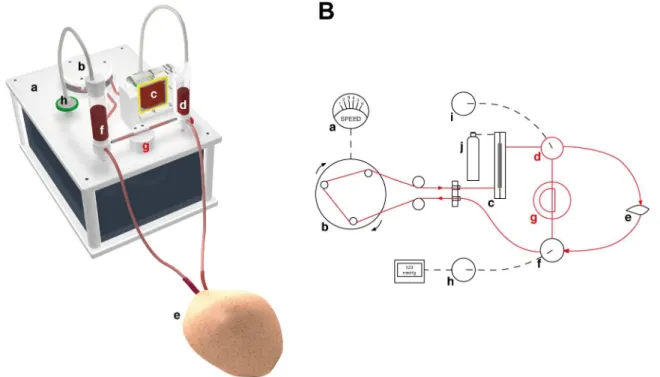
Fig 1. The perfusion system.(A) 3D rendering of the perfusion system used in this study. (B) Schematic drawing of the experimental setup. A motor (a) driven roller pump (b) pumps heparinized autologous blood through an oxygenator membrane (c), connected to an oxygen cylinder (j). The oxygenized blood is flows into the flap (e), while the venous return is collected in a reservoir (f) connected to a pressure gauge (h). If the pressure in the system surpasses a certain treshold, the pressure equalizes by opening a shunt (g) between arterial (d) and venous reservoirs. (i) Bubble trap.
To minimize a detrimental effect of progressive hemolysis, the perfusate was replaced after 4 hours and if (1) the filling volume reached a critical value of1 ml in the venous reservoir, or if (2) the closely monitored pH fell below 7.300.
Perfusate Composition and Temperature
To improve rheological properties and compensate for short supply, diluted (40% Ringer’s solution and 10% hydroxyethyl starch), heparinized (10 IE Heparin/ml blood) rodent whole blood was used as perfusate. A plasma expander (hydroxyethyl starch, HES, Volulyte 6%, Fa. Fresenius Kabi Deutschland GmbH, Bad Homburg v.d.H., Germany) was added to prevent excessive edema formation. The room temperature in the laboratory was controlled at 21 ± 1°C by an air-conditioning system. The perfusate temperature was allowed to equilibrate to room temperature (21 ± 1°C) prior to perfusion, where it remained constant throughout the extra-corporeal perfusion period without additional control mechanisms. The composition of the perfusate was kept unchanged throughout the experiment and was closely monitored during extracorporeal tissue perfusion. Therefore, blood samples were drawn immediately after prepa-ration of the perfusate (base value), as well as every 15–30 minutes during the 8-hour tissue perfusion. Blood gas and laboratory-chemical analysis (RAPIDLAB1348, Siemens Sector Healthcare, Erlangen) were performed with detection of pH-value, potassium (K+), oxygen sat-uration (SO2), oxygen partial pressure (pO2), carbon dioxide partial pressure (pCO2), sodium bicarbonate buffer (HCO3–) and hemoglobin level (Hb).
Surgical Groups
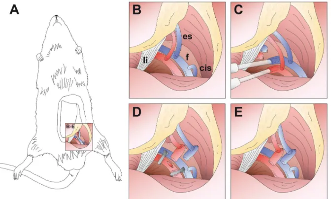
In 32 rats, the epigastric flap was raised based on the superficial epigastric vessels in the dimen-sions of 4 x 7 cm, as described in detail by Strauch and Murray [16]. The experimental groups are listed inTable 1. Another 32 rats served as either recipient animals for microvascular free flaps or as blood donors.
In group C1 (sham-operation), flaps were sutured back to the wound bed immediately after flap raise and animals were allowed to wake. In group C2 (ischemia), flaps were stored at room temperature for 8 hours. Subsequently, a second (recipient) rat was anesthetized and the epi-gastric flap was raised as described above and discarded. The ischemic flap was then transferred to the recipient animal, vessels were anastomosed end-to-side to the femoral vessels, the skin island was sutured into the wound bed and the recipient animals were allowed to wake. Donor animals were euthanised by intravenous lethal injection [2 ml per animal] of a combination of 200 mg Embutramid, 50 mg Mebezonium and 5 mg Tetracain per ml (T611, Intervet, Unters-chleißheim, Germany). In group C3,in vivoperfusion was maintained for 8 hours. Subse-quently, thein vivoperfused flaps were transferred to a recipient rat. The experimental setup of group ECP (extracorporeal perfusion) is depicted inFig 2. In this group, the nutritive vessels of the flap were connected to the perfusion device and extracorporeal perfusion was maintained for 8 hours. Subsequently, flaps were disconnected and transferred to a recipient rat. For post-operative analgesia, the rats received buprenorphine (50μg/kg s.c.; Temgesic1; Essex Pharma,
Germany) directly after waking up, as well as every 12 hours for 3 days.
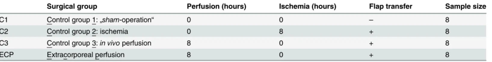
Table 1. Experimental protocol.
Surgical group Perfusion (hours) Ischemia (hours) Flap transfer Sample size
C1 Control group 1:„sham-operation“ 0 0 – 8
C2 Control group 2: ischemia 0 8 + 8
C3 Control group 3:in vivoperfusion 8 0 + 8
ECP Extracorporeal perfusion 8 0 + 8
Assessment of Flap Perfusion
In addition to observing flap color and capillary refill, measurement of tissue oxygen saturation (SO2in %), hemoglobin level (Hb, in AU, arbitrary units), blood flow (flow, in AU) and velocity (velocity, in AU) was noninvasively performed using comined laser Doppler flowmetry and remission spectroscopy (O2C, equipped with an LF-2 probe, Lea Medizintechnik, Giessen, Ger-many). This technique has been described in detail elsewhere and is an established procedure for the assessment of free flaps viability [17]. In all surgical groups, O2C was performed preopera-tively (base value) and at day 7. In group C2, C3 and ECP, additional O2C measurements were conducted after flap raise and after wound closure. Duringin vivo(C3) and extracorporeal perfu-sion (ECP), O2C was performed every 30 minutes. All measurements were performed in the cen-ter of the flap with equal surface pressure and full contact between probe and skin.
Indocyanine-Green (ICG) fluorescence angiography was performed in groups C3 and ECP duringin vivoand extracorporeal perfusion, respectively. The fluorescence signal was recorded using a mobile near infrared (NIR) fluorescence camera (PDE Photodynamic Eye, Pulsion Medical Systems SE, Feldkirchen, Germany). Flow analysis was performed with IC-CALC (Version 2.0, Pulsion Medical Systems SE, Feldkirchen, Germany).
Planimetric Measurement of Necrotic Areas
Flap healing was documented using a digital SLR camera (type Nikon D700, Fa. Nikon Corp., Chiyoda, Tokyo, Japan) mounted in a perpendicular direction to the flap with a tripod at day 7. Fig 2. Experimental setup (schematic drawing).(A) Overview image of a rat with raised epigastric flap perfused solely by the superficial epigastric vessels. (B-E) Detail images. (B) Operative situs with superficial epigastric vessels (es), femoral vessels (f), inguinal ligament (li) and superficial circumflex iliac vein (cis). (C) Canullated femoral vessels connected to the extracorporeal perfusion device. (D) After 8-hour continuous extracorporeal perfusion, femoral vessels are ligated distally to the epigastric vessels and severed proximally to the epigastric vessels. Subsequently, the flap is transferred to a recipient rat and the femoral vessel stumps are anastomosed end-to-side to the femoral vessels of the recipient animal. (E) Situs after successful microvascular flap transfer.
Pictures were analyzed with respect to vital and necrotic areas. Therefore, total flap area and necrotic areas were manually circumscribed with the help of a graphic tablet and pen and the cross-sectional area was calculated using ImageJ [18].
Structural Analysis
After documentation of the necrotic area, rats were euthanized as described above, while still in deep anesthesia. Epigastric flaps were harvested and fixed in a 4% formalin solution. The flap samples were then embedded in paraffin, sectioned at 5μm and stained with hematoxylin and
eosin for histological evaluation under light microscopy.
Statistics
The SPSS software package (SPSS 22, SPSS Inc., Chicago, IL, USA) was used for statistical anal-ysis. For rate of necrosis, the Kuskal-Wallis test was chosen to determine significant differences (P<0.05) between groups. If a significant difference was detected, the Mann-Whitney U test
was performed to compare the groups in pairs. Wilcoxon’s test was used to compare parame-ters inside a study group. The multiple test problem was not taken into account. All data are presented as mean ± standard deviation. Differences were considered statistically significant for a two-sided exactPvalue of less than 0.05. All observations were independently evaluated by two investigators blinded to the experimental groups.
Results
Assessment of the Perfusate Composition
Laboratory-chemical values measured after routine replacement of the perfusate after 4 hours (perfusion hours 4–8) closely resembled those measured in the first 4 hours of perfusion. Since in the second half of the perfusion period, laboratory-chemical assessment, however, was only sporadically performed these data were not considered for statistical analysis (Table 2). In 2 cases, the perfusate had to be changed prematurely at around 3.5 hours since the filling volume had sunk under the critical value and since the pH had fallen below 7.300, respectively.
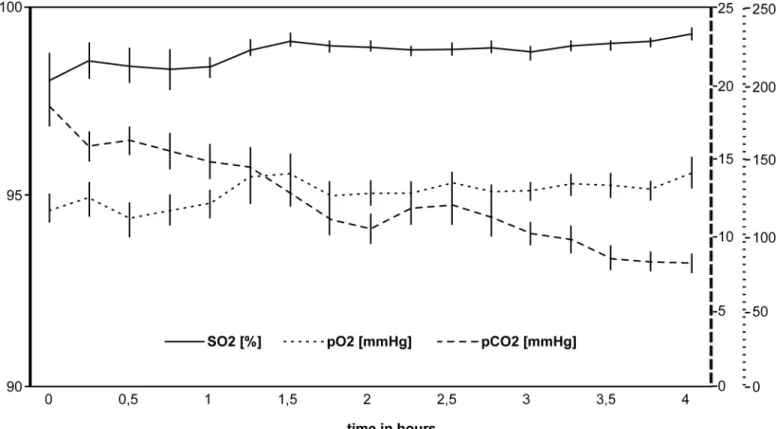
Changes in the blood gas parameters are illustrated inFig 3. Oxygen saturation and oxygen partial pressures showed high-normal values throughout the extracorporeal perfusion (base values SO298.7%, pO2118.9 mmHg). Both SO2(P= 0.016) and pO2(P= 0.008) increased sig-nificantly during the first 1.5 hours and stayed constantly high for the remaining time (SO2 99.3 mmHg, pO2131.7 mmHg after 4 hours). Carbon dioxide partial pressures (17.6 mmHg,
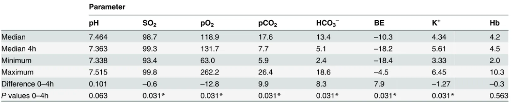
Table 2. Statistical analysis of laboratory-chemical changes in the perfusate.
Parameter
pH SO2 pO2 pCO2 HCO3– BE K+ Hb
Median 7.464 98.7 118.9 17.6 13.4 –10.3 4.34 4.2
Median 4h 7.363 99.3 131.7 7.7 5.1 –18.2 5.61 4.5
Minimum 7.338 93.4 63.0 5.9 2.4 –18.4 3.33 2.0
Maximum 7.515 99.8 262.2 26.4 18.6 –4.5 6.45 10.3
Difference 0–4h 0.101 –0.6 –12.8 9.9 8.3 7.9 –1.27 –0.3
Pvalues 0–4h 0.063 0.031* 0.031* 0.031* 0.031* 0.031* 0.031* 0.563
Comparison between preoperative base values (0) and values obained from the venous reservoir after 4 hours of extracorporeal tissue perfusion. Wilcoxon’s test was used for statistical analysis. Statistically significant differences (P<0.05) between time points are marked with an asterisk (*).
median) started below reference values (32.6–41.0 mmHg) and further decreased during the following 4 hours (7.7 mmHg,P= 0.031).
Oxygen partial pressures in the arterial reservoir (128.4 mmHg) were significantly higher than those measured in the venous reservoir (34.2 mmHg). The difference between arterial (98.9%) and venous (65.2%) oxygen saturation was 33.7%.
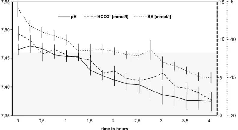
Changes in the acid-base balance of are illustrated inFig 4. The pH stayed stable within the reference range for 4 hours (P= 0.063). The sodium bicarbonate buffer (HCO3−13.4 mmol/l, median) and base excess (BE–10.3, median) base values were reduced due to hemodilution and further decreased significantly during the 4-hour period (HCO3−5.1 mmol/l, BE
–18.2,
P= 0.031 after 4 hours).
Although steadily increasing from 4.34 to 5.61 mmol/l (P= 0.031), potassium levels were kept well within the reference range for Fischer rats (3.8–6 mmol/l [20]). As could be expected following hemodilution, hemoglobin base levels were significantly decreased (5.1 ± 2.3 mg/dl) in comparison with the reference values (14.9 ± 1.3 [20]). Since the composition of the perfus-ate was not changed, hemoglobin levels remained constant throughout the 4-hour perfusion period (P= 0.563).
Assessment of Flap Perfusion
All 32 recipient rats survived the postoperative period and tolerated the anaesthesia and opera-tive procedure well. Autocannibalism was observed to different degrees in all laboratory ani-mals in group C2 (ischemia), but in no other study group. After commencing extracorporeal Fig 3. Changes in the blood gases during extracorporeal perfusion.The use of a membrane oxygenator yielded high oxygen levels (SO2) and oxygen
partial pressures (pO2). Carbon dioxide partial pressure (pCO2) decreased continuously during the first 4 hours of extracorporeal perfusion. The error bars
indicate the standard error.
perfusion (ECP), a healthy pink color returned to the flap, and a capillary refill could be observed in all cases. These signs of flap viability could be maintained throughout the 8-hour perfusion interval. ICG angiography confirmed perfusion of the skin island flaps in groups C3 and ECP during the perfusion period. Weight gain of extracorporeally perfused flaps was 10% during the perfusion interval as opposed to 0% in thein vivoperfused flaps.
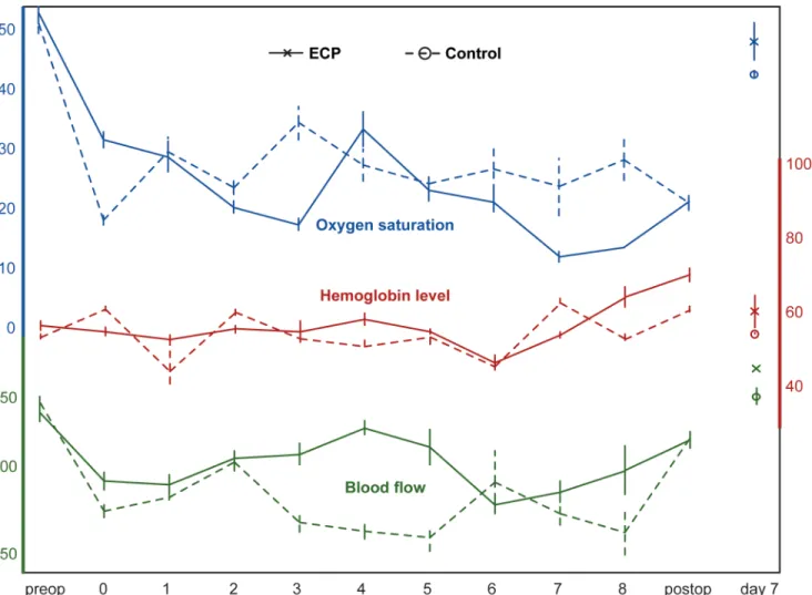
O2C results are depicted inFig 5and Tables3and4. The most comprehensive data were aquired in groups ECP and C3. Because of the analogous experimental setting, the comparison between these two groups also seems the most interesting to pursue. The curve progressions in both groups were remarkably similar. After flap raise and initiation of perfusion, a temporary decrease in oxygen saturation, blood flow and velocity was observed regardless of the perfusion method (extracorporeal orin vivo) in comparison with the initial values (before flap raise). In the further course of the perfusion, laser spectrophotometric values remained stable. Hemoglo-bin levels remained constant around 60 AU for 6 hours in both groups. Between hour 6 and 8, Hb raised slightly in both groups, but not statistically significant (ECPP= 0.375, K3
P= 0.999). At day 7, all laser spectrophotometric values were comparable with initial values in both groups. Due to high interindividual fluctuations of the method [21], no statistical compar-isons between groups was performed. In the sham group (C1), as could be expected, all values were comparable to initial values at day 7, with the exception of an elevated hemoglobin level (P= 0.008). Intraoperative measurements were not performed in this group. While after 8-hour ischemia (group C2) only the oxygen saturation was significantly decreased (P= 0.008), all laser spectrophotometric values had significantly decreased by day 7.
Fig 4. Changes in the acid-base balance during extracorporeal perfusion.Sodium bicarbonate buffer (HCO3–), base excess (BE) and pH-values all
showed a steady decrease during the first 4 hours of extracorporeal perfusion. The pH stayed within the reference range without substitution (reference range 7.33–7.46, according to Bakeret al. [19], grey area). The error bars indicate the standard error.
Assessment of Flap Viability at Day 7
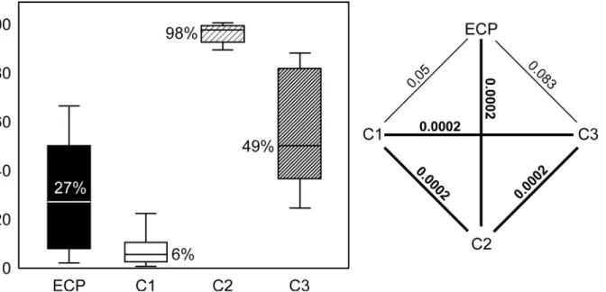
With 98%, 8-hour ischemia (C2) was associated with the highest rate of necrosis. Lowest rates were observed in in the sham group (C1), where only 6% of the transplanted tissue became necrotic. The rate of necrosis after 8-hour extracorporeal perfusion (27%) was significanly lower (P= 0.0002) than in group C2, while the difference between extracorporeal perfusion and both sham operation (P= 0.05) andin vivoperfusion (49%,P= 0.083) was not statistically different. All rates andPvalues are presented inFig 6.
Structural analysis corroborated the clinical findings and the planimetric assessment of the rate of necrosis. Clinically healed flaps showed regular tissue architecture with preservation of endothelial integrity and without degeneration of skin, skin appendages, fatty tissue, blood ves-sels, muscle or lymph nodes, while necrotic areas showed typical signs of degeneration like dis-ruption of the epithelium and fatty necrosis (Fig 7). Histologically, no signs of inflammation or bacterial contamination were observed.
Discussion
The objective of this study was to develop an extracorporeal perfusion device for the prolonged vital storage of microvascular free flaps that should serve as a fundamental research tool for the
Fig 5. Combined laser Doppler flowmetry and remission spectroscopy.Depicted are the mean (superficial) laser spectrophotometric values for oxygen saturation (in percent), blood flow (in AU) and hemoglobin levels (in AU) over the experimental course. Measurements were conducted preoperatively (preop), after flap raise (0), during the course of the extracorporeal (group ECP) orin vivo(group C3) perfusion (1–8), postoperatively (postop) as well as at day 7. The error bars indicate the standard error.
Table 3. Laser spectrophotometric data.
SO2
Group ECP C1 C2 C3
Time 0 8h 7d 0 7d 0 8h 7d 0 8h 7d
n 8 8 6 8 8 8 8 8 8 8 5
Med 53 12 48 42 24 24 6 16 54 9 38
Min 35 5 24 18 2 11 2 0 22 5 34
Max 67 29 63 67 58 53 10 36 68 39 51
Hb
Group ECP C1 C2 C3
Time 0 8h 7d 0 7d 0 8h 7d 0 8h 7d
n 8 8 6 8 8 8 8 8 8 8 5
Med 56 66 58 53 61 71 79 64 55 60 55
Min 48 51 46 45 54 54 26 0 42 49 49
Max 75 87 83 65 70 78 94 91 70 74 64
Flow
Group ECP C1 C2 C3
Time 0 8h 7d 0 7d 0 8h 7d 0 8h 7d
n 8 8 6 8 8 8 8 8 8 8 5
Med 133 125 184 188 86 113 86 7 137 114 156
Min 57 33 128 116 44 56 53 0 77 34 133
Max 200 170 222 300 140 148 121 72 211 222 173
Velocity
Group ECP C1 C2 C3
Time 0 8h 7d 0 7d 0 8h 7d 0 8h 7d
n 8 8 6 8 8 8 8 8 8 8 5
Med 21 16 24 24 16 22 19 11 20 18 21
Min 12 10 19 20 11 17 14 0 18 13 18
Max 25 21 27 36 19 49 53 52 30 29 25
Descriptive statistics of (superficial) combined laser Dopplerflowmetry and remission spectroscopy (O2C) values [oxygen saturation (SO2in %),
hemoglobin level (Hb in AU), bloodflow (Flow in AU) and bloodflow velocity (Velocity in AU)] of groups ECP (extracorporeal perfusion), C1 (sham operation), C2 (ischemia) and C3 (in vivoPerfusion) at the time points preoperatively (0), 8 hours (8h) and at day 7 (7d). The data are presented as total number of measured values (n), median (Med), minimum (Min) and maximum (Max) values.
doi:10.1371/journal.pone.0147755.t003
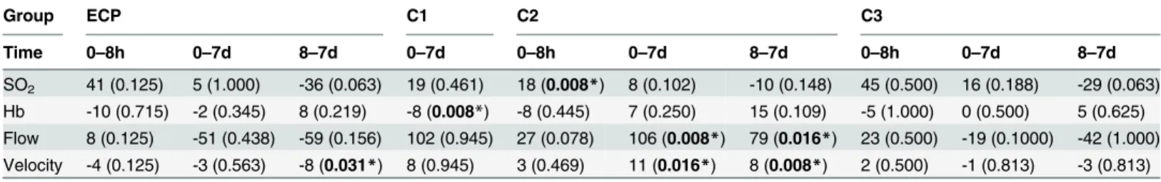
Table 4. Comparative statistics of laser spectrophotometric data.
Group ECP C1 C2 C3
Time 0–8h 0–7d 8–7d 0–7d 0–8h 0–7d 8–7d 0–8h 0–7d 8–7d
SO2 41 (0.125) 5 (1.000) -36 (0.063) 19 (0.461) 18 (0.008*) 8 (0.102) -10 (0.148) 45 (0.500) 16 (0.188) -29 (0.063)
Hb -10 (0.715) -2 (0.345) 8 (0.219) -8 (0.008*) -8 (0.445) 7 (0.250) 15 (0.109) -5 (1.000) 0 (0.500) 5 (0.625) Flow 8 (0.125) -51 (0.438) -59 (0.156) 102 (0.945) 27 (0.078) 106 (0.008*) 79 (0.016*) 23 (0.500) -19 (0.1000) -42 (1.000) Velocity -4 (0.125) -3 (0.563) -8 (0.031*) 8 (0.945) 3 (0.469) 11 (0.016*) 8 (0.008*) 2 (0.500) -1 (0.813) -3 (0.813)
Comparative statistical assessment of (superficial) combined laser Dopplerflowmetry and remission spectroscopy (O2C) values [oxygen saturation (SO2
in %), hemoglobin level (Hb in AU), bloodflow (Flow in AU) and bloodflow velocity (Velocity in AU)] of groups ECP (extracorporeal perfusion), C1 (sham operation), C2 (ischemia) and C3 (in vivoPerfusion). Wilcoxon’s test was used for statistical analysis between different time points [preoperatively (0), 8 hours (8h) and at day 7 (7d)];Pvalues<0.05 were considered statistically significant (*).
investigation of extracorporeal free flap perfusion. The rat epigastric flap was chosen as animal model, a fascio-lipo-cutaneous flap, we have gained experience with in the past [22–24]. We report the successful transplantation of rat epigastric flaps after 8-hour extracorporeal perfu-sion. After 8-hour ECP and subsequent transfer, the majority of free flaps had healed in by day 7 with only minor partial necrosis (27%). Notably, ECP yielded even better results thanin vivo
perfusion (49%), although not statistically significant (P= 0,083). Blood flow throughin vivo
perfused flaps may have been temporarily compromised by low systemic blood pressure during deep anaesthesia and by vasospasms caused by manipulation of the pedicle vessels during flap raise or vessel preparation. The principle of extracorporeal perfusion, on the other hand, allows specifying exactlywhen,what, andhow much of itenters a flap–without influencing the rest of the organism, and without being influenced by the rest of the organism. In our study, the use of an oxygenator and a controllable pump set at high physiological pressure and flow rates pro-vided a constant flow of oxygenated blood to the raised flaps. Perfusate temperature and per-fusate composition (heparinized, diluted blood) may also have helped to improve results in the ECP group: While maintaining the blood flow through the existing vascular network, oxygen consumption of the perfused flaps was reduced by allowing the perfusate to cool to room tem-perature (21 ± 1°C). In accordance with the law of Hagen-Poiseuille, dilution further improved the rheological properties of the perfusate. Finally, short-termex vivoperfusion of free flaps withheparinizedblood, as used in our study, is known to reduce the effect of ischemia-/ reper-fusion damage [25]. Even though it remains unclear if these factors can be successfully trans-ferred to long-term extracorporeal tissue perfusion, our results clearly indicate that short-term ECP can have a positive effect on flap viability.
Histological assessment confirmed the clinical observations, showing regular tissue architec-ture and intact vessel endothelium as well as intact epithelium in extracorporeally perfused flaps after one week. Considering the high success rate in the presented study, the cellular dam-age observed in other studies after prolonged perfusion [26–28] was either less substantial in our study or at least partially reversible.
Fig 6. Rate of necrosis.Left: median percentage of necrotic area in groups ECP (8-hour extracorporeal perfusion), C1 (sham operation), C2 (8-hour ischemia) and C3 (8-hourin vivoperfusion). Right: graphical representation of statistical differences between the study groups. All significant (<0.05) and
highly significant (<0.001)Pvalues are printed bold. Kruskal-Wallis test followed by Mann Whitney U test were used for statistical analysis.
Continuous evaluation of both perfusate and free flaps provided important insights on the pathophysiological processes during extracorporeal tissue perfusion. With initiation of the ECP, a clear capillary refill was observed. Venous return started about a minute later and the dark color of the blood impressively indicated the lower oxygen level of the venous return. As an indication for a maintained metabolism [29,30], arterio-venous difference was 33.7% in our study (arterial 98.9%, venous 65.2%) and remained constant throughout the perfusion interval. These results are substantiated by the O2C. After an initial decrease after flap raise, O2C val-ues remained constant throughout the 8-hour ECP and reached base valval-ues 1 week after flap transplantation. The observed initial decrease, however, was not statistically significant. The temporal change of O2C values during extracorporeal perfusion was remarkably similar toin vivoperfused flaps (Fig 4), indicating that the probable causes for the initial decrease of O2C values were vasospasms or a changed vascular anatomy of the flaps after flap raise rather than an issues directly related to ECP. Except for group C2 (ischemia), all O2C values reached base values in all groups after revascularization had occurred by day 7. Draguet al. [31] also observed a decrease of tissue oxygenation during extracorporeally perfused bovine rectus abdo-minis flaps, regardless of the perfusate used (crystalloid solution, whole blood). Since no active oxygenation was used in their study, however, oxygen levels were barely measurable (0–2%), underlining the importance of an active oxygenation [31].
Since no influence was taken on the composition of the perfusate, any changes in the com-position are representative for the pathophysiological and biochemical changes inside the flap.
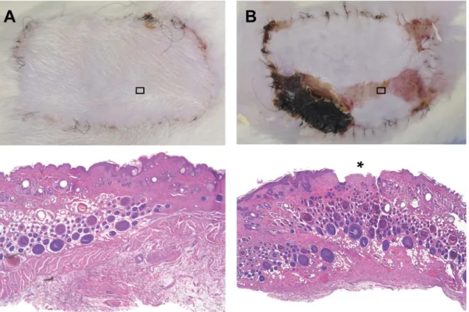
Fig 7. Clinical and histological images.(A) Completely healed in flap 7 days after 8-hour extracorporeal perfusion. Below: Histological image taken from the center of the same flap with normal tissue architecture and regular epithelium. (B) Partially necrotic epigastric flap 7 days after 8-hour extracorporeal perfusion. Below: Histologic image taken from an area with clinically apparent epitheliolyisis with histological disruption of the epithelium (*). HE staining, 30x magnification.
Analogous to Constantinescuet al. [8], constantly high oxygen saturations and oxygen partial pressures were measured during ECP despite significant hemodilution and the use of ambient air (21% oxygen) instead of pure oxygen. Apart from vasospasms and altered anatomy after flap raise, the discrepancy between constantly high oxygen values in theperfusateand a decreased oxygen saturation in the perfusedflapsmay be explained by the physical relationship between temperature and hemoglobin oxygen binding capacity: Hypothermia increases the affinity of hemoglobin to bind oxygen (hence the high oxygen levels in the perfusate) but also leads to a decreased tendency to release oxygen to the tissue, which may explain the decreased tissue oxygenation. Hypothermia decreases oxygen consumption and energy turnover in living tissues and is therefore commonly used for the vital storage of organs and tissues [8,32]. Like most authors who used whole blood for tissue perfusion [25,28,31,33–35], we allowed the perfusate temperature to drop to room temperature. The high success rate in our study suggests that the reduced oxygen delivery could at least partially be compensated by the simultaneous decrease of metabolic activity in the tissue. However, further scientific effort is needed to illu-minate the effect of temperature during extracorporeal tissue perfusion.
The high quality of the perfusion in our study was further confirmed by high, constant pH level and potassium levels that stayed within normal range throughout the measurement period. (Metabolic) Acidosis [36–38] and high potassium levels [8,38] are common signs of hypoxia observed during ECP. In our study, no supplementation of sodium bicarbonate [36–
38] was necessary to avoid acidosis and neither insulin nor glucose had to be administered to decrease potassium levels. Despite normal pH and potassium levels in our study, corbon diox-ide partial pressures, bicarbonate and base excess, however, decreased significantly during per-fusion. This acid-base inbalance, referred to asrespiratorically compensated metabolic acidosis, is usually caused by lactate acidodis in the course of ischemia- and hypoxia-related reduced perfusion. As the results from the preliminary study showed, lactate (and LDH) levels both increased significantly due to progressive hemolysis. It seems safe to assume that intravasal hemolysis has played a decicive role in the development of the observed acid-base dysbalance.
During extracorporeal tissue perfusion, basic bodily functions like respiration, removal of metabolites, acid-base balance and temperature regulation have to be maintained by a decou-pled circulation system. Apart from the“mechanical organs”(oxygenator, pump), theperfusate
pump system for the perfusion of small animal organs and composite tissues of which the authors claim it would lead to less mechanical damages to erythrocytes in comparison with conventional roller pumps. However, no data are presented with regard to hemolysis and it remains unclear on what scientific basis these assumptions were made. Although kidney perfu-sion was clearly in the focus of their study, the authors achieved to perfuse 5 out of 7 rabbit epi-gastric fat flaps over a period of 5 days. In 2 cases, excessive bleeding occurred in the first 24 hours and perfusion was terminated. In contrast to our study, however, Worner et al. did not attempt to re- or transplant the perfused tissue, so the clinical success of the perfusion can ulti-mately not be confirmed.
Perspectives
Despite numerous issues that still need to be addressed in further studies, the phenomenon of a decoupled circulation does offer some compelling advantages, like improved monitoring capa-bilities, flap conditioning by intermittend perfusion and the possibility to manipulate various pyhsiological aspects by changing pressure, flow or perfusate composition. Moreover, local administration of drugs, hormons or gene transfer becomes possible, without influencing the body’s circulation. This opens a wide field of new exciting clinical applications including pro-longed vital storage (as addressed in this study), bridging time to transplant [11], saving labora-tory animals [41], local chemotherapy in sarcomas [42] or melanomas [43], tissue engineering [44,45] or simulation of realistic preparation conditions for the training of reconstructive sur-geons [12]. Born out of necessity, we would like to address yet another theoretical clinical application of extracorporeal tissue perfusion. Founded on the knowledge that transplanted composite tissues eventually become independent from the supplying vessels [24,34,46], tem-porary ECP of transplanted free flaps may hold the key to free composite tissue transplantation without anastomosis. This would potentially allow complex reconstructions even in otherwise desolate cases, like in the irradiated, vessel depleted neck.
Conclusions
This study constitutes an important input to the understanding of the processes in living com-posite tissues during extracorporeal perfusion and offers a validated experimental model for further research. With this experimental setup, successful replantation after 8-hour real perfusion of rat epigastric flaps was possible. Notably, results after temporary extracorpo-real perfusion were superior toin vivoperfusion. This observation may be explained by maintaining a constant flow of oxygenated, heparinized blood to the raised flaps while provok-ing a hypothermia-induced downregulation of the flaps’oxygen consumption. Extracorporeal tissue perfusion remains a highly complex procedure, influenced by a myriad of paramters like the different ischemia tolerance of different tissues, the choice of perfusate, optimal oxygen/ carbondioxide ratio, perfusion pressures and temperature. The investigation of these funda-mental questions, however, is exacly what the described perfusion system and animal model was developed for.
Supporting Information
S1 Fig. Planimetric measurement of rate of necrosis.Upper row: Step-by-step assessment of the rate of necoris. ImageJ was used to assess the total flap area (in pixels), as well as the area of all viable parts of the flap and all necrotic parts. Dividing the pixel values (necrotic area/ total flap area100%) results in the percentage area of necrosis (= rate of necrosis).Lower row:
operation (C1), 8-hour ischemia (C2), 8-hourin vivoperfusion (C3)). (TIF)
S2 Fig. Indocyanine green angiography during extracorporeal perfusion.(A) Rat in supine position with femoral catheter (fc) in place. The asterisk (
) marks the reference area with max-imal indocyanine green (ICG) intensity. (B) ICG was administered directly into the venous res-ervoir, circulated through the tubing system, passed the membrane oxygenator (om) and accumulated in the arterial reservoir (ar). (C) Extracorporeally perfused epigastric flap (ef) with high ICG-signal in artery (a), vein (v) and throughout the whole skin island of the flap, indicating maintained flap perfusion. ICG is metabolized in the liver (li), which leads to a high ICG signal in this area.
(TIF)
Author Contributions
Conceived and designed the experiments: TM AMF. Performed the experiments: AMF TM AB LR. Analyzed the data: AMF AB. Contributed reagents/materials/analysis tools: PBL. Wrote the paper: AMF LMR TM KDW. Designed and developed the extracorporeal perfusion units: MH TM KDW AMF.
References
1. Carrel A, Lindbergh CA. The Culture of Whole Organs. Science. 1935; 81(2112):621–3. doi:10.1126/ science.81.2112.621PMID:17733174.
2. Hosgood SA, Nicholson ML. The first clinical case of intermediate ex vivo normothermic perfusion in renal transplantation. Am J Transplant. 2014; 14(7):1690–2. Epub 2014/05/13. doi:10.1111/ajt.12766 PMID:24816186.
3. Hosgood SA, Nicholson ML. First in man renal transplantation after ex vivo normothermic perfusion. Transplantation. 2011; 92(7):735–8. Epub 2011/08/16. doi:10.1097/TP.0b013e31822d4e04PMID: 21841540.
4. Nicholson ML, Hosgood SA. Renal transplantation after ex vivo normothermic perfusion: the first clini-cal study. Am J Transplant. 2013; 13(5):1246–52. Epub 2013/02/26. doi:10.1111/ajt.12179PMID: 23433047.
5. Blaisdell FW. The pathophysiology of skeletal muscle ischemia and the reperfusion syndrome: a review. Cardiovascular surgery. 2002; 10(6):620–30. Epub 2002/11/28. PMID:12453699.
6. Mayer B, von Baeyer H, Kaiser U. New dimensions for the vital storage of microsurgical free flaps: an experimental approach. Otolaryngol Head Neck Surg. 1993; 109(4):690–2. PMID:8233505.
7. Mayer B, Kaier U, Pundrich C, de Veer I, Lajous-Petter A- M, Kaempfer L. Die Entwicklung eines auton-omisch gesteuerten Lappencontainers zum Direkttransfer und zur warmen extrakorporalen Langzeit-konservierung freier, mikrochirurgischer Lappen, abgetrennter Extremitäten und isolierter Organe. Oto-Rhino-Laryngologia Nova. 1994; 4(4):190–3.
8. Constantinescu MA, Knall E, Xu X, Kiermeir DM, Jenni H, Gygax E, et al. Preservation of Amputated Extremities by Extracorporeal Blood Perfusion; a Feasibility Study in a Porcine Model. J Surg Res. 2010. Epub 2010/05/11. doi:10.1016/j.jss.2010.01.040PMID:20451920.
9. Dragu A, Birkholz T, Kleinmann JA, Schnurer S, Munch F, Cesnjevar R, et al. Extracorporeal perfusion of free muscle flaps in a porcine model using a miniaturized perfusion system. Arch Orthop Trauma Surg. 2010. Epub 2010/12/29. doi:10.1007/s00402-010-1251-8PMID:21188393.
10. Taeger CD, Friedrich O, Dragu A, Weigand A, Hobe F, Drechsler C, et al. Assessing viability of extra-corporeal preserved muscle transplants using external field stimulation: a novel tool to improve meth-ods prolonging bridge-to-transplantation time. Sci Rep. 2015; 5:11956. doi:10.1038/srep11956PMID: 26145230; PubMed Central PMCID: PMCPMC4491708.
11. Worner M, Poore S, Tilkorn D, Lokmic Z, Penington AJ. A low-cost, small volume circuit for autologous blood normothermic perfusion of rabbit organs. Artificial organs. 2014; 38(4):352–61. Epub 2013/08/29. doi:10.1111/aor.12155PMID:23981068.
publication of the European Association for Cranio-Maxillo-Facial Surgery. 2014. doi:10.1016/j.jcms. 2014.04.004PMID:24938642.
13. Ritschl LM, Fichter AM, Haberle S, von Bomhard A, Mitchell DA, Wolff KD, et al. Ketamine-Xylazine Anesthesia in Rats: Intraperitoneal versus Intravenous Administration Using a Microsurgical Femoral Vein Access. J Reconstr Microsurg. 2015; 31(5):343–7. doi:10.1055/s-0035-1546291PMID: 25702886.
14. Boehm O, Zur B, Koch A, Tran N, Freyenhagen R, Hartmann M, et al. Clinical chemistry reference data-base for Wistar rats and C57/BL6 mice. Biol Chem. 2007; 388(5):547–54. doi:10.1515/BC.2007.061 PMID:17516851.
15. Wolfensohn S, Lloyd M. Handbook of Laboratory Animal—Management and Welfare. 3. Auflage ed. Oxford, United Kingdom: Blackwell Publishing Ltd.; 2003.
16. Strauch B, Murray DE. Transfer of composite graft with immediate suture anastomosis of its vascular pedicle measuring less than 1 mm. in external diameter using microsurgical techniques. Plast Reconstr Surg. 1967; 40(4):325–9. Epub 1967/10/01. PMID:4863084.
17. Hölzle F, Rau A, Loeffelbein DJ, Mücke T, Kesting MR, Wolff KD. Results of monitoring fasciocuta-neous, myocutafasciocuta-neous, osteocutaneous and perforator flaps: 4-year experience with 166 cases. Inter-national journal of oral and maxillofacial surgery. 2010; 39(1):21–8. Epub 2009/12/01. doi:10.1016/j. ijom.2009.10.012PMID:19944567.
18. Rasband WS, inventorImageJ1997-2009.
19. Baker HJ, Lindsey R, Wesibroth SH. The Laboratory Rat: Biology and Diseases. London: Academic Press Inc.; 1979. 116 p.
20. Charles. Baseline Hematology and Clinical Chemistry Values for Charles River Fischer 344 Rats-CDF1(F-344)CrlBR as a Function of Sex and Age: Charles River; 1984 [cited 2014 22.08.2014]. Fa. Charles River, Kißlegg]. Available from:http://www.criver.com/techdocs/84jan_tb/t84tab04.htm12/19/ 2003.
21. Hölzle F, Loeffelbein DJ, Nolte D, Wolff KD. Free flap monitoring using simultaneous non-invasive laser Doppler flowmetry and tissue spectrophotometry. Journal of cranio-maxillo-facial surgery: official publi-cation of the European Association for Cranio-Maxillo-Facial Surgery. 2006; 34(1):25–33. Epub 2005/ 12/14. doi:10.1016/j.jcms.2005.07.010PMID:16343915.
22. Mücke T, Borgmann A, Fichter AM, Wagenpfeil S, Mitchell DA, Ritschl LM, et al. The influence of differ-ent VEGF administration protocols on the perfusion of epigastric flaps in rats. The British journal of oral & maxillofacial surgery. 2013; 51(6):555–62. doi:10.1016/j.bjoms.2012.09.007PMID:23041105.
23. Fichter AM, Borgmann A, Ritschl LM, Mitchell DA, Wagenpfeil S, Dornseifer U, et al. Perforator flaps—
how many perforators are necessary to keep a flap alive? The British journal of oral & maxillofacial sur-gery. 2014; 52(5):432–7. doi:10.1016/j.bjoms.2014.02.013PMID:24629454.
24. Mücke T, Borgmann A, Wagenpfeil S, Gunzinger R, Nobauer C, Lange R, et al. Autonomization of epi-gastric flaps in rats. Microsurgery. 2011. Epub 2011/04/20. doi:10.1002/micr.20892PMID:21503975.
25. Cooley BC, Tadych KL, Gould JS. Perfusion of free flaps with heparinized whole blood during ischemic storage. J Reconstr Microsurg. 1990; 6(1):49–53. Epub 1990/01/01. doi:10.1055/s-2007-1006802 PMID:2308128.
26. Yabe Y, Ishiguro N, Shimizu T, Tamura Y, Wakabayashi T, Miura T. Morphologic and metabolic study of the effect of oxygenated perfluorochemical perfusion on amputated rabbit limbs. J Reconstr Micro-surg. 1994; 10(3):185–91. Epub 1994/05/01. doi:10.1055/s-2007-1006586PMID:8071906.
27. Taeger CD, Muller-Seubert W, Horch RE, Prabst K, Munch F, Geppert CI, et al. Ischaemia-related cell damage in extracorporeal preserved tissue—new findings with a novel perfusion model. J Cell Mol Med. 2014; 18(5):885–94. Epub 2014/03/19. doi:10.1111/jcmm.12238PMID:24636195.
28. Dragu A, Kleinmann JA, Taeger CD, Birkholz T, Schmidt J, Geppert CI, et al. Immunohistochemical evaluation after ex vivo perfusion of rectus abdominis muscle flaps in a porcine model. Plast Reconstr Surg. 2012; 130(2):265e–73e. Epub 2012/07/31. doi:10.1097/PRS.0b013e3182589c2dPMID: 22842423.
29. Mayer B, von Baeyer H, Kaier U. Warme Vitalkonservierung freier, mikrochirurgischer Lappen über ein extrakorporales Kreislaufsystem. Oto-Rhino-Laryngologia Nova. 1992; 2(4):212–4. doi:10.1159/ 000312849
30. Mayer B. [Significance of extracorporally perfused microsurgical free flaps]. Laryngo- rhino- otologie. 2002; 81(9):640–3. doi:10.1055/s-2002-34452PMID:12357412.
32. Greaney PJ Jr, Cordisco M, Rodriguez D, Newberger J, Legatt AD, Garfein ES. Use of an extracorpo-real membrane oxygenation circuit as a bridge to salvage a major upper-extremity replant in a critically ill patient. J Reconstr Microsurg. 2010; 26(8):517–22. Epub 2010/08/11. doi:10.1055/s-0030-1262951 PMID:20697991.
33. Fowler JD, Li X, Cooley BC. Brief ex vivo perfusion with heparinized and/or citrated whole blood enhances tolerance of free muscle flaps to prolonged ischemia. Microsurgery. 1999; 19(3):135–40. Epub 1999/05/07. PMID:10231122.
34. Maeda M, Fukui A, Tamai S, Mii Y, Miura S. Extracorporeal circulation for tissue transplantation (in the case of venous flaps). Plast Reconstr Surg. 1993; 91(1):113–24; discussion 25–6. Epub 1993/01/01. PMID:8416516.
35. Li X, Cooley BC, Gould JS. Ex vivo perfusion with anticoagulated blood decreases ischemia/reperfu-sion injury. The Journal of hand surgery. 1993; 18(4):629–34. doi:10.1016/0363-5023(93)90306-N PMID:8349970.
36. Domingo-Pech J, Garriga JM, Toran N, Rusinol M, Girvent F, Rosines D, et al. Preservation of the amputated canine hind limb by extracorporeal perfusion. International orthopaedics. 1991; 15(4):289–
91. Epub 1991/01/01. PMID:1809705.
37. Usui M, Sakata H, Ishii S. Effect of fluorocarbon perfusion upon the preservation of amputated limbs. An experimental study. The Journal of bone and joint surgery British volume. 1985; 67(3):473–7. Epub 1985/05/01. PMID:3997959.
38. Müller S, Constantinescu MA, Kiermeir DM, Gajanayake T, Bongoni AK, Vollbach FH, et al. Ischemia/ reperfusion injury of porcine limbs after extracorporeal perfusion. J Surg Res. 2013; 181(1):170–82. doi:10.1016/j.jss.2012.05.088PMID:22748598.
39. Hassanein WH, Zellos L, Tyrrell TA, Healey NA, Crittenden MD, Birjiniuk V, et al. Continuous perfusion of donor hearts in the beating state extends preservation time and improves recovery of function. The Journal of thoracic and cardiovascular surgery. 1998; 116(5):821–30. Epub 1998/11/07. PMID: 9806389.
40. Mayer B, Kaempfer L. Der freie mikrochirurgische obere muskulokutane Trapezius-Lappen des Schweins: Ein ideales Trainingsmodell fìr mikrovaskuläre Rekonstruktionen und ein In-vitro-Modell für die experimentelle Mikrochirurgie. Laryngo-Rhino-Otologie. 1996; 75:175–7. PMID:8652035
41. Mayer B. Einsparung von Tierversuchen mit einem extrakorporalen Kreislaufsystem zur Vitalerhaltung von isolierten Gewebeblöcken. Der Tierschutzbeauftragte. 1998; 1:67–9.
42. Van Ginkel RJ, Van Berlo CL, Baas PC, Koops HS, Stuling RV, Elstrodt J, et al. Hyperthermic Isolated Limb Perfusion with TNF alpha and Cisplatin in the Treatment of Osteosarcoma of the Extremities: A Feasibility Study in Healthy Dogs. Sarcoma. 1999; 3(2):89–94. doi:10.1080/13577149977703PMID: 18521269; PubMed Central PMCID: PMC2395417.
43. McBride CM. Perfusion treatment for malignant melanoma of the extremity. Archivum chirurgicum Neerlandicum. 1970; 22(2):91–5. PMID:5449409.
44. Sekine H, Shimizu T, Sakaguchi K, Dobashi I, Wada M, Yamato M, et al. In vitro fabrication of functional three-dimensional tissues with perfusable blood vessels. Nat Commun. 2013; 4:1399. doi:10.1038/ ncomms2406PMID:23360990; PubMed Central PMCID: PMCPMC3660653.
45. Tee R, Morrison WA, Dusting GJ, Liu GS, Choi YS, Hsiao ST, et al. Transplantation of engineered car-diac muscle flaps in syngeneic rats. Tissue Eng Part A. 2012; 18(19–20):1992–9. doi:10.1089/ten. TEA.2012.0151PMID:22793168; PubMed Central PMCID: PMCPMC3463279.