Oxygen saturation and lactate concentration
gradient from the right atrium to the pulmonary
artery in the immediate postoperative following
cardiac surgery with extracorporeal circulation
INTRODUCTION
Several reports suggest that critically ill patients exhibit signiicant diferences
between their blood oxygen saturation (SaO2) values measured from blood
samples drawn either from the right atrium or from the pulmonary artery. he diferences span a range from 5 - 7%.(1-3) It was suggested that the discordance
may be explained by assuming that, in the right atrium of those patients, the blood is partially “de-saturated” by the very low oxygen content coming from the coronary sinus.(1) At the same time, the blood lactate concentration [Lac] of
coronary venous blood is very low because of the high rate of consumption by the myocardium in normal and even pathological situations, i.e., sepsis.(4) he
opposite occurs in conditions such as myocardial ischemia, where the glucose consumption is greater than the lactate consumption. Hence, in the ischemic condition, the concentration of lactate in the coronary sinus may be higher, Juan Carlos Pendino1,2, Leonardo Hess3, Sergio
Beltrame4, Gonzalo Aldamiz-Echevarría Castillo4, John Trujillo4
1. Unidad de Terapia Intensiva, Hospital Provincial del Centenario - Rosario, Argentina. 2. Facultad de Ciencias Medicas, Universidad Nacional de Rosario - Rosario, Argentina. 3. CIMA-Profisio, Facultad de Ciencias Medicas, Universidad Nacional de Rosario - Rosario, Argentina.
4. Servicio de Cirugía Cardíaca, Hospital IDCSalud - Albacete, España.
Objective: his prospective study aimed to characterize the changes in blood lactate concentration and blood oxygen saturation in patients during the immediate postoperative period of cardiac surgery with extracorporeal circulation.
Methods: Blood samples were
collected from 35 patients in a rapid and random order from the arterial line and from the proximal and distal port of a pulmonary artery catheter.
Results: he results showed no
statistically signiicant diferences between the blood oxygen saturation in the right atrium (72% ± 0.11%) and the blood oxygen saturation in the pulmonary artery (71% ± 0.08%). he blood lactate concentration in the right atrium was 1.7mmol/L ± 0.5mmol/L, and the blood lactate concentration in
Conflicts of interest: None.
Submitted on May 23, 2016 Accepted on March 27, 2017
Corresponding author: Juan Carlos Pendino Unidad de Terapia Intensiva Hospital Provincial del Centenario Rosario 2000, Argentina.
E-mail: [email protected]
Responsible editor: Gilberto Friedman
Gradiente de saturação de oxigênio e concentração de lactato
entre átrio direito e artéria pulmonar no pós-operatório imediato
de cirurgia cardíaca com circulação extracorpórea
ABSTRACT
Keywords: Oxygen/metabolism;
Oxygen consumption/physiology; Lactate; Postoperative period; horacic surgery; Extracorporeal circulation the pulmonary artery was 1.6mmol/L ± 0.5mmol/L (p < 0.0005).
Conclusion: he diference between the blood lactate concentration in the right atrium and the blood lactate concentration in the pulmonary artery might be a consequence of the low blood lactate concentration in the blood from the coronary sinus, as it constitutes an important substrate for the myocardium during this period. he lack of diferences between the blood oxygen saturation in the right atrium and the percentage of blood oxygen saturation in the pulmonary artery suggests a lower oxygen extraction by the myocardium given a lower oxygen consumption.
although the metabolic substrate is highly dependent on the degree of ischemia.(5)
In a group of critically ill patients, [Lac] and SaO2 are lower in the blood of the pulmonary artery than in the
blood of the right atrium.(6) he samples were obtained
through the proximal port (i.e., the right atrium) and distal port (i.e., the pulmonary artery) of a pulmonary
artery catheter (PAC).(7) Accordingly, this comparison
between the blood oxygen saturation in the right atrium (SraO2) and percentage of blood oxygen saturation in the
pulmonary artery (SpaO2) along with the comparison
between the blood lactate concentration in the right
atrium [Lac]ra and blood lactate concentration in the
pulmonary artery [Lac]pa has been used as a prognostic
factor in critically ill patients.(6)
he metabolic state of the heart can be dramatically modiied in conditions such as the postoperative period of cardiac surgery with extracorporeal circulation,
exhibiting changes in oxygen consumption (VO2) and
lactate metabolism. hese modiications have already been
highlighted by measurements of lactate, SaO2 and other
substrates in the blood from the coronary sinus.(8)
he present study was designed to evaluate these blood parameters sampled simultaneously using the two ports of a pulmonary artery catheter, giving access to blood from the pulmonary artery and from the right atrium.
he aim of this study to characterize the changes in [Lac]
and SaO2 in patients during the immediate postoperative
period of cardiac surgery with extracorporeal circulation.
METHODS
his prospective study was performed in the intensive care unit (ICU) from Hospital Idcsalud Albacete (Albacete, Spain). After approval from the Institutional Review Board of hospital (approval number 11/10), 35 patients were enrolled. he inclusion criteria were admission to the ICU shortly after cardiac surgery with extracorporeal circulation, age older than 18 years, PAC and arterial peripheral catheter insertion in the operating room. he exclusion criteria included uncorrected valve dysfunction and intracardiac communication. Informed consent was waived.
In all cases, the cardioplegia used was hematic with mild hypothermia (32°C), and the decision to use antegrade or retrograde cardioplegia depended on the type of surgery performed.
During the admission to the ICU, the medical team immediately veriied the correct positioning of the PAC. his was done by checking that a wedge pressure tracing
was obtained when the balloon was inlated with a volume between 0.5 and 0.8mL. Afterwards, the balloon was delated, and the normalization of the pulmonary artery pressure tracing was veriied. In addition, the right atrial pressure tracing was veriied to be obtained when the proximal port of the pulmonary artery catheter was connected to the transducer. Lastly, an X-ray examination conirmed that the catheter tip was at the pulmonary hilum, at a distance no more than 2cm from the cardiac silhouette.
Immediately after checking the pressure tracing, blood samples were drawn in a rapid succession and randomly from the arterial catheter and from the proximal and distal ports of the PAC. Under these conditions, blood drawn from the proximal port was assumed to be representative of right atrium blood, whereas distal port blood was regarded as representative of pulmonary artery blood. To avoid contamination of the blood with the continuous washing solution, the irst 2mL was discarded for the PAC, as well as the initial 5mL for the arterial line.
Samples for blood gas assessment were extracted in syringes speciic for this purpose (Pulsettm Westmed, Tucson, AZ, USA). Blood samples for assessing [Lac] were extracted using ad hoc tubes with sodium luoride and potassium oxalate. he blood samples were processed immediately. he oxygen saturation was measured using
the Nova Biomedical Phox Plus®
analyzer (Waltham, MA, USA). Lactate determination was performed with a standard clinical laboratory instrument (Dade Behring
Dimension®
RxL analyzer) (Deerield, IL, USA). he routine laboratory tests for the immediate postoperative control include hematocrit, white blood count, coagulation tests, blood glucose, plasma urea, plasma creatinine, serum sodium, serum potassium, serum chloride, serum magnesium, serum phosphorus, plasma creatine kinase (CK), plasma CK-MB and plasma troponin.
Statistical comparisons
Paired Student’s t-test was used to compare atrial versus
pulmonary artery measurements. Spearman correlation analysis was performed to compare [Lac]ra and [Lac]pa. he method of Bland & Altman was used to investigate the efect of [Lac] on the diferences between pairs of
observations. he relation between Δ[Lac] and ΔSaO2 and
other hemodynamic parameters (cardiac output, double
product, LVSWI, DO2, VO2 and O2ER) was analyzed
by employing the Spearman correlation test. Data are shown as the mean ± standard deviation (SD). he level of statistical signiicance was set at p < 0.05.
RESULTS
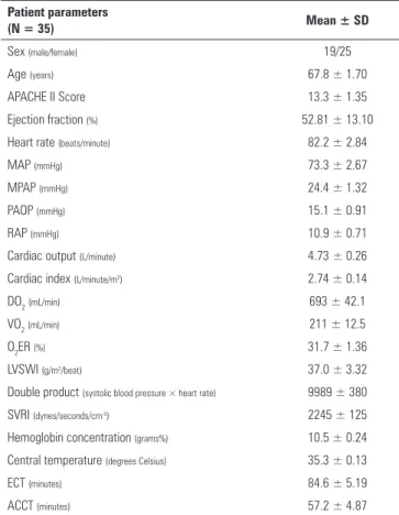
A group of 35 patients, 19 males and 16 females, aged 67.7 ± 10 years, was enrolled in the study. he preoperative left ventricular ejection fraction was 52.81% ± 13.1%. Ten of 35 patients underwent coronary artery bypass graft (CABG), 8 patients received valve prostheses at the mitral position (MVR), 10 patients received valve prostheses at the aortic position (AVR), 4 patients underwent concomitant MVR + AVR, 2 patients were subjected to simultaneous CABG + MVR, and the remaining patient received a Bentall prosthesis. he time elapsing between the aortic unclamping blood sample collection and hemodynamic measurements in the ICU was 59.4 minutes ± 11.2 minutes. Hemodynamic parameters, Hb values, cardiopulmonary bypass time, central temperature and demographics data are shown in table 1.
here were no statistically signiicant diferences between the SraO2 and SpaO2. he [Lac]ra was higher than
[Lac]pa (p < 0.0005) with a gradient of 0.1mmol/L ±
0.2mmol/L (Table 2). here was no signiicant correlation between Δ[Lac] and any of the following parameters:
cardiac output, double product, LVSWI, DO2, VO2 and
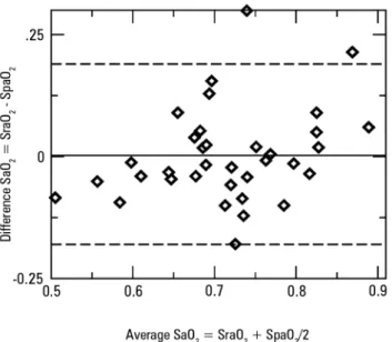
O2ER. he Bland & Altman test for SO2 and [Lac] showed a bias of 0.00061 (95%CI -0.185169 to 0.186391) and 0.1 for [Lac]ra (95%CI -0.25092 to 0.50092), respectively (Figures 1 and 2). Analysis on the relationship between the preoperative ejection fraction and age, extracorporeal circulation time, ICU stay, DO2, VO2 and IEO2, remained insigniicant.
DISCUSSION
Our irst working hypothesis was that there should
not be a SaO2 gradient between the blood of the right
atrium and pulmonary artery because there is expected to be a low oxygen extraction by the myocardium during
Table 1 - Demographic and hemodynamic parameters
Patient parameters
(N = 35) Mean ± SD
Sex (male/female) 19/25
Age (years) 67.8 ± 1.70
APACHE II Score 13.3 ± 1.35
Ejection fraction (%) 52.81 ± 13.10
Heart rate (beats/minute) 82.2 ± 2.84
MAP (mmHg) 73.3 ± 2.67
MPAP (mmHg) 24.4 ± 1.32
PAOP (mmHg) 15.1 ± 0.91
RAP (mmHg) 10.9 ± 0.71
Cardiac output (L/minute) 4.73 ± 0.26
Cardiac index (L/minute/m2) 2.74 ± 0.14
DO2(mL/min) 693 ± 42.1
VO2(mL/min) 211 ± 12.5
O2ER (%) 31.7 ± 1.36
LVSWI (g/m2/beat) 37.0 ± 3.32
Double product (systolic blood pressure × heart rate) 9989 ± 380
SVRI (dynes/seconds/cm-5) 2245 ± 125
Hemoglobin concentration (grams%) 10.5 ± 0.24
Central temperature (degrees Celsius) 35.3 ± 0.13
ECT (minutes) 84.6 ± 5.19
ACCT (minutes) 57.2 ± 4.87
SD - standard deviation; APACHE II - Acute Physiology and Chronic Health Evaluation II; MAP - mean arterial pressure; MPAP - mean pulmonary artery pressure; PAOP - pulmonary artery occlusion pressure; RAP - right atrial pressure; DO2 - oxygen delivery; VO2 - oxygen
consumption; O2ER - oxygen extraction index; LVSWI - left ventricular stroke work index;
SVRI - systemic vascular resistance index; ECT - extracorporeal circulation time; ACCT - aortic cross-clamp time.
the immediate postoperative period. Alternatively, there should be a gradient of lactate between the blood from the right atrium and the pulmonary artery because the myocardium, under these conditions, would be able to use additional lactate as the preferential substrate.
Two main indings are in line with our initial hypothesis. he irst one is the presence of a gradient in the concentration of lactate in paired samples obtained from the proximal and distal ports. he second one addresses the lack of diferences between SraO2 and SpaO2.
Table 2 - Oxygen saturation and lactate concentration of paired right atrium and pulmonary artery blood samples
Right atrium blood Pulmonary artery blood Gradient
(Δ right atrium - pulmonary artery)
SaO2 (%) 71.15 ± 1.88 71.09 ± 1.43 0.103 ± 1.59
[Lac] mmol/L 1.772 ± 0.1148 1.647 ± 0.1114* 0.125 ± 0.032
SaO2 - blood oxygen saturation; [Lac] - blood lactate concentration; * p < 0.001 when comparing atrial to mixed venous blood by paired t-test. Averages ± standard deviation.
Figure 1 - Bland & Altman plot comparing oxygen saturation in the right atrium and oxygen saturation pulmonary artery. SaO2 - blood oxygen saturation; SraO2 - oxygen saturation in the right atrium; SpaO2 - oxygen saturation pulmonary artery.
Figure 2 - Bland & Altman plot comparing lactate concentration in right atrium and lactate concentration in pulmonary artery. [Lac]ra - lactate concentration in right
atrium; [Lac]pa - lactate concentration in pulmonary artery. * Superposition of patients with similar values
corresponding to lactic acid measurements.
the myocardium oxidizes more glucose and less free fatty acids than in preoperative period and 6 hours after CABG surgery. When the aortic cross clamp is removed, the myocardium might extract more high energy substrates, presumably relecting its accumulation during
extracorporeal circulation.(8) his immediate period
following separation from extracorporeal circulation is accompanied by a hyperemic response evidenced by a progressive increase in coronary blood low and, furthermore, a decrease in the oxygen extraction ratio due to a limited ability to use oxygen.(8,9) In addition, the
lactate extraction ratio increases progressively from the time of release of the aortic cross-clamp.(8)
It seems sensible to expect a decrease in the myocardial oxidative activity as the protection exerted by hypothermic cardioplegia. his situation then allows for the accumulation of lactate in the context of myocardial oxygen deprivation. Because lactate can be easily metabolized to pyruvate; the former may be then the preferred substrate for aerobic metabolism after recovery post cardiac arrest, when surgery is accomplished. However, we are not able
to explain how the myocardium may use lactate without a corresponding increase in oxygen utilization. Sobrosa et al.(10) found positive gradients between arterial blood
lactate and blood from the coronary sinus, concomitantly
with a minimum diference between the SO2 of arterial
blood and the blood from the coronary sinus in patients undergoing CABG with cardiopulmonary bypass at the time of reperfusion.
hese changes may partly be explained by the transient depression of the myocardial capacity of extraction, described during early reperfusion.(11)
he variables analyzed by our group are the macrocirculatory ones assessing the overall metabolic status of tissues such as the myocardium, but it is unknown what occurs at the cellular level.
he presence of a cytoplasmic-to-mitochondrial lactate shuttle in the heart allows glycolysis to progress to lactate without the adverse consequence of acidosis or altered redox.(12) In isolated rat hearts with ischemia-reperfusion
was MCT isoform 1 expression during the early stages of reperfusion. Increased MCT4 expression may facilitate lactate extrusion during the ischemic period, while increased MCT1 may favor lactate transport into and out of cells simultaneously during early reperfusion.(13)
Rao et al. observed(14) that the persistence in the release
of lactate by the coronary sinus during reperfusion suggests a late recovery in myocardial aerobic metabolism likely related to inadequate protection during cardiopulmonary bypass as well as to impaired functional contractility of the heart and low cardiac output syndrome.
Caution should be exerted when comparing to the study by Gutierrez et al.,(7) in which blood samples were
obtained at diferent times, when it was decided that a PAC was necessary to guide luid therapy. Our study appears more homogeneous in terms of the underlying process and the time point at which the hemodynamic and laboratory tests were performed.
Right atrial blood is a mixture of superior vena cava and inferior vena cava blood. It is possible that the blood from the superior vena cava and inferior vena cava had not fully mixed at the proximal port of the PAC, and if so, the mixture of blood may have occurred distally to the proximal PAC sampling port. An answer to this question can only be achieved by direct measurement of [Lac] from the inferior vena cava inferior vena cava to the pulmonary artery.
Only four patients from our series had [Lac]pa higher
than [Lac]ra. his may be due to technical errors or
catheter position, or because they experienced myocardial ischemia. In this regard, the [Lac] in the coronary sinus was high because this condition was associated with the release of lactate by the coronary sinus serving to explain diferences. In these 4 patients who underwent heart valve replacement, there was no evidence of myocardial ischemia, in light of the usual methods for detecting ischemia at the bedside. Notably, 3 of them required a temporary pacemaker because they exhibited conduction disturbances during the immediate postoperative period. One of these four patients required norepinephrine at the end of surgery to maintain an adequate MAP, being the only one in need of vasoactive drugs among the total population.
Notably, the device employed for lactate measurements has a precision of ± 0.09mmol/L. Hence, when assuming a systematic instrumental bias, the diference in [Lac]
between the right atrium and the pulmonary artery would have remained statistically signiicant.
We found no diferences in the SaO2 between the right atrium and pulmonary artery, although the Bland-Altman test indicated that both variables were not interchangeable. he wide 95% limits of agreement between central venous
and pulmonary artery SaO2 might be accounted for by
dissimilar individual behaviors.
As above stated, subsequent to the release of the aortic cross-clamp, there may be a decrease in oxygen extraction by the myocardium, most likely related to mitochondrial dysfunction.
he oxygen heart demand is closely related to the
myocardial work.(4) he double product (an indirect
measure of VO2 by the myocardium) and LVSWI were
within normal ranges in our patient series.
Moreover, the total body metabolic demand is decreased during the immediate postoperative period of patients
undergoing hypothermic cardiopulmonary bypass,(15)
relected in a reduction in systemic VO2 and carbon
dioxide production. hese indings are related to deep sedation, mechanical ventilation and mild hypothermia.
Hence, the lack of diference between SraO2 and SpaO2
may be related to lower myocardial oxygen consumption in these patients.
CONCLUSION
Diferences were found in the blood lactate concentration between the right atrium and the pulmonary artery. his may be due to a low-lactate blood supply from the coronary sinus, in turn suggesting that the myocardium may preferentially use lactate as a substrate in that situation. he lack of diference between the blood oxygen saturation in the blood of the right atrium and pulmonary artery may be explained by a lower myocardial oxygen extraction.
ACKNOWLEDGMENT
Objetivo: Caracterizar as modiicações na concentração sanguínea do lactato e da saturação de oxigênio em pacientes no pós-operatório imediato de cirurgia cardíaca com circulação extracorpórea.
Métodos: Foram coletadas amostras de sangue de 35 pa-cientes, de forma rápida e aleatória, do acesso arterial e das por-tas proximal e distal de um cateter pulmonar.
Resultados: Não foram veriicadas diferenças estatistica-mente signiicantes entre saturação de oxigênio no átrio direito (72% ± 0,11%) e na artéria pulmonar (71% ± 0,08%). A con-centração sanguínea de lactato no átrio direito foi de 1,7mmol/L ± 0,5mmol/L, enquanto na artéria pulmonar esta concentração foi de 1,6mmol/L ± 0,5mmol/L (p < 0,0005).
Conclusão: A diferença entre as concentrações sanguíneas de lactato no átrio direito e na artéria pulmonar pode ser con-sequência da baixa concentração de lactato no sangue do seio coronário, já que o lactato é um importante substrato para o miocárdio durante este período. A ausência de diferenças entre saturação sanguínea de oxigênio no átrio direito e na artéria pul-monar sugere extração de oxigênio mais baixa pelo miocárdio, em razão do menor consumo de oxigênio.
RESUMO
Descritores: Oxigênio/metabolismo; Consumo de
oxigê-nio/isiologia; Lactato; Período pós-operatório; Cirurgia toráci-ca; Circulação extracorpórea
REFERENCES
1. Chawla LS, Zia H, Gutierrez G, Katz NM, Seneff MG, Shah M. Lack of equivalence between central and mixed venous oxygen saturation. Chest. 2004;126(6):1891-6.
2. Edwards JD, Mayall RM. Importance of the sampling site for measurement of mixed venous oxygen saturation in shock. Crit Care Med. 1998;26(8):1356-60.
3. Reinhart K, Kuhn HJ, Hartog C, Bredle DL. Continuous central venous and pulmonary artery oxygen saturation monitoring in the critically ill. Intensive Care Med. 2004;30(8):1572-8.
4. Dhainaut JF, Huyghebaert MF, Monsallier JF, Lefevre G, Dall’Ava-Santucci J, Brunet F, et al. Coronary hemodynamics and myocardial metabolism of lactate, free fatty acids, glucose, and ketones in patients with septic shock. Circulation. 1987;75(3):533-41.
5. Stanley WC, Lopaschuk GD, Hall JL, McCormack JG. Regulation of myocardial carbohydrate metabolism under normal and ischaemic conditions. Potential for pharmacological interventions. Cardiovasc Res. 1997;33(2):243-57.
6. Gutierrez G, Chawla LS, Seneff MG, Katz NM, Zia H. Lactate concentration gradient from right atrium to pulmonary artery. Crit Care. 2005;9(4):R425-9. 7. Gutierrez G, Comignani P, Huespe L, Hurtado FJ, Dubin A, Jha V, et al.
Central venous to mixed venous blood oxygen and lactate gradients are associated with outcome in critically ill patients. Intensive Care Med. 2008;34(9):1662-8.
8. Pietersen HG, Langenberg CJ, Geskes G, Kester A, de Lange S, Van der Vusse GJ, et al. Myocardial substrate uptake and oxidation during and after routine cardiac surgery. J Thorac Cardiovasc Surg. 1999;118(1):71-80.
9. Hskanson E, Svedjeholm R, Vanhanen I. Physiologic aspects in postoperative cardiac patients. Ann Thorac Surg. 1995;59(2 Suppl):S12-4. 10. Sobrosa CG, Jansson E, Kaijser L, Bomfim V. Myocardial metabolism after
hypothermic retrograde continuous blood cardioplegia with anterograde warm cardioplegic induction. Braz J Cardiovasc Surg. 2005;20(4):416-22. 11. Vanky FB, Hakanson E, Szabó Z, Jorfeldt L, Svedjeholm R. Myocardial
metabolism before and after valve replacement for aortic stenosis. J Cardiovasc Surg (Torino). 2006;47(3):305-13.
12. Brooks GA, Brown MA, Butz CE, Sicurello JP, Dubouchaud H. Cardiac and skeletal muscle mitochondria have a monocarboxylate transporter MCT1. J Appl Physiol. 1999;87(5):1713-8.
13. Zhu Y, Wu J, Yuan SY. MCT1 and MCT4 expression during myocardial ischemic-reperfusion injury in the isolated rat heart. Cell Physiol Biochem. 2013;32(3):663-74.
14. Rao V, Ivanov J, Weisel RD, Cohen G, Borger MA, Mickle DA. Lactate release during reperfusion predicts low cardiac output syndrome after coronary bypass surgery. Ann Thorac Surg. 2001;71(6):1925-30. 15. Sladen RN. Temperature and ventilation after hypothermic cardiopulmonary