ISOLATION
OF SEROTYPE HARDJO AND OTHER
LEPTOSPIRAE
FROM ARMADILLOS
IN ARGENTINA’
Donald M. My-er~,~ Albert0 Cuba Capam,
and Jaime Payan Moreno’
Thirteen Pathogenic Leptospira interrogans strains and two saprophytic Leptospira biflexa strains were isolated from 89 armadillos (Chaetophractus villosus) in Argentina. This is the first time that isolation and identification of serotype hardjo
from this animal species has been reported. Serologic,cultural, and histopathologicstudiesperformed in the course of this work indicate that the armadillo is an important reservoir-host for lefitospirae in Argentina.
Introduction
Wildlife populations widely infected with leptospires are an important source of leptospirosis in domestic animals and man. The epidemiologic importance of such carrier-hosts results from their ability to contaminate the environment with urine containing large numbers of infective organisms. In this regard, continually changing ecological conditions have made it necessary to search out new host-reservoirs for ever increasing numbers of known pathogenic Le@tospira interrogans sero- types.
In Argentina, the armadillo Chaeto- phractus villosus is abundant. The grass- land portions of the country that constitute its habitat are used extensively for raising cattle. The possible role of this armadillo as a potential natural reservoir for leptospires was first demonstrated by Cacchione et al.
*Also appearing in BoE Of Sanit Panam 83(l), 1977. ‘Leptospirosis and Pathology Units, Pan American Zoonoses Center, Pan American Health Organiza- tion, Casilla 3092, Correo Central, Buenos Aires, Argentina.
‘Colombian Agriculture and Livestock Institute, Cali, Colombia.
(1) through isolation of a canicola strain. Subsequently, Szyfres et al. (2) reported identifying and isolating two serogroup Bataviae serotypes (paidjan and argenti- niensis) from armadillos; one of these serotypes (paidjan) was also obtained from a cow.
A more recent armadillo study by Carillo et al. (3) obtained 24 leptospiral isolates from 438 captured animals. All of these leptospires proved to belong to Bataviae group serotypes argentiniensis, paidjan, or bataviae. However, examination of sera from the armadillos showed that their predominant antibodies were specific for the antigenically distinct Hebdomadis sero- group. Related serologic surveys of Argen- tine cattle have also found predominant agglutinins to the Hebdomadis group (4); these latter surveys made no cultural isola- tions. A later study, carried out on Argentine cattle by Myers and Jelambi (I) using improved culture techniques, suc- ceeded in isolating six serotype hardjo strains of the Hebdomadis group from bovine kidneys. Taken together, these findings suggested that the armadillo could be either a reservoir or a maintaining host for bovine leptospirosis.
The armadillo
Chaetophmctus zrihbsus.
The purpose of the present study was to
examine
armadillos
captured
from selected
cattle farms with
culture
techniques
ade-
quate for isolating
all leptospira
serotypes-
including
serotype hardjo.
Histopathologic
studies were also carried out on those arma-
dillos from which isolates were obtained,
in
an effort to further
determine
whether
the
armadillo
is an important
chronic
natural
reservoir
for leptospires
or is merely
an
accidental
host.
Materials and Methods
The 89 armadillos
examined
in this study
were hand-captured
on four cattle-raising
farms in the central area of the Province
of
Buenos Aires (Partido
de Azul).
They were
captured
in 13 batches of 6 to 10 animals
each from March
to July 1975.
Blood for serologic
testing was collected
from each animal by cardiac puncture.
The
animals were then killed
by ether anesthe-
sia, necropsied,
and examined
for gross
pathology.
An entire kidney of each animal
was removed
aseptically;
one portion
was
used for cultural
isolation
of leptospires,
and the rest was fixed in 10 per cent neutral
formalin
and embedded in paraffin.
Tissue
sections were cut and stained
using
both
conventional
staining
methods
(for histo-
pathologic
study) and Warthin-Starry
silver
stain (6) for detection
of leptospires.
Serologic
Testing
The sera were subjected to the microscop-
ic-agglutination
(MA)
test procedure
de-
scribed by Galton
et al. (7). The antigens
used in this
test were
live
Leptospira
interrogans
serotypes
grown
in
Stuart
(Difco)
liquid
medium
containing
10 per
cent pooled
normal
rabbit
serum.
The
specific
strains
used
were
as follows:
Pomona
strain
Pomona,
australis
strain
Ballico,
ballurn strain Castellon
3, Grippe-
typhosa
strain
Moskva
V, tarassoui
strain
Perepelicin,
hebdomadis
strain
H ebdoma-
dis, batauiae strain
Van Tienen,
canicola
strain
Hond
Utrecht
IV,
hardjo
strain
Hardjoprajitno,
wolffi
strain 3705, paidjan
strain
Paidjan,
argentiniensis
strain
LT
1019, Pyrogenes
strain
Salinem,
ictero-
haemorrhagiae
strain
RGA,
sejroe
strain
M 84, and autumnalis
strain Akiyami
A.
Myers et al. l LEPTOSPIRE ISOLATION FROM ARMADILLOS 133
Serum titers equalling or exceeding 1:50 were considered seropositive in this study. Although serum titers under 1:lOO are not routinely considered seropositive, negative MA titers were found for some animals from which leptospires were isolated-a finding which justified considering sera with lower titers positive.
Culture Procedures
For culture isolation attempts, kidney tissue was triturated and suspended in nine parts of sterile phosphate-buffered saline (pH 7.2). Serial ten-fold dilutions of the tissue suspensions were then made with the sterile buffered saline, and 0.5 ml of each 10e2 to 10m4 dilution was used to inoculate duplicate tubes to which 5.0 ml of Fletcher semisolid medium (containing 10 per cent pooled rabbit serum) had been added. The same dilutions of renal tissue were also used to inoculate duplicate tubes containing 5.0, ml of bovine albumin-polysorbate 80 (BA-
P80) medium-a medium using bovine
fraction V albumin (Pentex) that was pre- pared as described by Johnson and co-work- ers (8, 9).
obtained to make them satisfactory for use as antigen.
These leptospiral isolates were then tested against a battery of prepared hyperimmune rabbit antisera to pathogenic and saprophy- tic Le#tospira serotypes in order to deter- mine the serogroups of the isolates. Specific antisera were prepared for each isolate in accord with the methods of Galton et al. (7), and definitive identification was made of representative strains using the cross- agglutination-absorption procedures of Kmety et al. (II).
Results
Serologic examination of sera from 89 armadillos showed that 42 (47.1 per cent) had antibody titers of at least 1:50. Thirty-one of these positively reacting sera (34.8 per cent of all sera collected) yielded MA titers ranging between 1:lOO and 1:12,800 to one or more of the 16 leptospiral screening antigens. The 42 positive reac- tions and the predominant leptospiral agglutinins involved are shown in Table 1. As indicated, the most frequent reactions occurred against serotypes of the Hebdoma- For contamination control, other sets df, dis and Bataviae groups, which together cultures were made by exposing a 10-Z
saline dilution of renal tissue to the combined action of both neomycin and furazolidone (at a final concentration of 25
pg/ml each) for one hour prior to culture (IO), after which lo-’ and 10s4 dilutions were made in antimicrobial-free saline. A portion (0.5 ml) of each dilution was then added to two tubes of Fletcher and two tubes of BA-P80 semisolid media.
accounted for 78.6 per cent of the serologi- tally positive animals.
Cultures were incubated at 30°C and examined weekly for eight weeks by darkfield microscopy to detect leptospires. When growth was detected, successive transfers were made in semisolid media until growth was sufficiently abundant for adaptation to liquid media. Further sub- cultures were made in Stuart or BA-P80 liquid media until sufficient growth was
Leptospires were isolated from renal tissues of 15 armadillos (16.8 per cent of those examined). Reciprocal cross-aggluti- nation patterns against antisera to the different leptospiral serogroups showed that three of the isolates belonged to the Canicola group. In cross-agglutination- absorption tests these strains proved to be serologically homologous to serotype canico- la. Nine of the other isolates were identified as members of the Bataviae group, and their similar cross-reaction patterns with type strains showed them to be antigenically identical to serotype paidjan, serotype
argentiniensis, or serotype bataviae.
Table 1. Prevalence of predominant leptospiral se.rwn agglutinins in 89 armadillos captured on four cattle farms in Argentina.
Antigen I:50 l&O
Distribution of titersa Total positive sera 1:200 1:800 1:3,200 1:12,800
1:k”oo l:$OO 1:;:oo O”W Or No. %
Hebdomadis groupb 14 4 1 19 21.3
Bataviae group’ 2 3 4 4 1 14 15.7
Serotype canicola 1 1 2 2.2
Serotype fiomona 1 1 1.1
Miscellaneousd 6 6 6.7
Total 24 7 6 4 1 42 47.1
aReciprocal of the highest dilution showing 50 per cent or more agglutination. bHebdomadis group antigens: hardjo, wolffi, sejroe, and hebdomadis. cBataviae group antigens: argentiniensis, paidjan, and bataviae. dMultiple reactions to two or more antigens at the same titer.
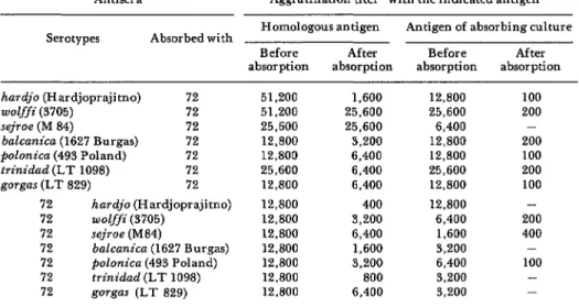
serogroup. Reciprocal-agglutination-ab- sorption tests were performed with this iso- late against various Hebdomadis group serotypes showing strong agglutinin activi- ty. The results of these tests are presented in Table 2. On the basis of current taxonomic criteria, which limit residual titers to 10 per cent or less of the pre-absorption homo-
logous titers, the tests show that this isolate is serologically homologous to serotype hardjo.
The remaining two isolates did not react with any of the leptospiral antisera and showed characteristics compatible with saprophytic strains of the species [email protected]$ira
biflexa.
Table 2. Results of cross-agglutination-absorption tests with the
L@os@n isolate from armadillo No. 72 and selected serotypes of the Hebdomadis group.
Antisera Agglutination titer” with the indicated antigen Serotypes Absorbed with Homologous antigen Antigen of absorbing culture
Before After Before After absorption absorption absorption absorption hardjo (Hardjoprajitno) 72 51,200 1,600 12,800 100 wolffi (3705) 72 51,200 25,600 25,600 200 sejroe (M 84) 72 25,600 25,600 6,400 - balcanica (1627 Burgas) 72 12,800 3,200 12,800 200 polonica (493 Poland) 72 12,800 6,400 12,800 100 trinidad (LT 1098) 72 25,600 6,400 25,600 200 gorgas (LT 829) 72 12,800 6,400 12,800 100 72 hardjo (Hardjoprajitno) 12,800 400 12,800 - 72 wolffi (3705) 12,800 3,200 6,400 200 72 sejroe(M84) 12,800 6,400 1,600 400 72 balcanica (1627 Burgas) 12,800 1,600 3,200 - 72 polonica (493 Poland) 12,800 3,200 6,400 100 72 trinidad (LT 1098) 12,800 800 3,200 - 72 gorgas (LT 829) 12,800 6,400 3,200 - =Reciprocal of the highest dilution showing 50 per cent or more agglutination.
Myers et al. l LEPTOSPIRE ISOLATION FROM ARMADILLOS 135
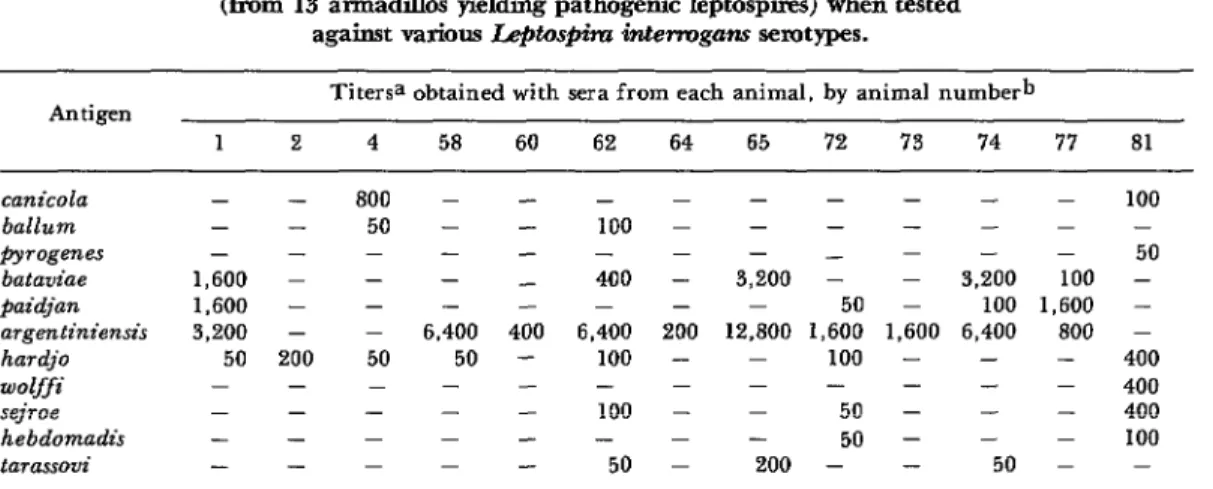
Table 3. Microscopic-agglutination titers obtained with Positive sera (from 13 armadihos yielding pathogenic Ieptospires) when tested
against various L#os@&rz interrogam semtypes.
Antigen
1
Titersa obtained with sera from each animal, by animal numberb
2 4 58 60 62 64 65 ‘72 ‘73 74 77 81 canida ballurn pyy7ogaes bataviae paidjan aTgf?ntiniensi~ hardjo wolffi sejroe hebdomadis tarassovi
- - 800 - - - - - 50 - - 100 - -
1,600 - - - _ 400 1,600 - - - - - 3,200 - - 6.400 400 6,400
50 200 50 50 - 100 100 - - -
- - - 50
- 3,200 -
- - 50
200 12,800 1,600 - - 100 - - - - - 50
50 - 200 -
- - - 100 - -
- 50 - 3,200 100 - - 100 1,600 - 1,600 6,400 800 - - - - 400 - - - 400 - - - 400 - - - 100 - 50 - - aTiters expressed as reciprocal serum dilution against antigen.
bAnimals with Canicola group strains: 2, 4, 81: with Bataviae group strains: 1, 58, 60, 62. 64, 65, 73, 74, 77: and with the Hebdomadis group strain hardjo: 72. No data are shown for two serologicaliy negative animals yielding strains of L. biflexa. No reactions occurred with the following L. interrogans antigens: autumnalis, grippotyphosa, Pomona, austnzlis, and icterohaemorrhagiae.
Of the 1.3 animals from which pathogenic
L. interrogans serotypes were isolated, 12 showed corresponding homologous group serum agglutinins with MA titers as high as 1:12,800 (see Table 3).
Auto&sy Findings
Macroscopic examination of the kidneys, heart, spleen and skeletal muscles of all the autopsied animals revealed no significant pathological alterations. Two armadillos from which leptospires were isolated had enlarged livers showing marked fatty meta- morphosis. Also, many of the animals had cyst-like formations 1 to 3 mm in diameter in their lungs. These latter incidental findings were diagnosed by microscopic examination to be unidentified parasitic nematode granulomas.
Histopathologic examination of the kid- neys from 11 of the 13 armadillos yielding pathogenic leptospiral isolates revealed aggregate alterations that permitted a diagnosis of interstitial nephritis. This characteristic interstitial nephritis was not observed in the two animals from which
Leptospira bz$exa strains were isolated. Lesions encountered in these two animals were scanty and consisted merely of hyaline casts in the collecting tubules plus limited deposits of material staining positively with periodic acid Shiff stain (PAS) around the glomerular tufts.
The diagnosis of interstitial nephritis was characterized by inflammatory lymphoid infiltration. This varied in intensity from slight to severe (see Plate 1). In all of the animals this lesion was accompanied by deposits of PAS positive material around the glomerular tufts and appeared more pro- nounced in the vicinity of the convoluted tubules. In animals whose inflammatory infiltration appeared more intense, these deposits formed thick layers around the wall of the Bowman’s capsules and convoluted tubules. Interstitial nephritis appeared intense in two animals, moderate in five, and only slight in four others.
Plate I-An
armadillo kidney section showing marked thickening of the Bowman’s
capsule (at left) and lymphoid interstitial infiltration
(at right). Stained with
hematoxilitxosin,
x 400.
Plate Z-Armadillo
kidney tissue treated with Warthin-Starry silver stain, showing
an abundance of leptospks within a convoluted tubule, x 1200.
Myers et al. l LEPTOSPIRE ISOLATION FROM ARMADILLOS 137
No evidence of interstitial nephritis was encountered in 60 kidneys from armadillos yielding no leptospiral isolates. However, lesions of minor pathological significance were observed in some of these animals. These lesions consisted mainly of hyaline casts and small areas of polymorphonuclear infiltration below the epithelium of the renal pelvis.
No histopathologic differences were ob- served with respect to differences in either the animals’ weight and sex or the infecting leptospiral strain.
Discussion
The findings in this study provide further evidence that a high percentage of armadillos in Argentina are infected with multiple leptospiral serotypes. A total of
16.8 per cent of the animals examined were found to have leptospires in their kidneys. Approximately 47 per cent of the sera from the captured animals showed predominant leptospiral agglutinins to serotypes of the Hebdomadis, Bgtaviae, Canicola, and Pom- ona sero-groups. This latter percentage was based upon our criterion that 50 per cent agglutination at a titer of 1:50 or more constituted a positive reaction. In this regard, it should be noted that serologic rates alone are not always valid indicators of the prevalence of infections in wildlife. For instance, when subjected to the MA test, sera from some animals may yield negative results for the same serotype with which the animals are actually infected.
The discovery of a high percentage of armadillos with Hebdomadis serogroup agglutinins in this study, similar to the findings reported by Carillo et al. (3), was interesting in view of that serogroup’s close ecological relationship to cattle. Reactor rates among Argentine cattle to Hebdomadis group antigens are known to be high, and six isolations of serotype hardjo have been made from bovine kidneys (5). The present study, employing improved culture meth-
ods, isolated serotype hardjo from one armadillo captured on a cattle farm. It is not possible at this time to assess the epidemiologic importance of this one isola- tion in relation to bovine leptospirosis; but the occurrence does represent the first known isolation of hardjo from this animal species, and shows the armadillo to be a host susceptible to infection with this serotype.
The significance of the isolation from armadillo renal tissues of two apparently saprophytic leptospiral strains having char- acteristics comparable to Leptospira biflexa
is unknown. Their appearance as possible contaminants is unlikely, since the kidneys were aseptically removed from the animals immediately before culture and the BA-P80 medium in which they grew had been prepared with distilled water and auto- claved. Furthermore, the Warthin-Starry silver staining technique detected lepto- spires in kidney sections from one of the two animals involved. Similar leptospires have previously been isolated from the frog by Diesch et al. (12) and from kidneys of apparently normal horses by Myers (13).
According to Turner (14) and Smith et al. (I$, a suitable carrier-host is one which is abundant, is easily infected, excretes lepto- spires, and still remains relatively unaffect- ed by the infection. In this vein, the efficiency of a carrier may be estimated by finding the ratio of the culture isolation rate to the positive serology rate. If the serology rate greatly exceeds the culture isolation rate, it would appear that infec-
tions are not persistent and that no leptospires are excreted for long periods of time. Using this criterion, the present study’s combined histopathologic findings of chronic nephritis and high rates of infection, based upon both serology and cultural isolations, suggest that the arma- dillo is an important natural reservoir-host for pathogenic leptospirae.
A serologic, bacteriologic, and histopathologic examination for leptospires was carried out on 89 armadillos (Chaetophractus villosus) from Ar _ gentina. Forty-seven per cent of the serum sam- ples yielded positive results when tested by micro-
scopic-agglutination. Predominant agglutina-
tion reactions were to the Hebdomadis and Bataviae serogroups.
A total of 15 Le$tosfiira isolations (from 16.8 per cent of the animals tested) w&e obtained from kidney tissue. Nine of the isolates were identified as belonging to the Bataviae group serotypes argentiniensis, @idjan, or batavitze; three other isolates proved to be the Canicola group serotype canicoZa; two others were Lefitosfiira biflexa strains; and the last isolate
was found to be serotype hardjo of the
Hebdomadis group. The latter finding repre- sents the first isolation of serotype hardjo from this animal species.
Histopathologic examination of kidneys from 11 of the animals yielding pathogenic leptospires
permitted a diagnosis of interstitial nephritis.
This interstitial nephritis, presenting the char- acteristic picture of lymphoid infiltration, appeared intense in two animals, moderate in five others, and only slight in the remaining four,
These histopathologic findings of chronic
nephritis, combined with the high positive sero- logic and cultural isolation rates, suggest that the
armadillo is an important natural reservoir-host for pathogenic leptospirae.
REFER.ENCE.3
(I) Cacchione, R. A., E. S. Cascelli, E. S.
Martinez, and J. M. Zuberbuhler. Leptospirosis
en animales silvestres: Aislamiento de una cepa de Lefitosfiiru cunicola de un peludo (Chaeto- phructus vill~sus). Revista de Znvestigaciones
Agropecuarias (Buenos Aires) 3:51-55, 1966.
(2) Szyfres, B., C. R. Sulzer, and M. M. Galton. A new leptospiral serotype in the Bataviae serogroup from Argentina. Trap Geogr
Med 19:344-346, 1967.
(3) Carillo, C. G., D. M. Myers, and B. Szyfres. Bataviae group leptospirae isolated from armadillos in Argentina. Tro$ GeogT Med
24:377-381, 1972.
(.4) Cacchione, R. Estado actual de la leptospi- rosis animal en la Republica Argentina. Revista de la Asociacio’n Midica Argentina 77133-39,
1963.
(5) Myers, D. M., and F. Jelambi. Isolation and identification of Leptospira hardjo from
cattle in Argentina. Trap Geog?’ Med 27:63-70,
1975.
(6) Sanders, B. J. Animal histology tech-
niques. In: E. C. Melby and N. H. Altman (eds.) Handbook of Laboratory Science. CRG Press, Cleveland, Ohio, p. 787, 1974.
(7) Galton, M. M., R. W. Menges, E. B. Shotts, A. J. Nahmias, and C. W. Heath. Lepto- spiTosis: Epidemiology, Clinical Manifestations in Man and Animals, and Methods in Luborato- ry Diagnosis. U. S. Public Health Service, Pub- lication No, 951, Washington, 1962.
(8) Johnson, R. C., and V. G. Harris. Dif- ferentiation of pathogenic and saprophytic leptospires. I. Growth at low temperatures. J
Bucteriol 94:27-31, 1967.
(9) Johnson, R. C., J. Walby, R. A. Henry, and N. E. Auran. Cultivation of parasitic leptospires: Effect of pyruvate. A# Microbial
Myers et al. l LEPTOSPIRE ISOLATION FROM ARMADILLOS 139
(10) Myers, D. M. Efficacy of combined furazolidone and neomycin in the control of contamination in Leptosfiira cultures. Anti-
micro6 Agents Chemother 7:666-671, 1975. (II) Kmety, E., M. M. Galton, and C. R. Sulzer. Further standardization of the agglu- tinin-absorption test in the serology of leptos- pires. Bull WHO 42~733-738, 1970.
(12) Diesch, S. L., W. F. McCulloch, J. L. Braun, and H. C. Ellinghausen. Leptospires isolated from a frog. Nature 209:939-940, 1966.
(13) Myers, D. M. Serological studies and isolations of serotype hardjo and Lefitospifa bifexa strains from horses of Argentina. J C&r
Microbial 3:548-555, 1976.
(14) Turner, L. H. Leptospirosis. Trans R Sot Trap Med Hyg 61:842-855, 1967.
(1.5) Smith, C.E.G., L. H. Turner, J. L. Harrison, and J , C. Broom. Animal leptospirosis in Malaya. I. Methods, zoogeographical back- ground, and broad analysis of results. Bull